|
Combined Educational & Scientific Session
Wednesday, 26 April 2017
| Room 315 |
16:15 - 18:15 |
Moderators: Arvind Pathak, Christopher Quarles |
Slack Channel: #s_cancer
Session Number: CE05
Overview
There is a dire need for new imaging tools for investigating the spatio-temporal heterogeneity of cancer as well as phenotypic alterations resulting from changes in the tumor microenvironment. MR methods are uniquely positioned to assess these factors non-invasively and in vivo. Therefore, this session will explore: (1) the role of imaging in assessing cancer heterogeneity, as well as (2) novel MR-based elastography methods to assess structural changes in cancer tissue. Both topics will be covered by leaders in their respective fields and will be followed by the presentation of the top-scoring scientific abstracts in each area.
Target Audience
Basic, translational and clinical researchers interested in expanding their knowledge on how to use state-of-the art MR-based approaches for assessing cancer heterogeneity and structure.
Educational Objectives
As a result of attending this course, participants should be able to:
-Explain the role of heterogeneity in cancer;
-Describe MRI methods that can be used to assess heterogeneity; and
-Assess the value of elastography in characterizing cancer tissue.
16:15
|
|
MR Elastography in Cancer 
Ralph Sinkus
|
16:45
 |
0972.
 |
Characterization of Renal Tumors: Integrating Biomechanical, Functional and Morphological Assessment Using 3 Tesla Magnetic Resonance Elastography
Davide Prezzi, Radhouene Neji, James Stirling, Sami Jeljeli, Hema Verma, Tim O'Brien, Benjamin Challacombe, Archana Fernando, Ashish Chandra, Vicky Goh, Ralph Sinkus
Incidentally detected renal tumors are overtreated surgically: up to 20% of them are benign, most frequently oncocytomas. We hypothesize that integrating biomechanical assessment with functional/morphological MRI can improve lesion characterization, precluding unnecessary surgery. Initial experience in 5 resected renal oncocytomas and 13 renal cell carcinomas (RCC) demonstrates that MR Elastography (MRE) at 30Hz with shear modulus parametric mapping is feasible and adds value within a multiparametric MRI assessment: oncocytoma displays higher median shear attenuation (α) and lower shear velocity (C) than RCC. MRE parameters appear to be stronger classifiers than quantitative DCE MRI parameters (Ktrans, kep), ADC and T2 signal intensity.
|
16:57
|
0973.
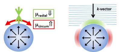 |
Using non-linear tissue biomechanics to infer static forces within tissue: towards quantifying IFP
Daniel Fovargue, Jack Lee, Adela Capilnasiu, Marco Fiorito, David Nordsletten, Ralph Sinkus
Quantifying static forces in the context of oncology, such as interstitial fluid pressure, would represent a valuable biomarker in therapy monitoring. Nonlinear tissue mechanics leads to distinct signatures of apparent anisotropic changes in mechanical shear properties in the vicinity of an object that exerts pressure onto its surroundings. Tissue nonlinearity can be modelled for instance via hyper elasticity. We show that the apparent modulation in tissue stiffness can be accounted for when incorporating the nonlinear anisotropic model into the estimation of the biomechanics via MR-Elastography. Knowledge of the deformation tensor enables direct quantification of underlying static forces, hence pressure.
|
17:09
 |
0974.
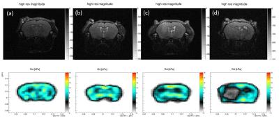 |
Monitoring Glioblastoma Progression in Mouse Brain with Magnetic Resonance Elastography
Navid Nazari, Michal Nowicki, Katharina Shregel, Sean Lawler, Ralph Sinkus, Paul Barbone, Samuel Patz
The longitudinal progression of glioblastoma was monitored in a cohort of mice with MRE and conventional RARE MRI. Increasing tumor size was easily seen with both modalities. In most cases, MRE maps showed a tumor margin that was sharper than RARE. Results were registered with histology, and variation of the shear modulus was compared with histology features. Both MRE and RARE demonstrated tumor regions with varying levels of heterogeneity, and in one animal, both homogeneous and heterogeneous parts were found to be growing separately as sub-populations of the same glioblastoma cell line within the brain.
|
17:21
|
|
Addressing Tumor Heterogeneity by Multi-Parametric MRI
Richard Carano
The presentation will focus on addressing tumor heterogeneity by a multi-parametric MRI approach.
|
17:51
|
0975.
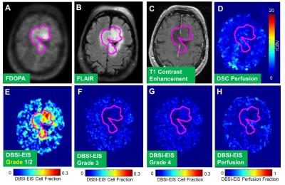 |
Quantitative characterization of brain tumor heterogeneity using diffusion basis spectrum imaging with extended isotropic spectrum (DBSI_EIS)
Qing Wang, Gloria Guzman, Yong Wang, Maria Ponisio, Yi Su, Pamela LaMontagne, Sheng-Kwei Song, Keith Rich, Sonika Dahiya, Jon McConathy, Tammie Benzinger
Tumors are typically heterogeneous, and may contain different grades of tumor cells, different types of tumor cells, edema and/or abnormal vascular structures. A noninvasive, non-radioactive technique to provide multiple parametric and quantitative images for better profiling the heterogeneity of tumors is highly needed. We demonstrated that diffusion basis spectrum imaging with extended isotropic spectrum (DBSI-EIS) is capable to identify structural heterogeneity in brain tumor lesions, including various grades of tumor cells and perfusion, which make it a new and unique technique to clinically evaluate tumors for comprehensive diagnosis and accurate treatment evaluation.
|
18:03
|
0976.
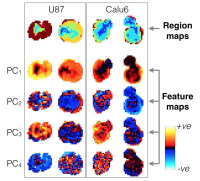 |
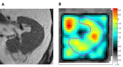
Automated identification of hypoxia-related regional variations in tumour microenvironment using dynamic contrast-enhanced MRI and oxygen-enhanced MRI
Adam Featherstone, James O'Connor, Ross Little, Yvonne Watson, Sue Cheung, Muhammad Babur, Victoria Tessyman, Roben Gieling, Kaye Williams, Julian Matthews, Geoff Parker
Hypoxia is an important prognostic indicator in most solid tumours. We present here automated, data-driven methods, using principal component analysis (PCA) and Gaussian mixture modelling (GMM), that consistently locate functionally distinct sub-regions in preclinical tumours, some of which are postulated to be relevant to hypoxia. Methods are based on dynamic contrast-enhanced (DCE)-MRI (reflecting perfusion) and oxygen-enhanced (OE)-MRI (reflecting oxygen delivery). We demonstrate the utility and stability of our methods through a combination of evaluation metrics, which may be incorporated in similar studies elsewhere.
|
|