|
Combined Educational & Scientific Session
ORGANIZERS: Stephan E. Maier, M.D., Ph.D & Jennifer A. McNab, Ph.D.
Thursday, 27 April 2017
| Room 315 |
15:30 - 17:30 |
Moderators: Petra Huppi, Christopher Kroenke |
Slack Channel: #s_diffusion
Session Number: CE09
Overview
Diffusion-related parameters in the maturing and aging brain.
Target Audience
The target audiences are: (1) clinicians who would like to know how ADC and FA change with age; (2) neuroscientists who would like to use diffusion imaging as a tool to analyze age-related changes in tissue diffusion; (3) image analysis experts who are interested in how to approach diffusion analysis in large subject cohorts with different age; and (4) physicists who would like to optimize diffusion imaging protocols for subject studies over a wide age range.
Educational Objectives
Upon completion of this course, participants should be able to:
-Identify diffusion parameters that change during brain maturation and aging and recall their typical changes;
-Plan suitable imaging protocols for studying brain maturation and aging; and
-Employ optimal image analysis tools to infer diffusion changes during brain maturation and aging.
15:30
|
|
Diffusion Imaging in Neurodevelopment 
C. Lebel
Diffusion imaging has been used extensively over the last decade or so to study healthy brain maturation during childhood and adolescence. Methods vary greatly across studies, but studies consistently report nonlinear maturation that continues into young adulthood, with the most protracted development occurring in frontal-temporal connections. These diffusion changes suggest increasing myelination, axonal packing, and/or coherence with age. Less consistent findings have been reported for specific timing of development (e.g., age at peak), and sex differences. Emerging new methods and large longitudinal or multi-site studies will greatly add to our understanding of brain development over the next few years.
|
16:00
|
|
Diffusion Imaging in Aging
Konstantinos Arfanakis
Diffusion MRI has an important role in research of the aging brain. Diffusion MRI can help elucidate the role of brain characteristics in the mechanisms supporting cognitive and motor health or leading to cognitive and motor decline in old age. Combining diffusion MRI with other imaging, clinical, neuropsychological, and most importantly neuropathologic information may provide invaluable insights towards the development of useful biomarkers of age-related diseases. To successfully accomplish the above, however, it is important to first realize the intricacies of conducting meaningful diffusion imaging investigations of the older adult brain.
|
16:30
|
1267.
 |
Differentiated maturation of white matter tracts in early developing brain aged 0-3 years
Qinlin Yu, Huiying Kang, Qinmu Peng, Minhui Ouyang, Michelle Slinger, Yun Peng, Fang Fang, Hao Huang
The brain development in the first several years after birth is perhaps most dynamic. However, the studies on white matter maturation of infants and toddlers with relatively evenly distributed ages in 0-3 years are rare. Here, we charted white matter development in subjects 0-3 years-of-age through measurements of DTI-derived metrics at the tract level and tract-group level. A 3-stage maturational pattern was revealed for all white matter tracts. The differentiated maturation among the white matter tracts and tract groups was found using DTI measurements.
|
16:42
 |
1268.
 |
Multi-shell neonatal brain HARDI template
Maximilian Pietsch, Jana Hutter, Anthony Price, Maria Kuklisova Murgasova, Emer Hughes, Johannes Steinweg, Nora Tusor, Jesper Andersson, Matteo Bastiani, Stamatios Sotiropoulos, Joseph Hajnal, J-Donald Tournier
We describe a method for creating a group template of the developing brain using advanced multi-shell high angular resolution diffusion (HARDI) data. We decompose the signal into an anisotropic CSF-like and a white matter-like directional component and build an unbiased template of those tissue types from 27 healthy term control babies acquired as part of the Developing Human Connectome Project (gestational age: 40.2+-1.4 weeks). This template will facilitate the analysis of microstructural features at a group level and allow longitudinal investigations into healthy and pathological brain maturation.
|
16:54
 |
1269.
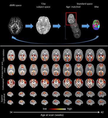 |
Automated pre-processing pipeline and quality control for neonatal diffusion MRI in the developing Human Connectome Project (dHCP)
Matteo Bastiani, Jesper Andersson, Lucilio Cordero-Grande, Maria Murgasova, Jana Hutter, Anthony Price, Antonios Makropoulos, Emer Hughes, Johannes Steinweg, Nora Tusor, Daniel Rueckert, A. David Edwards, Stephen Smith, Jacques-Donald Tournier, Joseph Hajnal, Saad Jbabdi, Stamatios Sotiropoulos
The developing Human Connectome Project (dHCP) is a collaborative 6-year project set to create a 4-dimensional map of structural and functional changes occurring throughout early development. Up to 1300 multi-modal MRI scans of foetuses and neonates (20 to 44 weeks gestational age) are currently being acquired. We present a fully automated pre-processing pipeline that allows us to efficiently analyse in-vivo diffusion MRI (dMRI) data despite the considerable technical challenges specific to neonatal imaging. We developed a quality control (QC) framework that allows us to identify issues or inconsistencies. This is especially useful when processing a very large number of subjects.
|
17:06
|
1270.
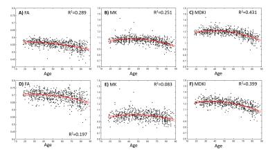 |
Interpreting age-related changes based on the mean signal diffusion kurtosis
Rafael Neto Henriques, Marta Correia
Previous studies have shown that measures of non-Gaussian diffusion from diffusion kurtosis images (DKI) provide unique information on age-related tissue changes. In this study, a novel non-Gaussian diffusion index invariant to the distribution of fibres is proposed and applied to 650 datasets from the Cam-CAN ageing project. The results show that the proposed biomarker is not only applicable to any tissue configuration but also less sensitive to noise and artefacts when compared to traditional DKI measures. Moreover, for white matter regions, age-related changes measured by this index seem to reflect axonal alterations likely related to axonal loss mechanisms.
|
17:18
|
1271.
 |
Registration-free analysis of diffusion MRI tractography data across subjects through the human lifespan
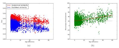
Viviana Siless, Juliet Davidow, Jared Nielsen, Qiuyun Fan, Trey Hedden, Marisa Hollinshead, Constanza Bustamante, Michelle Drews, Koene Van Dijk, Margaret Sheridan, Randy Buckner, Bruce Fischl, Leah Somerville, Anastasia Yendiki
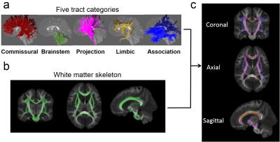
Diffusion MRI tractography produces massive sets of streamlines that need to be clustered into anatomically meaningful bundles. Conventional clustering techniques group streamlines based on their proximity in Euclidean space. We have developed an unsupervised method for clustering tractography streamlines based on their neighboring anatomical structures, rather than their coordinates in Euclidean space. In this work, we show how this approach can be extended to find corresponding clusters across subjects without inter-subject registration. We evaluate the approach on data from the MGH-Harvard-USC Lifespan Human Connectome Project, showing improved correspondence in tract clusters across subjects aged 8-90, without the need for registration.
|
|