|
Combined Educational & Scientific Session
ORGANIZERS: Vikas Gulani, M.D., Ph.D. & James G. Pipe, Ph.D.
Monday, 24 April 2017
| Room 315 |
13:45 - 15:45 |
Moderators: Vikas Gulani, James Pipe |
Slack Channel: #s_value
Session Number: CE12
Overview
This session continues the work started in the ISMRM Value Initiative, introduced in previous ISMRM meetings. The session will begin with an introduction, followed by two long format talks. One speaker will discuss how the value of MRI can be improved by utilizing super-fast imaging, with particular examples from clinical body imaging. A second speaker will discuss the Value of MR Imaging in the health-care system, touching on perspectives of patients, referring physicians, health system and payers. This talk will include the discussion of costs, impact of MR on treatment decisions, patient triage, and outcomes. The discussion will include a perspective on how MR can shift from being a revenue center in the present healthcare delivery model, to a value center (rather than a cost center) in the delivery model of the near future. After these two talks, the session will continue into proffered papers on studying the value of MRI from multiple perspectives.
Target Audience
Scientists and physicians interested in determining or improving the value of MRI in the healthcare system, either from a technical or a clinical perspective.
Learning Objectives
Upon completion of this course, participants should be able to:
-Describe examples of technology driven advances in MR imaging that have increased the value of MRI in clinical medicine;
-Identify definitions of value of MRI technology in the healthcare arena, from multiple perspectives such as that of patients, imaging scientists and physicians, referring physicians, payers, and the healthcare system;
-Recognize the coming challenges in medical imaging in general, and MRI in particular, as the perception of imaging in the health system shifts from that of a revenue center, to potentially a cost center;
-Evaluate potential strategies to redefine MRI as a value center in the future healthcare system; and
-Identify and evaluate ongoing, cutting edge technically driven and clinically driven strategies for maximizing value in MRI protocols in multiple body parts.
13:45
|
|
Opening Remarks |
13:50
|
|
Studying Value in MRI: A South Korean Perspective 
Jeong Min Lee
In summary, MR allows a high detection and characterization rate in the abdomen, without ionizing radiation. Also, abbreviated noncontract MRI protocol can be used for screening several abdominal diseases. To further increase the value of MRI, the additional effort for increasing compliance of patients for MR examination by incorporating free breathing sequences or shortening examination time, and increasing awareness of its superiority to CT in imaging many of the abdominal organs would be necessary.
|
14:05
|
|
Studying Value in MRI: A US Perspective
Yoshimi Anzai
The current healthcare transformation provides a perfect opportunity for the entire MR community to deliberate the value of MRI. It is essential to understand what each stakeholder wants or needs from MR or information obtained from MRI. A focused MR protocol with shorter scan time, reduced costs and accurate, timely, and actionable diagnosis are just a few examples. The challenges provide strong motivation and incentives for us to make strategies for developing high-value MRI. By doing so, imaging will become the value center, not the cost center, for the health system and patient care spectrum.
|
14:20
|
0222.
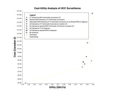 |
Cost-Utility Analysis of Ultrasound, Computed Tomography, Abbreviated and Standard MRI for Hepatocellular Carcinoma Surveillance
An Tang, Boyan Fan, Joshua Bérubé, Milena Cerny, Damien Olivié, Jeanne-Marie Giard, Luigi Lepanto, Jean Lachaine
Current clinical practice guidelines recommend ultrasound (US) every 6 months for surveillance of hepatocellular carcinoma (HCC) in at-risk patients. Despite higher sensitivity, there is uncertainty regarding the role of MRI for HCC surveillance, whether as an add-on or replacement test. Our results indicate that surveillance with standard MRI followed by CT if technically inconclusive provided the highest level of effectiveness. However, CT followed by MRI was more cost-effective than alternative surveillance strategies using a threshold of $50,000 per QALY gained. Further, lower cost of abbreviated MRI will be required to be used as a first-line imaging technique for surveillance.
|
14:24
|
0223.
 |
Optimizing MRI for Focal Liver Lesions: Are All Our Sequences Really Necessary?
Sara Dastmalchian, Nicholas Fulton, Majid Chalian, Ozden Kilinc, Mark Griswold, Vikas Gulani, Karin Herrmann
In this proof-of-concept study, our aim was to determine an optimal minimum number of MRI sequences which allow confident characterization of liver lesions into benign versus malignant categories with reasonable accuracy. We hypothesized that an abbreviated liver MRI protocol including single shot T2-weighted, pre and dynamic post-contrast T1-weighted images has the potential to reduce overall scan time and throughput. If this can be performed without significant loss in diagnostic accuracy it may improve efficacy and be beneficial to current practice.
|
14:28
|
0224.
 |
Short 15-min surveillance-protocol multiphasic contrast enhanced MRI for hepatocellular carcinoma detection in cirrhosis
Takeshi Yokoo, Gaurav Khatri, Lakshmi Ananthakrishnan, David Fetzer, Yin Xi, Ivan Pedrosa
Patient with cirrhosis are at increased risk of developing hepatocellular carcinoma (HCC) and routine surveillance imaging is recommended every 6 months. While MRI has high sensitivity for HCC detection, its routine use is controversial due to its long exam time, high cost, and limited access. In this prospective study, we demonstrated that a short 15-min surveillance MRI has similar HCC detection performance as the standard 45-min diagnostic MRI in patients with cirrhosis. Therefore, a short surveillance MRI may allow for a more efficient and cost-effective alternative to the current standard-of-care MRI for HCC surveillance.
|
14:32
|
0225.
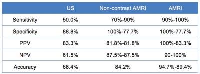 |
Diagnostic performance of an abbreviated gadoxetic acid enhanced-MRI (AMRI) vs. ultrasound for detection of small HCC: pilot study.
Cecilia Besa, Sara Lewis, Mathilde Wagner, Yujin Hoshida, Ruth Carlos, Claude Sirlin, Bachir Taouli
In this study, we aim to test the diagnostic value of an abbreviated MRI (AMRI) using gadoxetic acid (with patient injected outside the MRI room) compared to ultrasound (US) for hepatocellular carcinoma (HCC) detection in a population of patients with small HCC and controls. This study demonstrates that AMRI using T1WI obtained at the hepatobiliary phase (T1w-HBP), diffusion weighted imaging (DWI) and T2WI has superior diagnostic performance for HCC detection compared to US. This could serve as the basis for a future study assessing AMRI for HCC screening/surveillance in patients with cirrhosis.
|
14:36
|
0226.
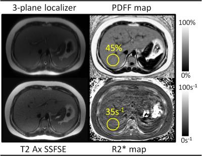 |
Demonstrating the Clinical Feasibility of a Rapid Non-Contrast MRI Protocol for Detection and Quantification of Hepatic Steatosis and Iron Overload
B. Dustin Pooler, Scott Reeder
Many clinical scenarios necessitate evaluation for hepatic steatosis or iron overload without indication for a complicated MR exam. Emerging confounder-corrected chemical shift encoded MRI (CSE-MRI) techniques can provide simultaneous estimation of liver proton density fat fraction (PDFF) and R2* as biomarkers of steatosis and iron overload, respectively. We have developed a highly focused CSE-MRI protocol which obtains these metrics in approximately 5 minutes of table time. Our initial clinical experience has shown this protocol to be feasible for evaluation of patients ranging from pediatric to geriatric, with clinically significant disease detected in a large fraction of patients scanned to date.
|
14:40
|
|
Discussion |
14:45
 |
0227.
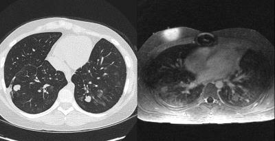 |
Contrast-enhanced Ultrashort Echo Time (UTE) MR of the chest to evaluate for metastatic nodules in pediatric patients with malignancy undergoing abdomen MR staging and surveillance: a high-value alternative or adjunct to CT
Anshul Haldipur, Evan Zucker, Joseph Cheng, Shreyas Vasanawala
Ultrashort Echo Time (UTE) MRI of the chest can be optimized for detection of metastatic lung nodules. In pediatric patients with a history of abdominal malignancies undergoing routine re-staging contrast-enhanced MRI exams, clinically significant metastatic lung nodules are detectable which, if diagnosed, could obviate or decrease the frequency of subsequent separate CT scans of the chest. Though cost of a chest CT is lower than an MRI, it is substantially more than the incremental cost of adding an additional sequence during already-scheduled MR Abdomen exams. Additionally, ionizing radiation is avoided, alleviating concerns about cumulative exposure following multiple serial follow-up examinations.
|
14:49
|
0228.
 |
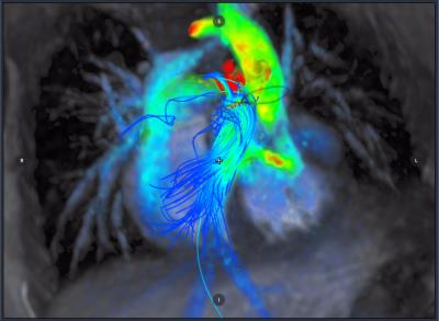
The use of MRI in the diagnosis of Chronic Thromboembolic Pulmonary Hypertension
Christopher Johns, Andy Swift, Jens Vogel-Claussen, David Kiely, Jim Wild
As surgical pulmonary endarterectomy significantly improves survival in patients with chronic thrombo-embolic pulmonary hypertension it is important to correctly identify patients. Using cardiopulmonary MRI it is possible to screen for the presence of chronic thombo-emboli in all cases who can tolerate MRI, reducing the requirement for SPECT (and therefore patient radiation exposure). The same scan can also predict the presence of pulmonary hypertension, and due to a high specificity we can reduce the reliance upon an invasive test (right heart catheterisation) by around 50%.
|
14:53
|
0229.
 |
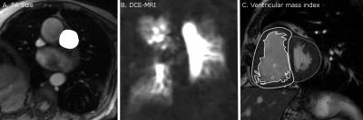
Combined 3D Cine/4D Flow Accelerated Cardiac Imaging with Cloud Computing. Toward Streamlined and Fast Comprehensive Cardiac MRI Exam
Haonan Wang, Peng Lai, Piero Ghedin, Shreyas Vasanawala, Anja Brau, El-Sayed Ibrahim
Currently, cine MRI is the gold standard for evaluating cardiac function. Nevertheless, in today’s practice, slices need to be acquired at different oblique, and 12-16 short-axis slices needs to be acquired for sufficient ventricular coverage. The same limits apply to hemodynamics-related assessment of valvular and vascular performance, for which multiple 2D oblique flow measurements need to be acquired. In this abstract, we present a combined 3D Cine/4D Flow accelerated cardiac imaging technique with cloud computing, which significantly reduces the scan and processing time, reduces the scan’s complexity, alleviates misregistration problems, and increases productivity.
|
14:57
|
0230.
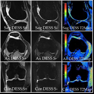 |
5 Minute Comprehensive Knee MRI with 3D Double-Echo Steady-State (DESS)
Akshay Chaudhari, Bragi Sveinsson, Jeff Wood, Dushyant Thakur, Kathryn Stevens, Chris Beaulieu, Marcus Alley, Curtis Abercrombie, Garry Gold, Brian Hargreaves
Knee MRI is performed commonly in the US for assessing acute injuries as well as degenerative diseases. However, current knee MRI protocols can require 25-30 minutes or more and cost approximately $1.1billion/year. In such instances, a short knee protocol could lower costs while increasing patient throughput, comfort, and access to care. In this study, we show that a five-minute double-echo steady-state (DESS) scan, with automatic T2 maps and fluid-nulled images, offers high efficacy and diagnostic utility compared to the standard knee protocol. These results suggest that a five-minute DESS scan could be used for comprehensive MRI of the knee.
|
15:01
|
0231.
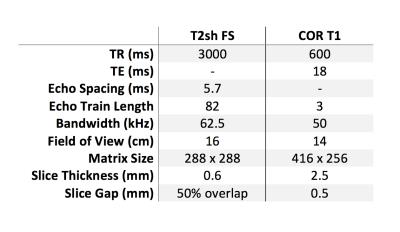 |
Targeted Rapid Knee MRI Exam using T2 Shuffling
Jonathan Tamir, Michael Lustig, Valentina Taviani, Marcus Alley, Becki Perkins, Lori Hart, Darla Mortensen, Shreyas Vasanawala
We investigate the effectiveness of a targeted rapid pediatric knee MRI exam with total exam time of about 10 minutes. We aim to enable same-day MRI access, accommodating the abbreviated protocol between other scheduled patients. The protocol is based on T2 Shuffling, a four-dimensional acquisition that permits volumetric reconstruction of images with variable T2-contrast. Preliminary data for ten subjects referred for the targeted knee MRI exam is presented. Mean time from registration to exam completion averaged 48.5 minutes, with one outlier of 269 minutes due to technologist error in documentation.
|
15:05
|
|
Discussion |
15:10
|
0232.
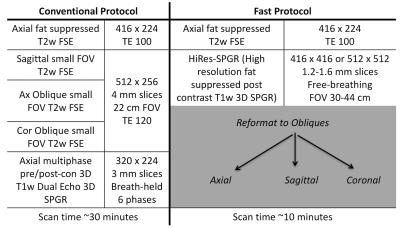 |
Faster and more accurate staging of rectal cancer through a two-sequence MR protocol based on high-resolution T1-weighted post-contrast 3D SPGR imaging
Andreas Loening, Marcus Alley, Shreyas Vasanawala
A fast 10-min MRI protocol for rectal cancer staging based on a high-resolution T1-weighted post-contrast sequence was compared to a conventional 30-40 min protocol based on multiple planes of T2-weighted imaging. With IRB approval, 37 consecutive patients were retrospectively identified whose MRI rectal cancer staging studies contained the necessary sequences allowing creation of conventional and hypothetical fast protocols. Two blinded readers assessed each protocol for findings determining cancer stage. The fast 10-min MRI staging protocol was significantly more accurate for assessment of nodal disease and rectal cancer stage, and allowed significantly more confidence in assessment for transmural extension of tumor.
|
15:14
|
0233.
 |
How Quick is our “Quick DWI” for Stroke?
Elaine Lui, David Wang, Nawaf Yassi, Bruce Campbell, Patricia Desmond
Stroke is an emergency. Although MRI is the imaging gold standard for the ischemic “core” in the hyperacute setting, MRI must be “quick” to remain relevant in the management pathway of this common disease. A “quick diffusion weighted imaging”(qDWI) protocol for acute stroke consisting of a single DWI sequence was implemented at our institution. qDWI reduced scan time by 84% compared to our conventional “Stroke” protocol, was 96.5% diagnostic, and with 90% not requiring further MRI. There was also a statistically significant quicker referral to scan time compared to the conventional “Stroke” MRIs.
|
15:18
|
0234.
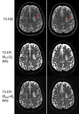 |
High-Resolution, Echo Planar Imaging on a Compact MRI Scanner: Applications in Stroke and Neuroimaging
Ek Tan, Paul Weavers, Erin Gray, Christopher Hardy, John Schenck, Matt Bernstein, Yunhong Shu, Thomas Foo, John Huston
Rapid (six minutes and under) neuroimaging protocols can improve patient throughput and scanner utilization, especially when accelerated using the relatively fast EPI readout. However, susceptibility-related image distortion in EPI has limited its useful spatial resolution (2-2.5mm) conventional, whole-body 3T MRI. A 3.5-fold faster slew-rate head-only gradient on a novel low-cryogen compact 3T scanner can provide an effective platform for twofold-faster EPI that achieves higher spatial resolution (1mm). The utility of rapid (under one minute) single- and multi-shot T2-weighted EPI on the compact 3T is evaluated and compared to routine T2-FSE imaging in patients with brain tumors and stroke.
|
15:22
|
0235.
 |
Accelerating multi-contrast imaging in neuro-exam with sharable information
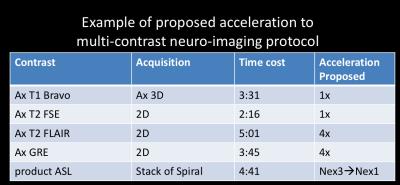
Enhao Gong, John Pauly, Greg Zaharchuk
Neurological disorders results in great clinical challenges and high societal burdens. Currently multi-contrast MRI exams are frequently used for diagnosis because of the various tissue contrasts provides complementary diagnosis information to distinguish normal tissue from pathology. However, the cost of acquiring these multiple sequences is extensive scanning time, which significantly increases both the diagnosis cost and patients’ discomfort. Here we proposed a new approach to accelerate multi-contrast imaging by using Parallel Imaging, Compressed Sensing and sharable information. We validated the new approach with experiments on both patients and healthy subjects. We demonstrate that we can reduce the multi-contrast MRI scanning time significantly while preserving the diagnostic information.
|
15:26
|
0236.
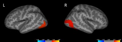 |
Prediction of treatment response using baseline structural and functional MRI in first-episode antipsychotic-naive schizophrenia
Su Lui, Lu Liu, Yuan Xiao, Bo Tao, Biqiu Tang, Qiyong Gong
Finding imaging biomarkers which could predict the treatment response is quite important to help the selection of therapy and save health resource.
|
15:30
|
|
Discussion |
15:35
|
|
Closing Remarks |
|