ISMRM 25th Annual Meeting & Exhibition • 22-27 April 2017 • Honolulu, HI, USA

Electronic Poster Session: Body
3164 -3187 Body: Diffusion
3188 -3211 Liver
3283 -3306 Genitourinary
3307 -3330 Thoracic MRI-2
 
Body: Diffusion
Electronic Poster
Body: Breast, Chest, Abdomen, Pelvis

 
Monday, 24 April 2017
Exhibition Hall  08:15 - 09:15


    Computer #

 
3164.   
73 Contribution of DKI and IVIM to liver T1rho imaging in the prediction of hepatitis B virus-related liver fibrosis
shuangshuang xie, qing li, zhizheng zhuo, yu zhang, yue cheng, wen shen
This study evaluated the individual T1rho and combined performances of T1rho, IVIM and DKI in predicting HBV related liver fibrosis. Twenty-two patients with HBV related liver fibrosis and twenty healthy control subjects were underwent whole-liver T1rho MR imaging, IVIM and DKI. The T1rho, D, D*, f , MD and MK values were compared between the two groups, and then the single and combined diagnostic efficiency of T1rho, IVIM and DKI was analyzed. Our results showed liver T1rho and MK increased and D, f, MD decreased in normal control group, and liver T1rho and MD had significantly difference between the two groups. T1rho had a moderate diagnostic efficiency, IVIM and DKI had a mild diagnostic efficiency to detect fibrosis. While, the combination of T1rho, IVIM and DKI improved the diagnostic efficiency significantly and had a good diagnostic efficiency to detect fibrosis. We conclude T1rho can be used to predicting liver fibrosis effectively, and IVIM and DKI can effectively complement existing T1rho MR imaging in predicting of liver fibrosis.


3165.   
74 Role of diffusion-weighted imaging in distinguishing thoracoabdominal neuroblastic tumors of various histological types and differentiation grades
Yang Wen, Yun Peng, Xiaomin Duan, Nan Zhang
Purpose: To evaluate whether DWI allow discrimination of thoracoabdominal neuroblastic tumors of various histological types and differentiation grades. Materials and Methods: The DWI scans of the thoracoabdominal neuroblastic tumors in twenty-five children were retrospectively evaluated. DWI was performed with 2 b values of 0 and 800s/mm2 on a 3.0T MR scanner.Results: In the 25 cases, ganglioneuroma (GN) was in 3 cases, ganglioneuroblastoma (GNB) -Intermixed in 4, GNB-Nodular in 3 and neuroblastoma (NB) in 15. The ADC values of the NBs were significantly lower than those of GNs/GNBs (P<.001). The ADC of GNB-Nodular/NB was significantly less than that of GN/GNB-Intermixed (p<0.0001). In GNB-Nodular and NB, the tumors with poorly differentiated and undifferentiated lesions (n=12) had significantly smaller ADC than those with differentiated composition (n=6) (P=.0012). Conclusion: ADC of DWI is highly valuable for discriminating thoracoabdominal neuroblastic tumors of different histological types and subtypes.


3166.   
75 Focus or Spread? An Investigation of the Effects of b-Value Acquisition Strategies on the Stability and Bias of Prostate Diffusion Imaging Results
Xiaodong Zhong, Phil Young, Peter Kollasch, Venkata Chebrolu, Brian Dale
In order to investigate the effects of b-value acquisition strategies on the stability and bias of the prostate DWI results, including calculated b-value images and ADC, a framework was developed. Using the DWI data from 8 prostate patients, this study revealed that acquiring many averages at a few b-values increases bias compared to acquiring few averages at many b-values, particularly when large numbers of averages have been removed. The framework and strategies proposed in this work may provide a useful tool to design b-value acquisition protocols to achieve the stability of prostate DWI results in clinic.


3167.   
76 The IASLC/ ITMIG Thymic Epithelial Tumor Staging: Comparison of Staging Capability among Whole-Body PET/MRI, MRI including DWI, PET/CT and Conventional Radiological Examination
Yoshiharu Ohno, Yuji Kishida, Sinichiro Seki, Kota Aoyagi, Masao Yui, Yoshimori Kassai, Wakiko Tani, Noriyuki Negi, Katsusuke Kyotani, Takeshi Yoshikawa
Accurate stage assessment is essential for choosing the appropriate treatment strategy for thymic epithelial tumor patients.  Recently, the International Association for the Study of Lung Cancer (IASLC) and International Thymic Malignancies Interest Group (ITMIG) proposed a new IASLC/ITMIG thymic epithelial tumor stage classification system.  The purpose of this study was to compare the diagnostic capability for the IASLC/ ITMIG thymic epithelial tumor staging among whole-body FDG-PET/MRI, MRI, FDG-PET/CT and conventional radiological examinations based on guidelines.  


3168.   
77 Motion Correction of Multi-b-value Diffusion-Weighted Images in the Kidney by Pyramidal Lucas-Kanade Registration
Jun Lv, Wenjian Huang, Jue Zhang, Xiaoying Wang, Jing Fang
Intravoxel incoherent motion diffusion weighted imaging (IVIM-DWI) is a novel functional MRI technique which has been demonstrated with excellent diagnostic capability. Respiratory motion artifacts are the main source of error in the acquisition and quanti?cation of parameters for multi-b-value DWI. This work develops a reliable approach to compensate for misalignments between multi-b-value images during free-breathing based on pyramidal Lucas-Kanade registration. Preliminary results show that our proposed approach can well correct motion-related artifacts misalignment. In addition, visual quality of the ADC, f, D and D* maps indicate that contours of kidney look better defined after registration.  


3169.   
78 Intravoxel Incoherent Motion Analysis for Perfusion Using Portal Vein Embolization Pig Model
Jia Ning, Tilman Schubert, Huijun Chen, Chun Yuan, Scott Reeder
Portal vein embolization (PVE) is often performed before liver tumor resection, to induce hypertrophy of the anticipated liver remnant and reduce the complications after partial liver resection. The perfusion changes of the embolized and non-embolized liver segments are of great interest. Intravoxel incoherent motion (IVIM) is a component of the diffusion weighted signal model that allows for separating estimation of perfusion. Estimation of IVIM from diffusion weighted imaging which may provide a unique way to measure the perfusion of liver tissue. This study aimed to test the intra-observer repeatability of IVIM and parameters change after PVE used a pig model.


3170.   
79 Free-breathing 3D Body Diffusion Imaging at 3T Using M1-compensated Diffusion Preparation and Stack-of-Stars Readout
Xiaoming Bi, Christopher Nguyen, Zhaoyang Fan, Yutaka Natsuaki, Rola Saouaf, Debiao Li, Gerhard Laub
Diffusion-weighted (DW) MRI enables qualitative and quantitative assessment of tissue diffusivity. Body diffusion imaging at 3T using conventional single-shot EPI sequence is challenged by respiratory and cardiac motions of subject, limited spatial resolution, and image distortion and ghosting. In this work, a new 3D imaging technique incorporating M1-compensated diffusion preparation and motion-robust stack-of-stars data acquisition was developed. Preliminary volunteer studies demonstrated its feasibility for free-breathing body diffusion imaging at 3T. From one patient underwent MR-PET scan, reduced ADC and increased FDG uptake was observed in the same focal lesion.


3171.   
80 A cardiac stationary phase based ECG trigger (CaspECG) of intravoxel incoherent motion diffusion MR in left liver lobe.
Zhiming Xiang, Zhu Ai, Jianke Liang, Guijin Li, Xiaolei Zhu, Xu Yan, Changhong Liang, Suzanne Palmer, ChiShing Zee
Intravoxel incoherent motion (IVIM) Diffusion weighted imaging (DWI) showed high clinical value in liver disease evaluation. However, it suffers from signal loss and motion artifacts due to cardiac and respiratory movement, especially in the left lobe. To improve robustness and reproducibility of parameter estimation for IVIM DWI, a novel DWI acqusition technique was introduced, which uses ECG trigger with delay time optimized by the periods of cardiac relative stationary phase. The results showed that the proposed acquisition method improves SNR of DWI data, and the repeatability and stability of IVIM-DWI derived parameters.


3172.   
81 Intravoxel incoherent motion diffusion-weighted imaging of hepatic warm ischemia-reperfusion injury in a rabbit model
Qian Ji, Zhi-qiang Chu, Pan-li Zuo, Wen Shen
Hepatic warm ischemia-reperfusion injury (WIRI) is clinically relevant in liver operation. We undertook this study to determine the feasibility of using IVIM for the early diagnosing and grading of hepatic WIRI. Fifty different grades of hepatic WIRI models and control rabbits were examined using a 3T clinical MR scanner, which followed by biochemical and histopathological analysis. There were significant differences of IVIM parameters between different groups. IVIM parameters corresponded well with biochemical parameters. ROC analysis showed the AUC of PF was the largest. This indicated that IVIM is a noninvasive and valuable technique for assessing and grading of hepatic WIRI.


3173.   
82 Elucidation of Male Urethral Sphincter Complex Using Diffusion Tensor Imaging (DTI) based Fiber-Tracking
Kyoko Sakamoto, Mahadevan Rajasekaran, Valmiki Bhargava, Vadim Malis, Shantanu Sinha
Urethral sphincters play an important role in urinary incontinence, a major clinical problem affecting the aging population. We elucidate the anatomy of the urethral sphincter muscles pertinent to urinary continence function using in vivo, non-invasive proton-density and diffusion tensor imaging and DTI-based fiber tracking in young adults. Muscle fiber tracking consistently revealed, perhaps for the first time, the existence of two sphincter like muscles, with one proximal near the bladder neck and the other more distal, supporting the two sphincter concept to constrict/close the urethral opening with important implications for the effect of prostatectomy on urethral closure function.


3174.   
83 Is voxel-wise ADC histogram repeatable?
Masamitsu Hatakenaka, Naomi Koyama, Koichi Onodera, Naoya Yama, Maki Onodera, Yoshifusa Kyuna, Mitsuhiro Nakanishi
Skewness and kurtosis of voxel-wise apparent diffusion coefficient show low repeatability. Radiologist should take this characteristic into account when interpreting DWI of the prostate.


3175.   
84 Segmentation of Bone Marrow of Pelvis in Multi-parametric MRI (T1-w/ADC-map) of Metastatic Breast Cancer Patients
Mahsa Rostamie, Anahita Fathi Kazerooni, Hamidreza Saligheh Rad
To assess treatment response through quantitative analysis, in metastatic breast cancer patients, computer-aided segmentation of bone marrows is beneficial. We propose a semi-automatic segmentation method based on level-set and region growing techniques applied to T1-W images to facilitate extraction of apparent diffusion coefficient (ADC) features for the purpose of treatment response assessment from bone marrows of pelvic region. The results of applying the method on T1w/ADC-map of 10 patients shows a dice score of 81%, suggestive of high agreement of our proposed segmentation approach with expert’s opinion.


3176.   
85 Early evaluation of liver fibrosis in radiation-induced liver fibrosis rat models using intra-voxel incoherent motion theory - video not available
Zhongping Zhang, Rong Ma, Dong Zhang, Changzheng Shi, Liangping Luo
This study found that IVIM-derived D could efficiently detect and differentiate the liver fibrosis in radiation-treaed rats at the early stage in vivo.  


3177.   
86 Prognostic value of the pretreatment apparent diffusion coefficient (ADC) for outcome prediction of chemorefractory colorectal liver metastases undergoing 90-Yttrium Radioembolization
Frederic Schmeel, Julian Luetkens, Frank Träber, Leonard Schmeel, Amir Sabet, Birgit Simon, Hans Schild, Dariusch Hadizadeh
Imaging-based prediction of therapeutic response is highly desireable for further therapy decisions in patients with advanced malignancies. Therefore, we investigated whether pre-treatment values of the apparent diffusion coefficient (ADC) on diffusion-weighted MRI could predict the outcome of patients with liver-predominant metastatic colorectal cancer prior to 90-Yttrium microspheres radioembolization. Uni- and multivariate analyses were performed comparing various variables with potential impact on progression-free and overall survival. Our results reveal that pathologic pre-treatment ADC, alongside with established clinical parameters, is a strong and independent predicor of both progression-free and overall survival before RE treatment.


3178.   
87 Clinical Robustness of Accelerated and Optimized Abdominal Diffusion Weighted Imaging
Jana Taron, Jakob Weiss, Petros Martirosian, Alto Stemmer, Konstantin Nikolaou, Mike Notohamiprodjo
We evaluated the robustness of an accelerated and optimized diffusion-weighted sequence using the simultaneous-multislice (SMS) technique for scan time reduction and a 3D Diagonal diffusion mode to optimize image quality in clinical routine. 152 patients received clinically indicated abdominal MRI including the optimized diffusion-weighted sequence (DWIOPT). A subgroup of 41 patients additionally received a standard diffusion-weighted sequence (DWISTD) as reference. Qualitative and quantitative image parameters were evaluated. In the interindividual comparison, DWIOPT proved equal to superior to DWISTD with comparable ADC-values. In the patients receiving DWIOPT only, image quality maintained substantial proving constant and stable results in a large cohort.


3179.   
88 Mean Kurtosis discriminates between low- and high-risk prostate cancer better than Mean diffusivity does
Maria Giovanna Di Trani, Alessandra Caporale, Marco Nezzo, Roberto Miano, Alessandro Mauriello, Pierluigi Bove, Guglielmo Manenti, Silvia Capuani
This work was finalized to compare the diagnostic potential of Diffusion Tensor and Diffusion Kurtosis Imaging in discriminating between low- and high-risk prostate cancer (Pca). Maps of Mean Diffusivity (MD), apparent Kurtosis (K) and apparent diffusion coefficient (D) were obtained from DWIs of 24 patients with different tumour grade. K maps better highlight differences between periferal PCa, PCa and benign tissue. In particular K discriminates between low- and high-risk PCa with a higher statistical significance compared to that of MD. DKI can improve the accuracy of the current PCa diagnosis providing a useful tool for PCa detection and grading.


3180.   
89 Unbiased diffusional kurtosis measurements in the pelvis at low signal-to-noise ratio: A new approach for noise-level estimations
Olaf Dietrich, Martina Brandlhuber, Moritz Schneider, Marco Armbruster, Melvin D'Anastasi, Maximilian Reiser
The purpose of this study was to propose a robust technique for the determination of unbiased kurtosis values in body applications; for this aim, a new strategy to pixelwise estimate the noise level in DKI acquisitions is described and its feasibility for pelvic DKI is demonstrated in prostate tissue.

The "naive" evaluation (without considering the image noise) resulted in significantly biased positive kurtosis values of urine of $$$K=0.42\,($$$standard deviation: $$$0.06)$$$, indicating non-monoexponential signal decay caused by the influence of noise at higher b-values. This bias is almost completely removed ($$$K=-0.01\,(0.32)$$$) after including the pre-calculated noise-level map in the evaluation.


3181.   
90 Gradient First Moment Dependence of ADC in Liver Diffusion Weighting Imaging.
Kévin Moulin, Eric Aliotta, Daniel Ennis
The effect of perfusion on Diffusion Weighting Imaging (DWI) and particularly the Apparent Diffusion Coefficient (ADC) at low b-value is a confounder in pathology assessment. In this study, in order to investigate the gradient first moment (M1) dependence of perfusion on the ADC, waveforms were generated using convex diffusion encoding (CODE) framework leading to a range ofM1 for a given b-value. After comparing ADC calculated with different M1 and different b-value, we found that motion compensated waveform (M1=0) ensures independence of the ADC to perfusion even at low b-value, which can improve SNR.


3182.   
91 Prognostic Value of Pretreatment Diffusion Weighted Magnetic Resonance Imaging based Texture in Concurrent Chemo-radiotherapy of Esophageal Squamous Cell Cancer - video not available
Zhenjiang Li, Chun Han, Hongsheng Li, Lan Wang, Jian Zhu, Weibo Chen, Baosheng Li
This study has important clinical significance. The pretreatment texture features combined with conventional prognostic factors may present a more accurate predictive tool for OS of ESCC patients. The parameters can be used to evaluate the prognosis of ESCC after CRT at an early time.


3183.   
92 Imaging non-enlarged abdominal lymph nodes and measuring their diffusion coefficient
Hannah Williams, Caroline Hoad, Luca Marciani, Gordon Moran, Giles Major, Robert Scott, Penny Gowland
Inflammatory conditions such as Crohn’s disease cause changes in the lymphatics including enlargement and necrosis, and so improved measures of number, size and function of lymph nodes could provide novel markers of local inflammatory response. Being able to identify small nodes would allow the effect of inflammatory diseases and response to therapy to be monitored. DWIBS was used here to isolate the signal from small abdominal lymph nodes. The diffusion coefficient of the pelvic and abdominal lymph nodes and the cisterna chyli with IVIM effects eliminated, were 0.9±0.2, 1.4±0.3 and 1.7±0.6 x10⁻³ mm2/s. 


3184.   
93 Towards development of tools for quantitative body DWI
Raj Attariwala, Amy Chamber, Wayne Picker, Mikko Maatta
Body DWI is a technique that is potentially quantifiable, and numerous clinical publications have demonstrated tissue variability of the ADC parameter. The variability of machine hardware and acquisition parameters impacts the signal from which ADC is calculated. To address the needs of standardization of machines, a body diffusion phantom and analysis software is being developed. Preliminary results are presented here.


3185.   
94 High Resolution Diffusion Weighted Imaging of the Liver using Readout Segmented EPI on 3T
Yishi Wang, Zhe Zhang, Xiaodong Ma, Ha-Kyu Jeong, Chun Yuan, Hua Guo
Readout segmented EPI (RS-EPI) can be used for high-resolution diffusion weighted imaging but there are still limited reports on its application to liver DWI. In this study, we reported to use RS-EPI for high resolution DWI of the liver.


3186.   
95 Training an Artificial Neural Network by Diffusion-Weighted MRI Data to Differentiate Between Prostate Cancer With High and With Low Gleason Score - permission withheld
Sebastiano Barbieri, Harriet Thoeny
We prospectively assess the feasibility of using DW-MRI data to train an artificial neural network which distinguishes between prostate cancer lesions with high (≥7) and with low (=6) Gleason scores in 84 patients. The accuracy of the artificial neural network is compared with the accuracy of classification based on apparent diffusion coefficient (ADC) values.


3187.   
96 Diffusion and perfusion parameters extracted by bi-exponential model are markers of healthy human placenta development. - permission withheld
Michele Guerreri, Silvia Capuani, Amanda Antonelli, Lucia Manganaro
The purpose was to investigate the potential of bi-exponential model of diffusion-weighted (DW) signal decay to quantify diffusion and perfusion in healthy human placentas. The relation between diffusion and perfusion parameters with microstructural changes occurring during placenta development was also investigated. 26 pregnant women underwent DW examination. Apparent diffusion coefficient D, pseudo-perfusion fraction f and pseudo-diffusion coefficient D* were obtained in specific placental regions. The Pearson correlations between D, D*,f and clinical data (Gestational Age, Body-Mass Index and basal Glycaemia) were evaluated. D and f show to be good indicators of placenta morphological changes due to Gestational Age.
 
Liver
Electronic Poster
Body: Breast, Chest, Abdomen, Pelvis

 
Monday, 24 April 2017
Exhibition Hall  08:15 - 09:15


    Computer #

 
3188.   
97 Hepatic MR Elastography (MRE) System Longitudinal Quality Assurance (QA) Protocol
Jun Chen, Phillip Rossman, Kevin Glaser, Richard Ehman
A suitable quality assurance (QA) phantom and image acquisition and processing procedures were developed for confirming the proper function and longitudinal stability of these MRE systems. The goals of this educational poster are to demonstrate the QA phantom, the longitudinal testing protocol, and the utility of detecting a problem caused by discontinuous motion. 


3189.   
98 Radiologic-Pathologic Correlation and MR Analysis of Hepatocellular Adenoma Subtypes.
Daniel Kehler, George Yang, Christine Zwart, Marcela Salomao, Alvin Silva
   Hepatocellular adenomas (HCA) present as four genetic subtypes that vary greatly in their clinical behavior and MR appearance.   Inflammatory HCA has the highest propensity for hemorrhage, is characterized by hyperintense T2 signal, and displays arterial hyper-enhancement that persists on portal venous and delayed phases.  HNF1-alpha mutated HCA portends a good prognosis, and is characterized by diffuse intracellular lipid.  Beta-catenin HCA  is less common and difficult to diagnose on imaging, though arguably the most important because of its high likelihood for malignant transformation. Unclassified HCA is not well understood in terms of imaging or clinical significance.  


3190.   
 
99 MRI Technique and Interpretation in the Evaluation of Hepatic Steatosis
Zachary Borden, Scott Reeder
Hepatic steatosis is a common affliction with important prognostic implications.  Conventionally, liver biopsy has been required for the diagnosis of steatosis although this may result in inadequate spatial sampling and significant associated complications.  The non-interventional imaging methods of ultrasound and computed tomography may be used but are limited in accuracy.  MRI offers an ideal method to globally and accurately interrogate for liver fat.  Multiple MRI techniques including spectroscopy, in-phase/out-phase, conventional fat suppression, complex and magnitude-based CSE-MRI methods have been used in the evaluation of hepatic steatosis.   These techniques possess unique advantages and disadvantages which must be understood to optimize patient care. 


3192.   
101 Dynamic contrast enhanced 3D T1WI at upper abdomen using combination of parallel imaging and compressed sensing on a wide-bore 3T unit: Comparison of effects with Gd-DTPA and Gd-EOB-DTPA
Motoyuki Katayama, Takayuki Masui, Kei Tsukamoto, Mitsuteru Tsuchiya, Yuki Hayashi, Masako Sasaki, Takahiro Yamada, Yuji Iwadate, Naoyuki Takei, Kang Wang, Kevin King, Harumi Sakahara
Consecutive four phases of dynamic contrast enhanced 3D T1WI in the upper abdomen could be obtained during one breath-hold in combined use of parallel imaging and compressed sensing at wide-bore 3T system. The imaging protocols with Gd-DTPA and Gd-EOB-DTPA provided good image quality. Although image contrasts with Gd-EOB-DTPA might be inferior to those with Gd-DTPA, patterns of time intensity curves with study with dynamic contrast of each protocol were similar to each other. 


3194.   
103 Gadoxetic acid-enhanced MRI findings as a risk of multicentric recurrence of hepatocellular carcinoma after hepatectomy
Masaki Matsuda, Shintaro Ichikawa, Utaroh Motosugi, Masanori Matsuda, Hiroshi Onishi
Nodules of non-hypervascular and low signal intensity during hepatobiliary phase (NLH nodules) detected by gadoxetic acid-enhanced magnetic resonance imaging (EOB-MRI), also known as hypovascular hypointense nodules, is important MR features that indicate the hepatocellular carcinoma (HCC) development in the future. The presence of NLH nodules is known as at high risk of HCC development in the liver. Hence, we hypothesized that NLH nodules can be also a risk of recurrence after surgical resection of HCCs. We evaluated the prognostic value of NLH nodules in patients with HCC. Presence of NLH nodules is at high risk of multicentric recurrence after hepatectomy.


3193.   
102 T2* measurements in liver at 1.5, 3 and 7T
E. Doran, S. Bawden, P. Glover, A. Peters, S. Francis, R. Bowtell, P. Gowland
Whole body 7T MRI has the potential to improve monitoring of chronic liver disease by building on advances made in parallel transmit technology. This abstract outlays the measurement of T2* in the liver at 7T and compares with measurements on the same subjects at 3 and 1.5T. Acceptable values were obtained across all field strengths and T2* maps of the liver were obtained from analysis of multi-gradient echo imaging sequences. This work forms the start of a normative 7T data set that will provide vital information for developing other 7T MR liver sequences.


3191.   
100 Feasibility of measuring the T1 relaxation times before and after Gd-EOB-DTPA administration for characterization of liver tumors
Yoshihiko Fukukura, Takashi Iwanaga, Yuichi Kumagae, Hiroto Hakamada, Koji Takumi, Kiyohisa Kamimura, Masanoari Nakajo, Hiroshi Imai, Takashi Yoshiura
This study focused on the potential of T1 relaxation times measurement before and 20 min after Gd-EOB-DTPA administration to characterize liver tumors. T1 relaxation before Gd-EOB-DTPA administration showed the highest area under the receiver operating characteristic (ROC) curve for distinguishing hemangiomas and HCCs. T1 relaxation times 20 min after Gd-EOB-DTPA administration showed the highest the area under the ROC curve for differentiating HCCs from metastases. ΔR1 was considered to be useful for differentiation between hemangiomas and metastases because of the highest the area under the ROC curve.


3195.   
104 IDEAL-IQ MR Imaging at 3 T for the Quantification of Fat: A Phantom Study - permission withheld
Masatoshi Hori, Nicoline Post, Hiromitsu Onishi, Hiroyuki Tarewaki, Aliou Dia, Takashi Ota, Paul Sijens, Noriyuki Tomiyama
The purpose of this study was to evaluate the accuracy of fat quantification and the effects of iron on the measurement in IDEAL-IQ MR imaging at 3 Tesla by comparing the results of MRS using a fat-water-iron phantom. A gel phantom comprised of twenty-eight vials containing various proportions of fat and iron was constructed. Fat fraction and R2* value were measured by IDEAL-IQ and MRS on a 3-T scanner. This study showed that IDEAL-IQ yielded an accurate quantification of fat content with a smaller degree of error due to susceptibility effects of iron compared to MRS at 3 Tesla.


3196.   
105 Accurate hepatic MRI proton density fat fraction assessment can be achieved with four regions-of-interest
Cheng Hong, Tanya Wolfson, Ethan Sy, Alexandra Schlein, Soudabeh Fazeli Dehkordy, Adrija Mamidipalli, Scott Reeder, Rohit Loomba, Claude Sirlin
A common approach to estimating a composite proton density fat fraction (PDFF) on MRI-PDFF maps is to draw a region-of-interest (ROI) in each of the nine Couinaud segments. This is laborious and technically challenging, however. In this secondary analysis of 398 patients, we demonstrate that 4-ROI sampling strategies that sample 2 ROIs in each hepatic lobe achieve close agreement with the 9-ROI composite. With further validation, a simple 4-ROI sampling strategy may become the new standard for measuring PDFF in clinical trials.


3197.   
106 Quantitative Estimation of Liver Function using Gadoxetic Acid-Enhanced MR Imaging: in Vitro and in Vivo Comparison between R1 and R2* Relaxometry - permission withheld
Keitaro Sofue, Masakatsu Tsurusaki, Takamichi Murakami, Katsusuke Kyotani, Yu Ueda, Tomoyuki Okuaki, Satoru Takahashi, Mustafa Bashir, Kazuro Sugimura
R2* relaxometry that is simultaneously obtained on proton density fat fraction map in gadxetic acid-enhanced MRI can quantitatively estimate liver function.  PDFF sequence can potentially quantify steatosis, iron overload, and liver function simultaneously.


3199.   
108 Water-fat separated T1 mapping in the liver and correlation to hepatic fat fraction
Claudia Fellner, Philipp Wiggermann, Dominik Nickel, Niklas Verloh, Stephan Kannengießer, Christian Stroszczynski, Michael Haimerl
Fat signal fraction (FF) and R2* mapping as well as MRS of the liver were performed in 201 patients. Results for FF from imaging and MRS were compared to a newly defined parameter T1_FF. T1_FF was calculated from a variable flip angle 3D GRE technique with 2 echo times. Based on the Dixon method, T1 maps from in-phase and from water signal were calculated and T1_FF was deduced. T1_FF correlated well with FF and might be used as a novel estimation of fat fraction. Relevant discrepancies were seen in cases with high R2*.


3200.   
109 Quantitative Assessment of Liver Fibrosis using MR-Phase Information
Motohira Mio, Tetsuya Yoneda, Kazuki Tani, Tatsuo Toyofuku, Toshihiro Maeda, Syoichi Morimoto
The aim of this study was to evaluate a potential of MR-phase information for a quantification of liver fibrosis. We measured phase value of the liver and examined the correlation between that value and the stage of fibrosis. Additionally, we also evaluate the correlations among phase value and the serum biomarkers of fibrosis and cirrhosis. The phase value showed high statistical correlation to stages of fibrosis, and to be linearly proportional to the following biomarkers; PLT, APRI, FIB-4 index. In conclusion, MR-phase information may is to be a noninvasive quantitative tool for liver fibrosis and its progression.


3201.   
110 Weighted k-t SPIRiT with Golden Angle Radial Sampling for Dynamic Contrast-Enhanced Liver Imaging
Junyu Wang, Fuyixue Wang, Yajie Wang, Jia Ning, Zijing Dong, Kui Ying, Huijun Chen
Acquiring high spatial-temporal resolution images is important for dynamic contrast-enhanced (DCE) imaging. In this work, we proposed a weighted k-t SPIRiT method and tested it in both simulation and in-vivo liver imaging studies based on motion-insensitive golden angle radial stack-of-stars sampling. In this study, feasibility of the weighted k-t SPIRiT has been validated. The results showed that, weighted k-t SPIRiT can improve the image quality compared with SPIRiT, while preserve highly accurate temporal information.


3202.   
111 Assessment of early stage of liver fibrosis using MRI T1rho
Qing Li, Shuangshuang Xie, Zhizheng Zhuo, Yue Cheng, Wen Shen
This study assessed the potential of MR T1rho for early liver fibrosis. Ten normal contrast (N), twelve patients with liver fibrosis stage F1 and seven with F2 took the MR T1rho scan. With the stage of liver fibrosis raising, T1rho value increased. And T1rho value of N vs. F1, N vs. F2 and N vs. F1-2 showed significant difference. And the AUC of N vs. F1, N vs. F2 and N vs. F1-2 were 0.858, 0.810, and 0.838. We conclude that MR T1rho has potential to diagnose the early stage of liver fibrosis.


3198.   
107 Estimation of Lobar Liver Function using Gadoxetic Acid-Enhanced MR Imaging: Comparison with 99mTc-GSA SPECT Imaging - permission withheld
Keitaro Sofue, Masakatsu Tsurusaki, Takamichi Murakami, Kazuhiro Kitajima, Utaru Tanaka, Masato Yamaguchi, Koji Sugimoto, Satoru Takahashi, Kazuro Sugimura
Hepatobiliary phase image on gadoxetic acid-enhanced magnetic resonance (MR) imaging has been described as a way to quantify liver function and potential to estimate regional liver function.  The purpose of this study was to investigate whether gadoxetic acid-enhanced MR imaging can estimate lobar liver function by comparing with 99mTc-GSA SPECT imaging.

The results of this study showed that hepatic lobar function significantly differs according to the presence of biliary obstruction.  Combined volumetric and functional assessment calculated by hepatobiliary phase images on gadoxetic acid-enhanced MR images can estimate lobar liver function.


3203.   
112 Use of intravoxel incoherent motion (IVIM) derived texture parameters in prediction of microvascular invasion of hepatocellular carcinoma (HCC) in patients with hepatitis B virus infection
Qungang Shan, Jingbiao Chen, Ronghua Yan, Yao Zhang, Hao Yang, Xin Li, Zhongping Zhang, Yunhong Shu, Churong Lin, Tianhui Zhang, Bingjun He, Zhuang Kang, Xi Long, Jin Wang
Hepatocellular carcinoma (HCC) is the most common primary malignancy of the liver worldwide. Microvascular invasion (MVI) is a significant predictor of prognosis and preoperative prediction of MVI is useful for deciding treatment strategy. We assessed the value of ADC and IVIM texture parameters in predicting the MVI of HBV-related HCCs by whole tumor analysis. Our results showed that ADC and IVIM derived texture parameters were useful for the prediction of MVI of HCCs. Texture analysis of ADC and IVIM is a promising method for predicting MVI of HBV-related HCC.


3204.   
113 Liver R2 Quantification at 3 Tesla in Patients with Iron Overload - Interim Validation Result
Ali Pirasteh, Qing Yuan, Changqing Wang, Diego Hernando, Scott Reeder, Ivan Pedrosa, Takeshi Yokoo
Determination of liver iron concentration (LIC) by R2-relaxometry has been extensively validated at 1.5T but feasibility of translating this technology to 3T remains uncertain. The purpose of this study was to assess the feasibility of a R2 quantification at 3T against the standard 1.5T R2 in subjects with known or suspected iron overload. In 13 subjects enrolled thus far, we found that liver R2 at 3T linearly correlated with R2 at 1.5T over observed LIC range of 0.3-32.3 mg iron/g liver. We conclude that noninvasive liver LIC determination may be feasible by R2-relaxometry at 3T.


3205.   
114 Assessment of magnetic resonance imaging markers in the event of drug induced liver toxicity
Abigail Chahil , Dan Antoine , Atul Minhas, Thomas Leather , Anja Kipar, Kevin Park, Harish Poptani
Challenges faced in diagnosis and treatment of drug induced liver injury (DILI) have formed the aim of this project to quantify non-invasive MRI and MRS markers to aide research into future therapeutics of DILI. Using an acetaminophen overdose model, we have managed to quantify 4 different MRI biomarkers: liver volume, T2 mapping, ADC and water/fat ratio. Significant changes were observed with an increase in liver volume and T2 relaxation and water/fat ratio, with a decrease in diffusion coefficient. Some degree of normalization in T2 and ADC values was noted after NAC treatment indicating that MRI and MRS can play an important role in the diagnosis of DILI. 


3206.   
115 Quantitative characterization of disease progression in a mouse model of non-alcoholic steatohepatitis using molecular MR and non-contrast MR imaging - permission withheld
Philip Waghorn, Diego Ferreira, Chloe Jones, Nicholas Rotile, Iris Chen, Chuantao Tu, Bryan Fuchs, Peter Caravan
To deliver a non-invasive quantitative measure of patients with nonalcoholic fatty liver disease (NAFLD) who are likely to develop a fibrotic pathology, we characterized the natural history of a nonalcoholic steatohepatitis (NASH) mouse model using MRI. Characterization included fat quantification and MR relaxometry measurements by imaging at 4 time points and comparing with histology and biochemical markers. Fibrogenesis was assessed using the novel Gd-based MR probe, Gd-Hyd which was previously shown to detect fibrogenesis in a mouse CCl4 model of liver fibrosis


3207.   
116 Using MRI to assess alterations in liver blood flow and oxygenation in response to physiological stress tests: meal challenge, hypercapnia and hyperoxia.
Eleanor Cox, Naveenthan Palaniyappan, Richard Dury, Robert Scott, Guruprasad Aithal, I Guha, Susan Francis
Assessment of the capacity for dynamic changes in liver blood flow and oxygenation may provide a mechanism to improve the stratification of chronic liver injury. Here we assess dynamic hepatic blood flow and liver T2* alterations in response to postprandial hyperaemia following a meal, and hypercapnia and hyperoxia gas challenges. We show significant changes in blood flow and T2* in response to these challenges, and highlight that both the mode and FWHM of the T2* distribution should be assessed. However, such stress tests can only be applied in participants with higher baseline T2* for any change to be evident. 


3208.   
117 MR ARTS-GROWL: A Non-Iterative Motion-Resistant Technique for High Spatiotemporal Liver DCE Imaging
Zhifeng Chen, Liyi Kang, Zhongbiao Xu, Chenguang Zhao, Feng Huang, Feng Liu, Ling Xia
Motion, mainly caused by respiration, is an unavoidable problem in abdomen MR imaging, which often leads to image blurring and edge ghosting. We propose to combine motion-sorted information with dynamic artificial sparsity approach for radial dynamic contrast-enhanced (DCE) MR imaging. The results demonstrate that better image quality including SNR and low image bluring and more diagnostic information can be generated compared to non-motion-resistant scheme incorporated method.


3209.   
118 Variable Density CAIPIRINHA for Highly Accelerated Volumetric Dynamic Contrast-Enhanced Imaging of Liver
Fuyixue Wang, Zijing Dong, Feiyu Chen, Yuxin Hu, Jia Ning, Feng Huang, Huijun Chen
Dynamic Contrast-Enhanced (DCE) imaging is widely used in detection and characterization of many liver diseases. In clinical practice, breath holding is commonly used to overcome respiratory motion and get high quality images. However, long breath holds can be painful or unavailable for some patients. To address this problem, we present a variable density 2D CAIPIRINHA sampling technique with a novel reconstruction framework for highly accelerated volumetric DCE liver imaging. Simulation and in-vivo experiments were performed to evaluate the performance of the proposed method compared with other current techniques. The results show great improvement of image quality within shorter acquisition time.


3210.   
119 Factors related to the failure of MR elastography of the liver: multivariate analysis - permission withheld
Kengo Yoshimitsu
 Seventy consecutive patients with chronic liver diseases and whose R2* values of the liver exceeded 100 s-1 were retrospectively recruited, and factors related to MR elastography failure were analyzed. Iron accumulation which corresponds to R2* value over 200 s-1 and Child-Pugh score 10 are found to be the two independently significant factors that are related to the failure of MR elastography of the liver. For the assessment of the degree of liver fibrosis in those associated with these factors, other modality, such as ultrasonic elastography, may be considered.


3211.   
120 Diagnosis of Liver Cirrhosis Using Morphological Score with Magnetic Resonance Laparoscopy
Satoshi Funayama, Utaroh Motosugi, Shintaro Ichikawa, Hiroshi Onishi
Liver biopsy is a gold standard for the diagnosis of liver cirrhosis. However, biopsy can be false negative because of small amount of tissue sampled. Laparoscopy has been also used as another gold standard for the diagnosis of cirrhosis by directly assessing the liver surface, which compensate for the limitation of biopsy. Laparoscopy like 3D image can be obtained by 3D reconstruction of gadoxetic acid-enhanced hepatobiliary phase images (MR laparoscopy). The two MR laparoscopy findings, i.e. the rib pitting of liver surface and the sharpness of edge, showed good performance for discriminating liver cirrhosis from non-cirrhosis.
 
Genitourinary
Electronic Poster
Body: Breast, Chest, Abdomen, Pelvis

 
Monday, 24 April 2017
Exhibition Hall  09:15 - 10:15


    Computer #

 
3283.   
73 Impact of temporal resolution on quantitative renal perfusion MRI: Assessment using a single contrast injection and a continuous golden-angle radial sampling technique with iterative reconstruction.
Johannes Budjan, Philipp Riffel, Melissa Ong, Robert Grimm, Kai Tobias Block, Frank Zöllner, Stefan Schoenberg, Ulrike Attenberger, Daniel Hausmann
To evaluate the impact of temporal resolution on quantitative renal perfusion MRI, an intra-individual comparison of retrospectively reconstructed datasets with 4 different temporal resolutions (1.5s to 10.1s) was performed in 22 patients. This was achieved using a continuously acquired sequence that uses a combination of radial sampling, sparse imaging, iterative reconstruction, parallel imaging, and a single contrast injection. No statistically significant differences in renal plasma flow were found between the groups. This suggests that the effect of temporal resolution plays a subordinated role in quantitative renal perfusion MRI.


3284.   
74 Optimization of inversion-time sampling for precise estimation of renal perfusion with ASL
Christopher Conlin, Jeff Zhang
This study outlines an approach for selecting optimal TIs at which to sample renal ASL data. We present an error-propagation factor for a model of the ASL signal and propose to optimize TI sampling through minimization of this factor. Using FAIR ASL data from 7 human subjects, we show that renal perfusion estimates obtained with optimal TI sampling are more accurate and precise than estimates obtained with uniform TI sampling, particularly when ASL data is acquired at only a few TIs.


3285.   
75 MR renography shows that serum-clearance methods overestimate GFR in patients with ascites
Christopher Conlin, Jeff Zhang, Kristi Carlston, Daniel Kim, Kathryn Morton, Vivian Lee
In this study, the impact of ascites on clearance-based GFR estimation was examined by comparing GFR estimates from 99mTc-DTPA clearance and MR renography in cirrhosis patients with varying degrees of ascites. 99mTc-DTPA clearance significantly overestimated GFR relative to MR renography in patients with moderate-to-severe ascites, likely because of extra-renal clearance of tracer into abdominal ascites fluid. Conversely, MR renography was unaffected by the presence of ascites because it tracked uptake and excretion of tracer specifically by the kidneys. This ascites-insensitivity makes MR renography a promising technique for GFR assessment in cirrhosis patients, a population with a high incidence of ascites.


3286.   
76 Influence of hip position on oxygenation and perfusion of renal allografts using BOLD, DWI and ASL MRI
Maryam Seif, Laila Mani, Florence Nikles, Chris Boesch, Gaelle Diserens, Bruno Vogt, Peter Vermathen
Functional kinking due to tethering of iliac arteries by adjacent fibrotic tissue may occur in kidney graft recipients when sitting, and in turn lead to repetitive graft hypoperfusion. The aim was to investigate if perfusion and oxygenation were changed in transplanted kidneys during leg flexion (>90°) compared to the straight-leg position by employing fMRI (DWI, BOLD, ASL). Contradicting our hypothesis, perfusion increased in flexed leg compared to straight-leg position. Furthermore, furosemide had a significantly lower impact on R2*-values in flexed than in straight leg position. In conclusion, results demonstrated an acute impact of strong leg flexion on functional renal parameters.


3287.   
77 Reproducibilty of Multiparametric Assessment of Chronic Kidney Disease - permission withheld
Charlotte Buchanan, Huda Mahmoud, Eleanor Cox, Benjamin Prestwich, Nicholas Selby, Maarten Taal, Susan Francis
Chronic kidney disease (CKD) is a heterogeneous disease, with previous studies showing conflicting changes in MR parameters. Here, we use multi-parametric MRI of DWI, T1 and T2* mapping and ASL to assess haemodynamic and structural changes in Stage 3 and 4 CKD patients and examine the reproducibility of these measures. A significant reduction in renal cortex ADC and perfusion was found between CKD patients and healthy volunteers. In contrast, renal cortex and medulla T1 values increased in CKD, with a reduction in corticomedullary differentiation. MR measures in CKD patients were found to be highly reproducible between scan sessions. 


3288.   
78 Multiparametric Assessment of Acute Kidney Injury
Charlotte Buchanan, Huda Mahmoud, Eleanor Cox, Benjamin Prestwich, Maarten Taal, Nicholas Selby, Susan Francis
Acute Kidney Injury (AKI) is a sudden reduction in kidney function, with causes and degree of renal recovery varying widely between individuals. MRI provides a method to assess changes associated with AKI. Here, we use multi-parametric MRI to monitor renal changes in AKI at time of injury and recovery. At peak AKI an increase was seen in renal volumes and T2* values while cortical ADC and perfusion was lower during the AKI phase. T1 maps showed an increase at time of AKI with a reduction in corticomedullary differentiation. At 3 months post AKI, T1 remained higher than HVs. 


3289.   
79 Reliable estimation of kidney filtration rate with DCE-MRI using motion-robust high spatiotemporal resolution Radial VIBE
Sila Kurugol, Onur Afacan, Deborah Stein, Michael Ferguson, Richard Lee, Reid Nichols, Ravi Seethamraju, Jeanne Chow, Simon Warfield
Chronic kidney disease poses a significant health burden, and patients benefit from early detection of kidney function. Serum-creatinine based estimation is insensitive to early changes and nuclear medicine studies expose patients to radiation. In this work, we evaluated a novel technique, motion-robust high spatiotemporal resolution Dynamic Radial VIBE (DRV) with compressed sensing, to reconstruct high-quality dynamic image series, and to precisely estimate filtration rate per kidney and per voxel. Our results suggest that, compared to conventional Cartesian VIBE, DRV reconstructs higher quality motion-robust images and results in improved the goodness-of-fit to the tracer kinetic model, reducing RMSE and increasing the precision of filtration rate parameter.


3290.   
80 Multiparametric MRI of renal transplant: preliminary results and repeatability study in patients with stable renal function.
Octavia Bane, Sonja Gordic, Stefanie Hectors, Paul Kennedy , Mathilde Wagner, Jeff Zhang, Rafael Khaim, Fadi Salem, Vinay Nair, Madhav Menon, Sara Lewis, Bachir Taouli
Intrinsic conditions leading to renal graft dysfunction have so far been difficult to diagnose non-invasively because of the overlap in symptoms and laboratory metrics. MRI provides an accurate assessment of the morphology of the transplanted kidney, as well as of vascular or obstructive renal disorders. The long-term goal of our study is to validate functional MRI as a “virtual biopsy” by developing a multiparametric MRI protocol using advanced quantitative MRI sequences in renal transplant patients. We report initial results and test-retest repeatability of quantitative mpMRI parameters of diffusion, perfusion and hypoxia in renal allografts.


3291.   
81 Treating male infertility: Can spectroscopy supplant biopsy in the search for sperm?
Pippa Storey, Oded Gonen, Andrew Rosenkrantz, Kiranpreet Khurana, Tiejun Zhao, Rajesh Bhatta, Joseph Alukal
Many men who wish to start a family but lack sperm in their semen undergo surgical exploration of the testes in the hope of extracting sperm at source. However, there are currently no tests that can accurately predict whether sperm will be found. We performed proton spectroscopy at 3T in the testes of eight infertile patients and nine controls. Choline concentrations were significantly lower in patients (mean 1.4mM, range 0.8 – 1.9mM) than controls (mean 4.1mM, range 3.0 – 5.1mM), p<0.0001. This suggests that choline, which is a marker of membrane synthesis and cell proliferation, may be useful for detecting spermatogenesis noninvasively.


3292.   
82 Machine Learning to Identify Sarcomatoid De-Differentiation in Renal Cell Carcinoma by Multiparametric MRI - permission withheld
Nicolas Rognin, Daniel Jeong, Jasreman Dhillon, Michael Poch, Natarajan Raghunand
We developed a machine learning application to detect renal cell carcinoma (RCC) tumors with sarcomatoid de-differentiation, a rare form of aggressive cancer with poor prognosis. Proof-of-concept was demonstrated by analyzing multiparametric MRI volumetric data of 24 tumors, of which 11 were sarcomatoid RCC and 13 were non-sarcomatoid clear cell RCC. Our machine correctly classified 10 out of 11 sarcomatoid RCC cases (91% sensitivity) and 10 out of 13 clear cell RCC cases (77% specificity), with an overall classification accuracy of 20 out of 24 tumors (83%).


3293.   
83 Apparent diffusion coefficient histogram shape analysis for monitoring early response in patients with advanced cervical cancers undergoing concurrent chemoradiotherapy
Zhengyang Zhou, Jian He, Weibo Chen, Lijing Zhu
Thirty-two patients with advanced cervical cancer underwent DWI before CCRT, at the end of 2nd and 4th week during CCRT and immediately after CCRT completion to explore whether ADC histogram shape could assess the treatment response. Whole lesion ADC histogram analysis generated several histogram shape including skewness, kurtosis, s-sDav, width, standard deviation, as well as first-order entropy and second-order entropies. Skewness and kurtosis both showed high early decline rate at the end of 2nd week of CCRT. All entropies kept decreasing since 2 weeks after CCRT. ADC histogram shape analysis held the potential in monitoring early tumor response during CCRT.


3294.   
84 Evaluation of renal allograft function after transplantation using diffusion kurtosis imaging
Wangxing Fu, Dandan Zheng, Jingliang Cheng, Yingyu Che, Chunxiao Bu
To evaluate the feasibility of DKI in assessment of renal allograft function after transplantation, 13 patients with renal allograft underwent DKI of kidneys,which were divided into two groups according to eGFR. Maps of fractional anisotropy FA, MK, Ka, Kr and MD were generated. There was significant differences in FA, MK, Ka, Kr values of both the cortex and medulla of kidney between two groups. There was significant correlation between eGFR and cortical FA, MK, Ka, Kr, medullary FA, Ka. DKI could be a useful tool in the evaluation of renal function in allograft after transplantation.


3295.   
85 Whole-Tumor Quantitative Apparent Diffusion Coefficient Histogram and Texture Analysis to Differentiation of Minimal Fat Angiomyolipoma from Clear Cell Renal Cell Carcinoma
Anqin Li, Zhen Li, Haojie Li, Daoyu Hu
The purpose of this study was to determine whether whole-tumor ADC histogram and texture analysis is helpful for distinguishing MFAML from ccRCC. The differences of ADC histogram and texture parameters between MFAML and ccRCC were compared using the independent-sample t test. There were significant differences on ADCmean, ADCmedian, ADC25%, ADC75%, ADC90% and skewness between MFAML and ccRCC. The ADC90% derived from the whole-tumor ADC histogram analysis showed the best diagnostic value in discriminating MFAML and ccRCC. Therefore, whole-tumor ADC histogram and texture parameters can be used as a quantitative tool to distinguish MFAML from ccRCC.


3296.   
86 Free breathing prospectively navigated renal BOLD for improved SNR and T2* accuracy
Glen Morrell, Vivian Lee
A prospectively gated free-breathing renal BOLD sequence was compared to conventional breath hold renal BOLD.  The new sequence gives consistently better SNR than the conventional sequence.  RMS error of T2* estimation was investigated as a measure of accuracy.  The free-breathing renal BOLD sequence allows flexible tradeoff of imaging time with SNR and can improve the accuracy of renal BOLD T2* estimation.


3297.   
87 Prospective study of intravoxel incoherent motion (IVIM) MRI of bladder as a biomarker for prediction of bladder cancer aggressiveness - video not available
Miaomiao Zhang, Yan Chen, Xinying Cong, Lizhi Xie
Comparing the IVIM-MRI parameters with postoperative histopathological findings, we found significant correlations between quantitative parameters derived from IVIM-MRI and histological grade, as well as the depth of invasion. We concluded that IVIM-MRI quantitative parameter could be a promising imaging biomarker for prediction of bladder cancer aggressiveness.


3298.   
88 The Cortico-Medullary ADC Difference reduces inter-system variability in Renal Diffusion-Weighted Imaging
Iris Friedli, Lindsey Crowe, Bénédicte Delattre, Thomas De Perrot, Pierre-Yves Martin, Sophie De Seigneux, Jean-Paul Vallée
Our goal was to determine if the cortico-medullary Apparent Diffusion Coefficient difference (ΔADC), that previously exhibited a strong correlation with renal fibrosis, is independent of MR system. Comparison of ADC (cortex, medulla and Δ) over Siemens (1.5T AERA, 3T PRISMA, 3T SKYRA) and Philips (1.5T INGENIA, 3T PET-MR) systems was carried out in eight volunteers. Significant ADC differences were measured for the cortex and medulla independently using PRISMA and AERA and, for cortex of AERA and INGENIA (p<0.05). ΔADC corrected inter-scanner variability with no significant differences across all MR systems (p>0.05). 


3299.   
89 Kidney tumor characterization with diffusion-MRI: diffusion-tensor and tri-exponential modeling
Sophie van Baalen, Marino Asselman, Caroline Klazen, Martijn Froeling, Frank Simonis, Bart Vroling, Bennie ten Haken
We present preliminary results of our study into the characterization of kidney tumors using diffusion derived parameters. We have acquired DTI and IVIM sequences and fitted the diffusion tensor and tri-exponential model to obtain parameters FA, MD, ffast, fintermediate and D. Among the first four patients planned for nephrectomy for suspected kidney tumor we found three RCCs, one cyst and one hemangioma. ffast, the fraction of the diffusion signal that reflects fast fluid motion, is lower in non-malignant lesions, whereas D is lower in the two cc-RCCs. The differences in parameter values between lesion types are reflected in parameter maps.


3300.   
90 Diffusion Tensor Imaging (DTI) in the Differential Diagnosis of Clear Cell Renal Cell Carcinoma (ccRCC) and infiltrative Transitional Cell Carcinoma (TCC)
mingzhe Xu, ailian Liu, meiyu Sun, lihua Chen, bing Wu
It’s difficult to differentiate intrarenal transitional cell carcinoma from centrally located renal cell carcinoma by contrast enhanced CT when the tumor composition is complex. In this study, DTI were performed to investigate the utility of the apparent diffusion coefficient (ADC) and fractional anathtropy (FA) values for differential diagnosis of ccRCC and infiltrative TCC, thus assessing the sensitivity and specificity of ADC and FA values. The FA value from DTI, which reveals the different structure of tumors, provides an effectively non-invasive means to distinguish ccRCC from TCC because of its good sensitivity.


3301.   
91 Does PROPELLER acquisition improve bladder imaging by substantially reducing motion artifacts?
Huyen Nguyen, Zarine Shah, Lai Wei, Michael Knopp
This study is to evaluate the clinical application of PROPELLER reconstruction technique to reduce breathing-induced motion artifacts in bladder imaging. High resolution T2W images were acquired with and without multivane (PROPELLER technique) in fourteen patient scans. Image quality was assessed by a radiologist in terms of motion artifacts and the visualization of tumor margins. Scores were given in the range of 1 to 4. The results showed that the application of multivane significantly improved both motion artifacts and tumor visualization (both P<0.001). In conclusion, PROPERLLER reconstruction is a robust technique to substantially reduce the artifacts caused by unavoidable breathing motion to provide the delineation of bladder tumors against surrounding tissues.


3302.   
92 Phosphorescence Lifetime Imaging to validate BOLD MRI Derived blood PO2
Jon Thacker, Anthony Felder, Pottumarthi Prasad, Mahnaz Shahidi
BOLD MRI measurements are the only non-invasive method sensitive to renal oxygenation. In a previous study we showed that BOLD MRI measurements can be combined with a statistical model to estimate renal oxygenation in rat kidneys. In this study, we examine the use of phosphorescence lifetime imaging (PLI) as a potential method for refining the model. It is important to differentiate between blood and tissue PO2, which is a key benefit of PLI. We found that the PLI is sensitive to changes following LNAME. However, the absolute PO2 values were lower than those estimated by BOLD MRI and possible reasons are discussed.


3303.   
93 Detectability of oxygen saturation in renal blood using intravoxel incoherent motion (IVIM) imaging
Tatsuo Nagasaka, Hideki Ota, Hitoshi Nemoto, Hajime Tamura
IVIM imaging acquired with two echo times may provide transverse relaxation time of blood fraction, which can be converted to blood oxygen saturation. We confirmed consistent increases in the renal blood oxygen saturation estimated by this method after an oral water load. IVIM imaging may become a non-invasive method to estimate blood oxygen saturation in kidneys.


3304.   
94 Metabolomic Analysis by HRMAS-MRS: Preliminary Study for Tumor Diagnosis
Francesco Palmas, Sarah Prophet, Lindsey Vandergrift, Taylor Fuss, Shulin Wu, Chin-Lee Wu, Adam Feldman, Leo Cheng
Kidney cancer is the third most common genitourinary malignancy in the US and leads to over 14,000 deaths and over 61,000 new cancer diagnoses per year. Since metabolic pathways affect the development of several malignancies, MRS was applied to highlight the existing alteration between adjacent benign and cancer tissues. Furthermore, comparison of fresh-frozen paired samples was performed to assess whether such strategy may interfere and deliver different results. This approach was able to discriminate according to pathological condition (benign-tumor) and showed no significant differences in fresh-frozen pairs.


3305.   
95 In-Vivo semi-LASER Renal Magnetic Resonance Spectroscopy (MRS): Pilot Study in Healthy Volunteers
Kartik Jhaveri, Timothy DeVito
MR Spectroscopy (MRS) can provide chemical composition of in vivo tissue non-invasively. Detection of signature chemical composition in tumors which are expected based on knowledge of histopathology could enable discrimination between tumor subtypes and grade by MRS. In this feasibility study we demonstrate ability to perform with success renal MRS in health volunteers mapping normal signature composition of renal tissue and provide a platform to expand the work to renal cancer tumor composition mapping and discrimination therewith based on metabolite content.


3306.   
96 Arterial Spin Labeling Imaging for Evaluation of Renal Changes in Remaining and Donated Kidneys Early after Living Renal Allograft Transplantation
Fan Mao, LiHua Chen, Tao Ren, ChengLong Wen, Zhen Wang, Wen Shen
 Arterial Spin Labeling (ASL) MRI is a noninvasive approach for assess renal function,which provides a quantitative measure of perfusion without the use of an exogenous contrast. Our study detected cortex perfusion changes in remaining and donated Kidneys. The result showed ASL can be used for detecting renal changes in remaining and donated kidneys early after living renal allograft transplantation.
 
Thoracic MRI-2
Electronic Poster
Body: Breast, Chest, Abdomen, Pelvis

 
Monday, 24 April 2017
Exhibition Hall  09:15 - 10:15


    Computer #

 
3307.   
97 
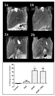
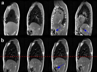
Respiratory motion-resolved MRI of the lung and liver for evaluation of cystic fibrosis
Alexander B. Hilario T., Eduardo Baum, Alexandre Franco, Li Feng, Marilisa Baldissera, Ricardo Soder, Bruno Hochhegger, Matteo Baldisserotto, Ricardo Otazo
Free-breathing MRI with reconstruction of respiratory phases using the XD-GRASP technique is demonstrated for evaluation of cystic fibrosis patients. A platform for automated data processing is developed for robust utilization in a clinical setting. XD-GRASP is validated in a cohort of 9 patients against the conventional technique.


3308.   
98 
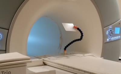
Optimized 3D ultrashort echo-time lung imaging of young children without sedation
Wingchi Kwok, Jacqueline Wameling, Mitchell Chess, Clement Ren, Gloria Pryhuber, Jason Woods
Our purpose was to develop an optimized study design for 3D ultrashort TE lung imaging of young children without sedation. Eight preterm born subjects all at age 4 were recruited. Siemens work-in-progress PETRA_D sequence was used with respiratory triggering. Repeated short scans were acquired to reduce motion artifacts. Various techniques, including video watching, practicing lying still at home and gift incentive, were employed to help achieve subject compliance. Five subjects were scanned successfully. The images revealed abnormalities including peribronchial thickening, pneumatocele and atelectasis. Our study design allows the monitoring of lung development and evaluation of lung diseases in young children.


3309.   
99 A preliminary study of in-vivo visualization of oxide oxide enhancement in focal inflammatory lesion of lung parenchyma using a 3D radial gradient-echo-based UTE sequence (CODE) - permission withheld
Soon Ho Yoon, Suh-Young Lee, Chanhee Lee, Jinil Park, Jin Mo Goo, Jang-Yeon Park
Dual-echo ultrashort echo-time CODE pulmonary MRI preliminarily succeeded in generating a positive contrast of iron oxide in rabbits with granulomatous lung disease, by using clinically-usable superparamagnetic iron-oxide nanoparticles, ferumoxytol, which was hardly achievable via conventional T2* MR sequence. This new pulmonary MR imaging biomarker possibly gives a chance to differentiate a benign inflammatory lesion from malignancy.


3310.   
100 
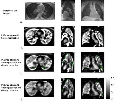
Repeatability of global percent enhancement and regional defect quantification in oxygen-enhanced 3D radial ultrashort echo time MRI - permission withheld
Wei Zha, Stanley Kruger, Kevin Johnson, Robert Cadman, Andrew Hahn, Scott Nagle, Sean Fain
Oxygen-enhanced 3D radial UTE MRI (OE-MRI) shows promise as an alternative to hyperpolarized gas MRI for evaluation of ventilation abnormalities. Ten subjects (2 normal, 2 asthmatics and 6 cystic fibrosis) underwent OE-MRI for multiple scans (test/re-test) during visits separated ≤15 days apart. The intra-subject percent signal enhancement (PSE) maps from OE-MRI were compared for median whole-lung PSE, ventilation defect percent (VDP) and spatial agreement between defects. The results suggest good agreement on two global measures, Median PSE and VDP, with low-to-moderate spatial alignment on the inter-visit segmented defects. Improvement of spatial defect repeatability will be a goal of future work.


3311.   
101 
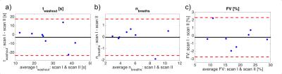
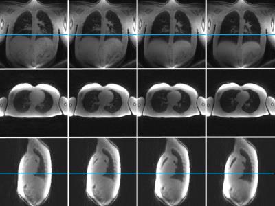
Repeatability of regional lung ventilation quantification using 19F fluorinated gas washout magnetic resonance imaging in free breathing
Marcel Gutberlet, Andreas Voskrebenzev, Agilo Kern, Till Kaireit, Jens Hohlfeld, Frank Wacker, Jens Vogel-Claussen
Since quantification of regional lung ventilation using 19F fluorinated gas washout imaging in free breathing is feasible even in obstructed lungs, it may improve diagnosis, monitoring and therapy of obstructive lung diseases like asthma and COPD. However, for application in clinical studies the knowledge of the accuracy of this technique is important.  Repeatability of the 19F gas washout parameters washout time, number of breaths and fractional ventilation between to scans was assessed in eight healthy volunteers. Due to the excellent repeatability of the number of breaths and fractional ventilation, regional lung ventilation can be accurately quantified using 19F gas washout MRI in free breathing.


3312.   
102 
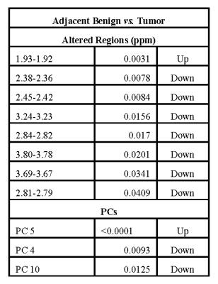
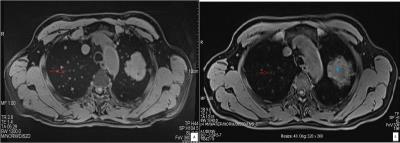
MR Elastography vs. Diffusion-weighted MRI in Characterization of Anterior Mediastinal Solid Tumours - permission withheld
Wei Tang, Ning Wu, Yao Huang
Anterior mediastinal solid tumours were routinely interpreted on CT and MR imaging. However, imaging features of these tumours could be non-specific; evidence suggestive of definitive diagnosis was needed and obtained with invasive procedure of biopsy for some cases. In the present study, we attempted to extent the application of MR elastography to the mediastinum in characterization of anterior mediastinal solid tumours, with the comparison to diffusion-weighted MRI. Thirty-four patients histologically-confirmed with thymic carcinoma in 10, thymoma in 10 and lymphoma in 14 were evaluated. It was found that stiffness value measured on elastogram was significantly higher in thymic carcinoma than that of thymoma and lymphoma. However, measurements of apparent diffusion coefficient (ADC) were not significantly different among three groups. It was demonstrated that MR elastography reflecting the mechanical properties of tumours can be used to characterize anterior mediastinal solid tumours, particularly in distinguishing thymic carcinoma from lymphoma.


3313.   
103 
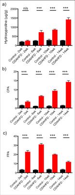
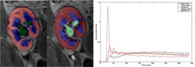
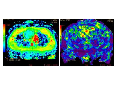
Mapping Vascular Behaviour in Lung Perfusion of two-year old children after Congenital Diaphragmatic Hernia Repair using  Tissue Similarity Maps
Dimitrios Markellos, Meike Weis, Thomas Schaible, Stefan Schoenberg, Lothar Schad, Frank Zöllner
Congenital Diaphragmatic Hernia (CDH) is associated with lung hypoplasia reflected in decreased pulmonary microcirculation and secondary pulmonary hypertension. Using tissue similarity maps (TSM), Haacke et al. demonstrated mapping vascular behaviour and calculating relative pulmonary blood volume (rPBV) maps in perfusion weighted imaging of the brain. In this study we have investigated the significance of the TSM technqiue for lung perfusion imaging in two-year old children after CDH repair.


3314.   
104 
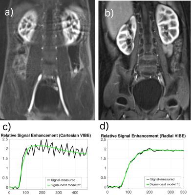
Diagnosing Lung Nodules on various MRI Imaging: comparison of T1-Weighted-3D VIBE-dixon sequence and T1-weighted 3D star vibe sequence
Chuangbo Yang, Nan Yu, Qi Yang, Shaoyu Yu, Youmin Guo, Liya Ma
T1-weighted 3D Star VIBE sequence obtaining scan under free breathing can provide high-resolution imaging. Therefore, our study was to assess the accuracy of various MRI sequences for the diagnosis of pulmonary nodules and to estimate the ability of MRI for display the morphology of pulmonary nodules. We concluded that T1-weighted 3D Star VIBE sequence allow for identification of patients with pulmonary nodules with high detection rate. It can also provide more information of nodules than routine T1-weighted 3D VIBE sequence dose.


3315.   
105 
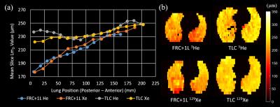
Exploring Lung Inflation Mechanisms with 3D 3He and 129Xe Whole Lung Morphometry Mapping
Ho-Fung Chan, Juan Parra-Robles, Guilhem Collier, Jim Wild
Alveolar dimensions change with lung inflation, however, there is currently no consensus on whether this change is primarily due to expansion and if alveolar recruitment plays a role. Here, multiple b-value DW-MRI was used to investigate lung inflation mechanisms in-vivo, and 3D 3He and 129Xe whole lung morphometry (LmD) maps were acquired in five healthy volunteers at lung inflation states of FRC+1L and TLC. Decreases in the anterior to posterior gravitational gradient were observed between FRC+1L and TLC with both nuclei. Smaller than predicted LmDvalues at TLC suggests that both alveolar expansion and recruitment may occur during lung inflation.


3316.   
106 
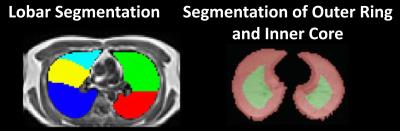
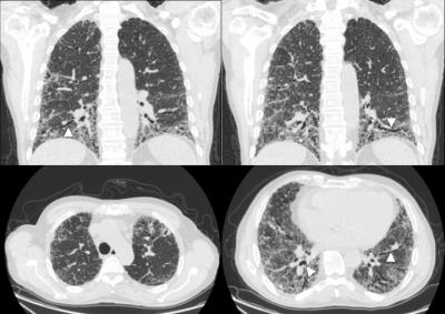
Analysis of Regional Lung Function Detected by Hyperpolarized Xenon-129 MRI in Subjects with Interstitial Lung Diseases
Kun Qing, Talissa Altes, John Mugler, III, Nicholas Tustison, Kai Ruppert, Jaime Mata, Yun Shim, G.Wilson Miller, Iulian Ruset, F.William Hersman, Borna Mehrad
Previous study showed that hyperpolarized xenon-129 MRI is highly sensitive in detecting functional changes in lungs with interstitial lung diseases (ILD). The degree to which these changes vary regionally in the lung has not been determined, however. In this work, we compared abnormalities in lung function in different regions of the lung, and found significant differences in xenon-129 gas uptake between subjects with ILD and controls. These results support that xenon-129 MRI may provide unique information about lung physiology associated with lung fibrosis.


3317.   
107 Self-gated ultra-short echo time lung MRI for quantitative ventilation assessment
Lenon Pereira, Tobias Wech, Andreas Weng, Andreas Kunz, Simon Veldhoen, Thorsten Bley, Herbert Köstler
Self-gated Non-Contrast-enhanced Functional Lung MRI (SENCEFUL) is a technique based on a FLASH sequence that can assess ventilation and perfusion in free-breathing without the use of contrast agents. However, SENCEFUL suffers from the fast T2-induced signal decay in the lung parenchyma and offers no real 3D coverage. Thus, this work presents the use of a 3D ultra-short echo time (UTE) sequence in SENCEFUL MRI for the generation of ventilation-weighted maps.


3318.   
108 
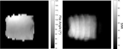
Steady-state Free Precession for Improved Signal to Noise in Lung Ventilation Imaging with 19F Perfluoropropane at 1.5 T
Adam Maunder, Neil Stewart, Madhwesha Rao, Fraser Robb, Jim Wild
Fluorinated gas MRI is a promising method for pulmonary ventilation imaging that does not require additional polarization equipment. To date, short echo time spoiled gradient echo sequences with long repetition times relative to T1 have been employed for lung ventilation imaging in humans. Here, we present an optimization of steady state free precession sequences for imaging of C3F8 gas in lungs, and demonstrate that image signal-to-noise ratio may be improved by exploiting the short T1, and relatively long T2.


3319.   
109 
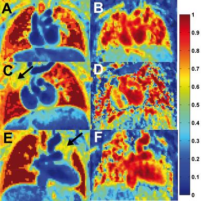
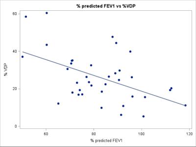
Hyperpolarized Helium-3 MRI Insights into Subtypes of Emphysema in Chronic Obstructive Pulmonary Disease (COPD)
Yanping Sun, Christian Lo Cascio, Pallavi Balte, Jia Guo, Emilia Hermann, Firas Ahmed, Belinda Dsouza, Robert Steiner, Jay Leb, Casandra Almonte, Paul Hughes, Stephen Dashnaw, James Wild, Martin Prince, Emlyn Hughes, Benjamin Smith, Eric Hoffman, R. Graham Barr
To examine the association of ventilation defects on 3He MRI with emphysema subtypes, participants (n=41) between 60 and 85 years of age with 10 or more packyears of smoking were studied.  Ventilation defect percentage (VDP) was associated with percent emphysema (r =0.43; p=0.008) and, with borderline statistical significance, extent of visual emphysema (r=0.31; p=0.07). There was no relationship between VDP and extent of centrilobular (p=0.61) or panlobular (P=0.98) emphysema. VDP was associated with extent of paraseptal emphysema (PSE) (r=0.42; p=0.01). These findings suggest that small airways disease may be a component of PSE but not other subtypes of emphysema.


3320.   
110 Temporally-resolved volumetric imaging (4DMRI) of the lungs
Zarko Celicanin, Alina Giger, Grzegorz Bauman, Philippe Cattin, Oliver Bieri
Treatment planning relies on accurate organ motion modeling. Temporally-resolved volumetric imaging (4DMRI) of the lungs using a recently developed ultra fast steady state free precession sequence was attempted. No artifacts were present in the lungs, while image stacking was accurate with no significant image sorting issues.


3321.   
111 
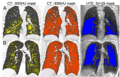
Ultra-Short Echo Time MRI Quantification of Airspace Enlargement in Bronchopulmonary Dysplasia and Alpha-1 Antitrypsin Deficiency: Parenchyma Destruction, Air trapping or Both?
Heather Young, Dante Capaldi, Khadija Sheikh, David McCormack, Cory Yamashita, Grace Parraga
UTE MRI signal-intensity has not yet been evaluated in young patients with AATD and BPD, where there may be different mechanisms of parenchyma and airway destruction.  There is the potential to demonstrate UTE MRI as a quantitative-measurement-tool for longitudinal and treatment-response evaluations in these vulnerable patients.  We evaluated UTE MRI and CT using -950HU and -856HU radiodensity-thresholds, and a ‘sliding-threshold’ for the UTE image, identifying regions with low-signal-intensity for multiple threshold values. Regions of normalized UTE signal-intensity <29 suggest airspace enlargement, and demonstrate the potential utility of UTE MRI in quantifying this without ionizing-radiation in AATD and BPD subjects.


3322.   
112 T2* Quantification of the Lung Using 3D Ultrashort Echo Time Cones Sequences - permission withheld
Amin Nazaran, Michael Carl, Yanchun Zhu, Yajun Ma, Eric Chang, Jiang Du
Imaging of the lungs without a contrast agent is a challenging task due to the low SNR. The main challenge stems from the air in the lungs, which causes low proton density and high magnetic susceptibility at interfaces with tissue. Ultra short echo time (UTE) techniques have the potential to acquire high signal-to-noise ratio (SNR) images due to their short achievable TEs, much less than 1ms.  The lung imaging based on 3D radial UTE sequences were previously reported. In this work, we applied three-dimensional UTE with Cones trajectories (3D UTE-Cones) to quantify lungs’ tissue.


3323.   
113 Self-gated Non–Contrast-Enhanced Functional Lung (SENCEFUL) MRI for Evaluation of Endoscopic Lung Volume Reduction in Patients with Lung Emphysema
Simon Veldhoen, Andreas Weng, Tobias Wech, Andreas Kunz, Stephanie Sauer, Thorsten Bley, Herbert Köstler
Purpose of the study was to assess the feasibility of SENCEFUL ventilation imaging for evaluation of therapy success after endoscopic lung volume reduction. Two patients with lung emphysema who underwent bronchial valve implantation were scanned before and after the procedure using SENCEFUL-MRI. Color-coded lung ventilation maps and quantitative ventilation values were pre- and postinterventionally compared. The first patient showed a ventilation defect corresponding to the location of the implanted valves with reduction of the quantitative ventilation. The second patient did not present such alterations and was later classified as non-responder to therapy. SENCEFUL-MRI detected clinically successful and non-successful outcomes correctly.


3324.   
114 Single Breath CSSR-DWI: A New Method to Simultaneously and Quantitatively Assess the Changes of Respiratory Membrane and Pulmonary Microstructure in Human
Junshuai Xie, Huiting Zhang, Xiuchao Zhao, Haidong Li, Sa Xiao, Ke Wang, Hao Yang, Xianping Sun, Guangyao Wu, Chaohui Ye, Xin Zhou
The blood-gas exchange function and the pulmonary microstructure are generally affected by the lung inflation levels. Here We developed a new method Single Breath CSSR-DWI to simultaneously quantify the respiratory membrane and the pulmonary microstructure via hyperpolarized 129Xe in a single breath. A new parameter SVRd/g was defined from ADC and SVRd to characterize the ‘%-predicted dissolved SVR’. Human pulmonary functional and structure information were successfully obtained in a single breath via hyperpolarized 129Xe. Compared to the healthy young subjects, SVRd/g of the asymptomatic aged subjects decreases while the size of the pulmonary microstructure increases.


3325.   
115 Quantify Pulmonary Gas-Exchange Function with Hyperpolarized 129Xe CEST MRI
Haidong Li, Zhiying Zhang, Xiuchao Zhao, Yeqing Han, Xianping Sun, Chaohui Ye, Xin Zhou
In this study, the pulmonary gas-exchange function was quantitatively evaluated globally and regionally by hyperpolarized 129Xe MRI using the method of chemical exchange saturation transfer (CEST). A new parameter, named as pulmonary gas consumption time constant (Toc), was proposed to characterize the gas exchange function. The parameter showed significant difference between the COPD and healthy rats, and we believe it will be a useful parameter in evaluating the pulmonary function.


3326.   
116 
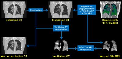
A spatial comparison of CT-based surrogates of ventilation with hyperpolarized Helium-3 & Xenon-129 MRI
Bilal Tahir, Paul Hughes, Helen Marshall, Neil Stewart, Felix Horn, Guilhem Collier, Graham Norquay, Kerry Hart, James Swinscoe, Matthew Hatton, Jim Wild, Rob Ireland
Synopsis: Image registration of lung CT images acquired at different inflation levels has been proposed as a surrogate method to map lung ‘ventilation’. However, this technique requires validation against established ventilation modalities such as hyperpolarised gas MRI. Here, we develop an image acquisition and analysis strategy to facilitate direct spatial correlation of ventilation CT with both hyperpolarised 3He & 129Xe MRI and apply our method to a cohort of lung cancer patients.


3327.   
117 
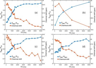
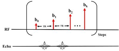
Evaluation of 129Xe-RBC signal dynamics and chemical shift in the cardiopulmonary circuit using hyperpolarized 129Xe NMR
Graham Norquay, Neil Stewart, Jim Wild
The signal dynamics of hyperpolarized 129Xe in the pulmonary capillaries and veins was evaluated by employing multi-TR pulse sequences, with TR values ranging from 100-6000ms. The ratio of signal from 129Xe dissolved in red blood cells (RBC) and tissue/plasma (TP) was found to increase for longer TRs whilst the 129Xe-RBC chemical shift was observed to decrease with increasing TR. This observed chemical shift difference is unprecedented, and understanding the nature of the underlying mechanisms causing this shift is crucial for future in vivo experiments using 129Xe-RBC chemical shift as a measure of blood oxygenation.


3328.   
118 Assessment of acinar destruction in idiopathic pulmonary fibrosis with hyperpolarised 3He gas diffusion-weighted MRI: reproducibility of ADC metrics and correlation with physiological parameters of disease severity.
Nicholas Weatherley, Ho-Fung Chan, Neil Stewart, Laura Saunders, Guilhem Collier, Graham Norquay, Madhwesha Rao, Laurie Smith, Matthew Austin, Stephen Renshaw, Stephen Bianchi, Jim Wild
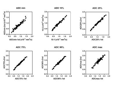
Outcome metrics from diffusion-weighted MRI (DW-MRI) were evaluated for reproducibility and clinical correlation in a cohort of patients with idiopathic pulmonary fibrosis (IPF). It was hypothesized that fibrotic lung tissue in IPF undergoes changes that increase the rate of intra-acinar Brownian diffusion.

Apparent diffusion coefficient (ADC) mean, median and histogram metrics are highly reproducible and correlate both quantitatively with regional fibrosis on CT and qualitatively with carbon monoxide transfer coefficient (KCO) from pulmonary function tests. DW-MRI imaging metrics may provide novel insights into microstructural disease severity in IPF and may prove to be a useful non-ionising imaging biomarker of disease.


3329.   
119 
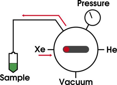
Hyperpolarized xenon by d-DNP using the clinical GE SpinLab polarizer system
Christian Mariager, Steffen Ringgaard, Jan Ardenkjær-Larsen, Christoffer Laustsen
Hyperpolarized (HP) 129Xe have been demonstrated as a useful probe for magnetic resonance (MR) lung imaging and show promise for in vivo perfusion imaging and brown adipose tissue characterization. Reports of large polarization enhancements for 129Xe using dynamic nuclear polarization (DNP) have raised expectations that DNP can be an alternative to the standard spin exchange optical pumping (SEOP) method. We show that it is possible to produce HP 129Xe gas using the clinical GE SpinLab polarizer, thus extending the practical use of the system beyond the primary purpose of hyperpolarizing liquid biomolecules.


3330.   
120 Combination of Variable and Constant Flip Angles for Hyperpolarized 129Xe Multi-b Diffusion MRI in a Single Breath-hold
Weiwei Ruan, Jianping Zhong, Ming Zhang, He Deng, Yeqing Han, Chaohui Ye, Xin Zhou
To propose a novel flip angle scheme for hyperpolarized gas multi-b diffusion MRI. It combined the variable and constant flip angles and was named as the combination of variable and constant flip angle (CVCFA). Computer simulation was used to systematically compare the proposed flip angle scheme with the common-used flip angle schemes, including the interleaved constant flip angle (ICFA) and the variable flip angle (VFA) schemes. The CVCFA scheme was used for hyperpolarized xenon diffusion MRI to measure the pulmonary morphology in rats, noninvasively. The results showed the CVCFA was suited for the hyperpolarized gas multi-b diffusion MRI.
 

The International Society for Magnetic Resonance in Medicine is accredited by the Accreditation Council for
Continuing Medical Education to provide continuing medical education for physicians.