CEST/MT/NOE: Animal Models & Human Translation
Electronic Poster
Contrast Mechanisms
Monday, 24 April 2017
| Exhibition Hall |
17:15 - 18:15 |
| |
|
Computer # |
|
3735.
 |
73 |
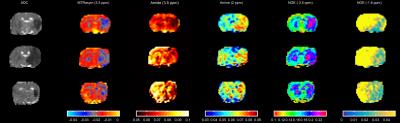
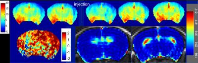
CEST imaging of an unlabelled chemotherapy agent (gemcitabine) 30 minutes after administration in a mouse model of colorectal cancer 
Thomas Roberts, May Zaw-Thin, Angela D'Esposito, Yanan Zhu, John Connell, Mark Lythgoe, Simon Walker-Samuel
Imaging of drug delivery is useful for improving our understanding of the physical factors that contribute to the efficacy of anticancer therapies. Gemcitabine, a standard chemotherapeutic agent, has two hydroxyl groups and one amine group, making it potentially amenable to CEST imaging via proton exchange with tissue water. Recently, Li et al. (2016) showed that many anticancer drugs can induce CEST contrast and demonstrated that liposome-encapsulated gemcitabine can be imaged 5-hours post-administration in a pre-clinical model of cancer1. Here, we extend this exciting work and investigate the use of CEST to image acute gemcitabine uptake within 30 minutes of administration.
|
|
3736.
 |
74 |
In Vivo Tracking of Hyaluronic Acid Hydrogel Degradation Using Temporal Evolution of Chemical Exchange Saturation Transfer Signal in a Mouse Subcutaneous Injection Model
Mohammed Salman Shazeeb, Rubina Corazzini, Dinesh Bangari, Robert Fogle, Jennifer Johnson, Paul Konowicz, Xiaoyou Ying, Pradeep Dhal
Hyaluronic acid (HA) hydrogels have a wide range of applications in biomedicine from regenerative medicine to drug delivery applications. In vivo quantitative assessment of these hydrogels using magnetic resonance imaging (MRI) provides a powerful technique to assess the biodegradability of HA hydrogels. This study investigated the potential of chemical exchange saturation transfer (CEST) MRI in tracking HA hydrogels with varying degradation profiles in vivo in a mouse subcutaneous injection model over 77 days. Since CEST-MRI provides a unique chemical signature to visualize HA hydrogels, this technique can be used as a guide in hydrogel optimization process for drug delivery applications.
|
|
3737.
 |
75 |
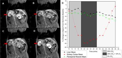
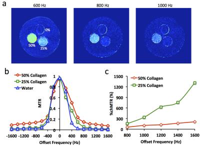
Magnetization Transfer MRI Noninvasively Detects Renal Fibrosis Swine Atherosclerotic Renal Artery Stenosis at 3.0 T - permission withheld
Kai Jiang, Christopher Ferguson, John Woollard, Roger Grimm, James Krier, Xiangyang Zhu, Lilach Lerman
In this study, we tested the capability of magnetization transfer (MT) imaging for measuring renal fibrosis in a swine model of atherosclerotic renal artery stenosis (ARAS) at 3.0 T. A collagen phantom study was performed to select appropriate offset frequencies of MT pulses for collagen detection. In an in vivo study, the MT ratio (MTR) and percent change in MTR between two offset frequencies were quantified, and both showed a good correlation with renal fibrosis measured ex vivo by Picro-Sirius red staining, supporting the use of MT to assess renal fibrosis in swine ARAS.
|
|
3738.
 |
76 |
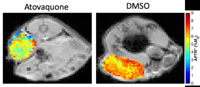
Modification of tumour hypoxia with Atovaquone measured by CEST MRI
Kevin Ray, James Coates, Rathi Puliyadi, Thomas Ashton, Paul Kinchesh, Sean Smart, Michael Chappell, Geoff Higgins, Nicola Sibson
Tumours often have areas of hypoxia, which renders cancer cells resistant to many therapies. Recently the anti-malarial drug Atovaquone has been shown to modify the oxygen consumption rate of cancer cells, reducing tumour hypoxia. Non-invasive mapping of tumour hypoxia would be particularly useful for translation of these preclinical findings into a clinical environment. Here, we tested the hypothesis that CEST MRI could be used to visualise changes in tumour hypoxia.
|
|
3740.
 |
78 |
In Vivo Detection of Reactive Oxygen Species Using MRI with Endogenous Contrast - video not available
Rong-Wen Tain, Alessandro Scotti, Weiguo Li, Xiaohong Joe Zhou, Riya Thomas, Leon Tai, Kejia Cai
Reactive oxygen species (ROS) contribute to pathogenesis of many human diseases including Parkinson’s and Alzheimer's diseases, cancer, and diabetes. There is a crucial need for using fully noninvasive imaging to further evaluate the role of ROS in pathogenesis and the potential treatment strategies. Our previous phantom studies demonstrated that ROS containing unpaired electrons can be detected with endogenous CEST and T1 weighted contrasts. However, in vivo detection of ROS using MRI has not yet been demonstrated. This study therefore aimed to demonstrate the feasibility of in vivo ROS detection using endogenous MRI.
|
|
3741.
 |
79 |
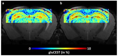
Imaging of neuronal compartment using gluCEST method
Jérémy Pépin, Pierrick Jego, Julien Valette, Gilles Bonvento, Julien Flament
GluCEST imaging has been proposed to image brain glutamate distribution with a better resolution than spectroscopic methods and has many potential applications for the study of neurodegenerative diseases. In this study, we pushed further the limits of gluCEST imaging by combining high magnetic field and high performance cryoprobe to acquire gluCEST images with the best resolution so far. Thanks to the organization of hippocampal cell layers and the high resolution, we acquired gluCEST data in regions mostly reflecting the neuronal compartment.
|
|
3742.
 |
80 |
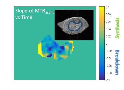
Assessment of hepatic glycogen metabolism ex vivo and in vivo in mice using chemical exchange saturation transfer MRI
Corin Miller, Jin Cao, Chunlian Zhang, Eduard Chekmenev, Bruce Damon, Alan Cherrington, John Gore
Despite being integral to whole body glucose homeostasis, liver glycogen remains difficult to measure non-invasively. Recent works have demonstrated the feasibility of detecting liver glycogen using CEST MRI. In this presentation we present data that builds upon this observation and investigate whether CEST can be used to monitor glycogen synthesis and breakdown in mice in real time both ex vivo in a perfused liver system, and in vivo. Treatment with hyperglycemia or glucagon resulted in increases or decreases, respectively, in the CEST MTRasym AUC over time. This demonstrates that CEST-based approaches can be used to non-invasively monitor glycogen metabolism.
|
|
3743.
 |
81 |
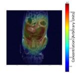
GlucoCEST MRI and brain blood barrier permeability in the mouse brain
Maria Yanez Lopez, Nicoleta Baxan, Miriam Ries, David Sharp, Magdalena Sastre
The aim of the study is to examine the feasibility of using GlucoCEST to evaluate subtle BBB dysfunction in the mouse brain, by comparing it with conventional gadolinium DCE. DCE and GlucoCEST showed no significant differences between WT and 5XFAD mice (3 months). However, the degree of the response after injection was similar for DCE and GlucoCEST for all animals except one, indicating shared contributions to the signal and supporting the potential for biodegradable d-glucose as a cheap, low-risk alternative/complement to DCE MRI. More work is required to assess GlucoCEST sensitivity to low-level BBB dysfunction, such as in Alzheimer’s disease.
|
|
3744.
 |
82 |
2-Deoxyglucose-Weighted MR Imaging in Rodent Brain Using Inverse Z-Spectrum Analytic Scheme
Ping-Huei Tsai, Fei-Ting Hsu, Hua-Shan Liu, Hsiao-Wen Chung, Yu-Chieh Kao, Chia-Feng Lu, Huai-Lu Chen, Paul Blakeley, Gilbert Aaron Lee, Cheng-Yu Chen
Our proposed method provides an alternative to extract glucose profile and could be more robust to the field drift, which may be helpful in the implementation of in vivo brain glucoCEST imaging for further application.
|
|
3745.
 |
83 |
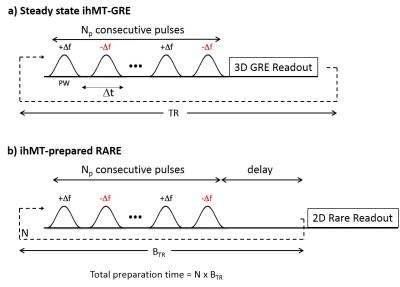
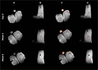
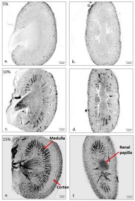
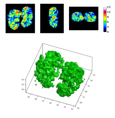
3D-CEST Imaging of a Mouse Model of Polycystic Kidney Disease
Shanrong Zhang, Matanel Yheskel, Vishal Patel, Masaya Takahashi, A. Dean Sherry
The objective is to develop a new 3D-CEST imaging method (3-dimensional chemical exchange saturation transfer) to investigate a mouse model of polycystic kidney disease (PKD). It is based on 3D magnetization prepared rapid acquisition gradient echo sequence (3D MPRAGE) by applying a pre-saturation pulse consisting of three continuous Gauss shaped pulses.
|
|
3739.
 |
77 |
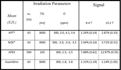
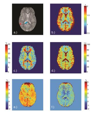
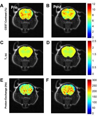
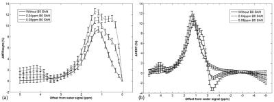
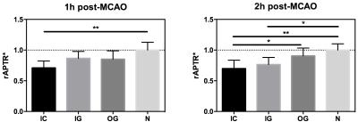
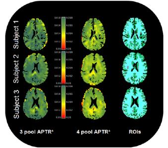
Investigation into the origin of the APT MRI signal in ischemic stroke
Kevin Ray, James Larkin, Brad Sutherland, George Harston, Andrew Baldwin, Alastair Buchan, Peter Jezzard, James Kennedy, Michael Chappell, Nicola Sibson
Studies employing CEST MRI to study ischemic stroke focus on the sensitivity of amide proton transfer (APT) MRI signals to tissue pH, assuming identical intracellular protein concentration as healthy tissue. This study shows that whilst cytoplasmic protein concentration remains stable in penumbral stroke regions, it decreases in the infarct core. By analysing APT MRI data with APTR*, which is specifically sensitive to amide proton exchange effects, we demonstrate that the APT signal change in infarct core is dominated by decreased protein concentration, whilst penumbral APT changes can be attributed to decreased tissue pH.
|
|
3746.
 |
84 |
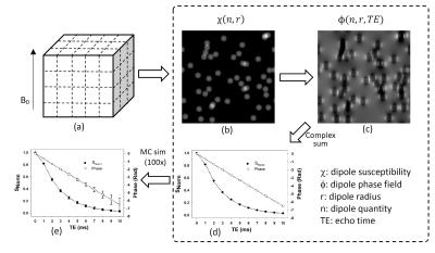
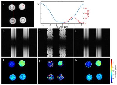
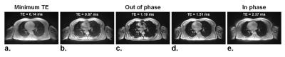
Z-spectrum acquisition and interpretation in the presence of fat: influence of imaging parameters
Shu Zhang, Jochen Keupp, Ivan Dimitrov, Robert Lenkinski, Elena Vinogradov
CEST-MRI is increasingly evolving from brain to body applications. One of the known problems in body imaging is the presence of strong lipid signals. Their influence on the CEST signal is acknowledged but underexplored. The goal is to investigate the effects of lipids on the Z-spectrum taking TE into account. We performed simulations and verified the results in phantoms and in vivo. We demonstrate the mutual influence of fat fraction and TE on the Z-spectrum for gradient echo based sequences. This study provides a systematic understanding of lipid artifacts in CEST imaging and lays the foundations for their efficient removal.
|
|
3747.
 |
85 |
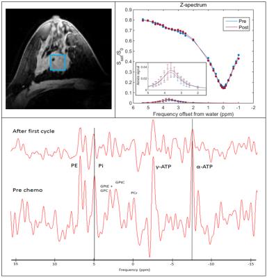
Monitoring neoadjuvant chemotherapy in breast cancer patients using CEST and 31P-MRS at 7 tesla
Erwin Krikken, Vitaliy Khlebnikov, Moritz Zaiss, Wybe J.M. van der Kemp, Tijl A. van der Velden, Hanneke W.M. van Laarhoven, Dennis W.J. Klomp, Jannie P. Wijnen
Treatment monitoring is of importance for breast cancer patients receiving systemic therapy. Metabolic imaging methods such as CEST and 31P-MRS may have potential to predict treatment efficacy in an early stage of the treatment. In this study we assessed the amide proton transfer (APT) signal and the pH change in breast cancer patients before and after the first cycle of neoadjuvant chemotherapy to explore the relation between APT and pH. We observed changes in both the APT signal and the pH between the two measurements. These changes may serve as biomarkers for predicting treatment response to NAC in an early stage.
|
|
3748.
 |
86 |
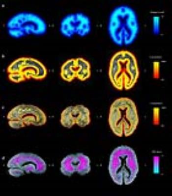
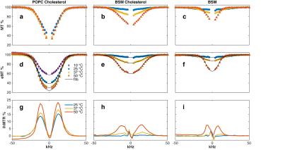
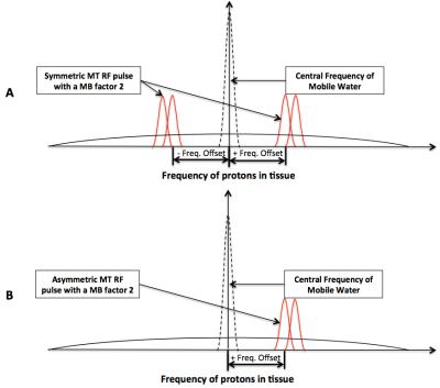
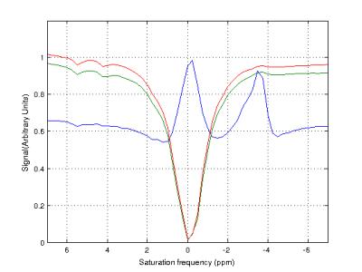
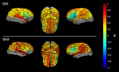
Are MT and NOE (at -3.5 ppm) in z-spectroscopy coupled in the brain?
Nicolas Geades, Olivier Mougin, Simon Shah, Penny Gowland
The origin of the Nuclear Overhauser Enhancement (NOE) signal observed in the CEST spectrum of the brain is still under debate. The effect is detected upfield from water, in the frequency range of non-exchangeable aliphatic/olefinic protons, indicating that the transfer of magnetization is not occurring via proton or chemical exchange; furthermore the lineshape is relatively narrow, suggesting the signal is coming from mobile protons with T2 of the order of 300μs. This study investigates the correlation between NOE and MT in the human brain at 7T, and shows the two effects are strongly coupled, across a wide age range.
|
|
3749.
 |
87 |
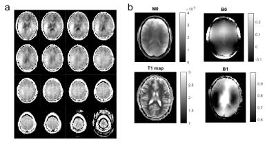
3D Quantitative CEST MRI of the human brain at 9.4T
Moritz Zaiss, Philipp Ehses, Klaus Scheffler
Selective quantitative CEST MRI was shown to be feasible at 7T and for single-slice readout. In this study we extend qCEST MRI to a field strength of 9.4 T making use of the increase spectral resolution, and by employing a single-shot 3D CEST approach, more coverage of the human brain was realized.
|
|
3750.
 |
88 |
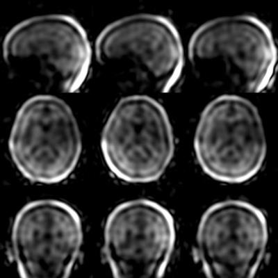
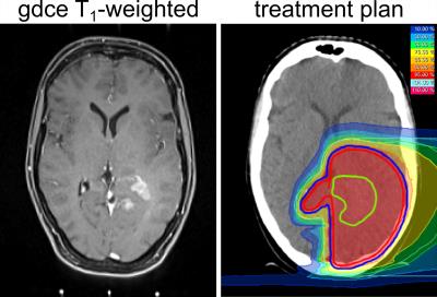
A longitudinal study of brain tumors in the course of radiotherapy using protein CEST MRI at 7T
Jan-Eric Meissner, Andreas Korzowski, Sebastian Adeberg, Steffen Goerke, Nicolas Behl, Heinz-Peter Schlemmer, Jürgen Debus, Mark Ladd, Peter Bachert, Daniel Paech
Chemical Exchange Saturation Transfer (CEST) offers unique contrasts sensitive to micro environmental information such as protein concentration and pH. Thus CEST could be a promising biomarker to investigate radiotherapy-induced changes in tissue. In this study we examine brain tumor patients in the course of definitive radiotherapy on a 7 T whole-body scanner using the relaxation compensated contrast AREX. We compare the results of CEST imaging to Single Voxel Spectroscopy and high-resolution T2-weighted imaging.
|
|
3751.
 |
89 |
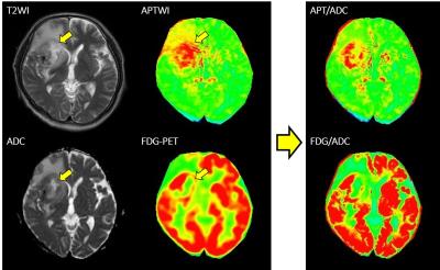
Voxel-wise comparison of amide proton transfer (APT) weighted image and fluorodeoxyglucose (FDG)-PET in brain tumors with a PET/MR system
Koji Sagiyama, Yuji Watanabe, Ryotaro Kamei, Sungtak Hong, Jochen Keupp, Hiroshi Honda
Amide proton transfer (APT) imaging has been reported to be useful for assessing malignancy or evaluating treatment efficacy. In this study, we evaluated the validity of APT signals in brain tumors by direct voxel-wise comparison with standardized uptake values (SUVs) from fluorodeoxyglucose positron emission tomography (FDG-PET) on a PET-magnetic resonance (PET/MR) system. APT imaging showed discrepancies with FDG-PET due to structural inhomogeneity, and the correlation between APT signals and SUVs was poor. The correlation was significantly improved after correcting for the apparent diffusion coefficient (ADC). APT/ADC could be a reliable metabolic marker with better correlation with SUVs.
|
|
3752.
 |
90 |
Repeatability study of APT CEST quantification techniques for identification of ischaemic penumbra in stroke
Yunus Msayib, George Harston, Yee Tee, Fintan Sheerin, Nicholas Blockley, Thomas Okell, Peter Jezzard, Stephen Payne, James Kennedy, Michael Chappell
The aim of this study was to identify the analysis technique with the optimum repeatability for quantifying APT CEST imaging for use in the clinical setting. The repeatability of eight quantification techniques was assessed across imaging time points in healthy subjects, and between the contralateral hemispheres of stroke patients. Model-based techniques exhibited better repeatability compared to simpler model-free methods, and are thus better-suited for use in clinical imaging at 3T.
|
|
3753.
 |
91 |
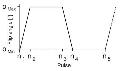
Chemical exchange rotation transfer (CERT) of human brain at 3 T
Eugene Lin, Hua Li, Zhongliang Zu, Elizabeth Louie, Xiaoyu Jiang, Daniel Gochberg
It has been shown that the changes in chemical exchange saturation transfer (CEST), and specifically amide proton transfer (APT) and nuclear Overhauser effect (NOE), reflect abnormal tissues in tumor, stroke and other diseases. However, quantitative and specific imaging of these effects is challenging due to the influences from asymmetric magnetization transfer and direct water saturation. These obstacles can be avoided with chemical exchange rotation transfer (CERT), which is a pulsed version of CEST with the constraint of constant average power and varying rotation angle. In this study, we present initial CERT results in human brain at 3 T, with the goal of quantifying APT and NOE.
|
|
3754.
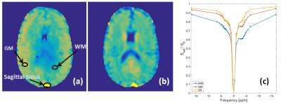 |
92 |
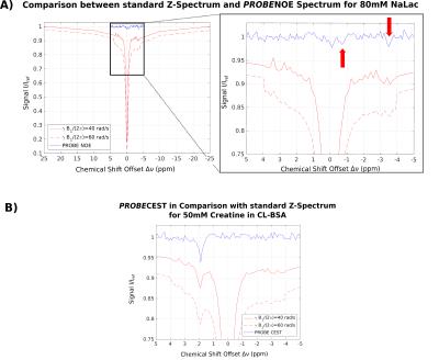
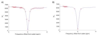
The Chemical Exchange Saturation Transfer (CEST) and Nuclear Overhauser Enhancement (NOE) effects observed in human blood at 7 T under different physiological conditions.
Simon Shah, Olivier Mougin, Andrew Carradus, Nicolas Geades, William Morley, Penny Gowland
CEST maps at 7T display higher APT and amine signal from the sagittal-sinus compared to elsewhere in the brain. We investigate CEST and NOE signals detected in ex-vivo blood under with varying oxygenation, haematocrit levels, pH and cell structure. Showing that human blood produces significant amounts of CEST and NOE, dominated by the cellular components of blood and independent of blood oxygenation. Clotting and lysing red-blood-cells has no impact on the observed CEST and NOE. We also observed increased APT and NOE with increasing pH. These result are important when interpreting exchange in conditions associated with blood volume change.
|
|
3755.
 |
93 |
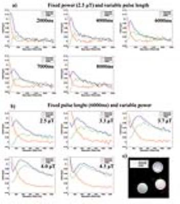
Optimization of Amide Proton Transfer (APT) Imaging Experimental Parameters for Brain Tumors: Does RF Saturation Length Always Increase APT Contrast?
Hye-Young Heo, Yi Zhang, Shanshan Jiang, Jinyuan Zhou
Current APT imaging studies have used a moderate repetition time or relaxation delay to reduce the scan time. However, when a relatively short relaxation delay compared with T1 is applied, the steady-state water longitudinal magnetization is reduced, resulting in a decrease in APT effect. Therefore, experimental parameters must be optimized on a combination of the RF saturation power, saturation length, and relaxation delay. Here, we quantitatively investigated the dependence of APT and NOE signals on the experimental parameters using Bloch simulations and rat brain tumor models at 4.7 T.
|
|
3756.
 |
94 |
Development of Glutamate-Sensitive CEST at Clinical Field Strength for In Vivo Application
Kristin O'Grady, Samantha By, Bailey Box, Seth Smith
Cognitive impairment (CI) is a significant symptom of multiple sclerosis (MS), is the strongest predictor of unemployment in MS patients, and is critical to the decline of quality of life. There is an unmet need for imaging techniques that probe the pathological substrate of CI at a clinically relevant field strength. To address this need, we have investigated the translation of glutamate-sensitive chemical exchange saturation transfer (GluCEST) MRI to 3T, as glutamate abnormalities have been linked to CI in MS. Our results demonstrate the clinical feasibility of GluCEST imaging for application to studying cortical gray matter glutamate signals in vivo.
|
|
3757.
 |
95 |
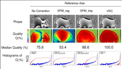
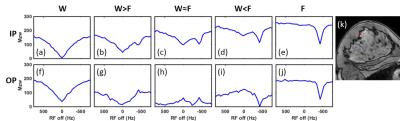
Correction of fat artifacts for unbiased CEST-MRI of the human breast at 7 T
Ferdinand Zimmermann, Steffen Görke, Johannes Breitling, Kerstin Klopries, Johannes Windschuh, Heinz-Peter Schlemmer, Mark Ladd, Daniel Paech, Peter Bachert
Chemical Exchange Saturation Transfer (CEST) MRI in the mammary gland is affected by the high fat content in the human breast. Chemical shift induced artifacts are visible in the Z-Spectrum. We showed a method to test and verify water-fat separation techniques in vitro. Transfer of the gained insights to realize water-only CEST-MRI in the human breast is currently under investigation.
|
|
3758.
 |
96 |
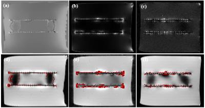
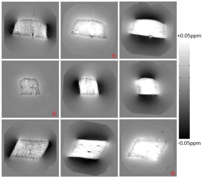
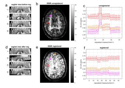
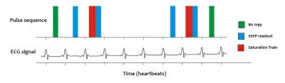
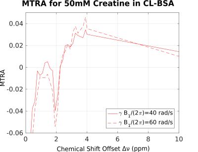
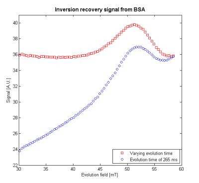
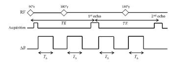
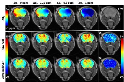
Interleaved B0-mapping during dynamic Creatine-CEST for correction of temporarily fluctuating B0 inhomogeneities during plantar flexion exercise at 7T
Esau Poblador Rodriguez, Philipp Moser, Barbara Dymerska, Siegfried Trattnig, Wolfgang Bogner
Once the time resolution and specificity of Cr-CEST become comparable with 31P-MRS, it may replace it for the diagnosis of muscular disorders and treatment assessment due to its superior spatial resolution and sensitivity. The need of a complete Z-spectrum acquisition for B0 inhomogeneity correction limits the achievable time resolution and is unable to track temporal ?B0 due to subject movement or B0 field drifts that occur during a CEST experiment. Temporal B0 field tracking allows independent B0 inhomogeneity correction for each z-spectral point, which may substantially improve the reliability of spectral CEST asymmetry analysis.
|
|