Tuesday, 25 April 2017
| Room 314 |
16:15 - 18:15 |
Moderators: Geon-Ho Jahng, Toshiaki Taoka |
Slack Channel: #s_neuro
Session Number: O04
16:15
|
0610.
 |
Diffusion tensor and restriction spectrum imaging reflect different aspects of neurodegeneration in Parkinson’s disease 
Tuva Hope, Per Selnes, Irena Rektorová, Zuzana Balážová, Anders Dale, Atle Bjørnerud, Tormod Fladby
Diffusion measures within the brain are assumed to be associated with neuronal and glial structure and integrity. In this study, we compare how diffusion tensor imaging (DTI) and restriction spectrum imaging (RSI) detect micro-structural changes within brain regions associated with motor function in Parkinson's Disease.
|
16:27
 |
0611.
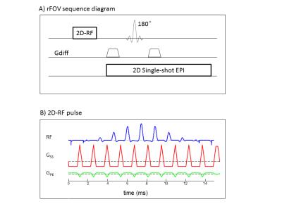 |
Lateral Dependence of Brainstem Structural Abnormalities in Parkinson’s Disease as Revealed by High-Resolution Non-Gaussian Diffusion MR Imaging
Zheng Zhong, Douglas Merkitch, Muge Karaman, Yi Sui, Jennifer Goldman, Xiaohong Zhou
Parkinson’s disease (PD) is a neurodegenerative disorder characterized by progressive degeneration of dopaminergic neurons in the substantia nigra (SN). With the ability to reveal tissue microstructural changes, non-Gaussian diffusion models with high b-values can provide a wealth of information related to the neurodegenerative process and complement the conventional Gaussian diffusion model. Non-Gaussian diffusion imaging is typically performed with limited spatial resolution and subject to image distortion. In this study, we have combined a high-resolution, distortion-free diffusion sequence with a non-Gaussian diffusion model to analyze the lateral dependence of tissue abnormalities in the SN of PD patients compared to healthy controls.
|
16:39
|
0612.
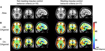 |
Pharmacological arterial spin labeling reveals distinct mesocorticolimbic blood flow in Parkinson’s disease patients with compulsive reward-driven behaviors
Daniel Claassen, Adam Stark, Charis Spears, Kalen Petersen, Scott Wylie, Nelleke van Wouwe, Robert Kessler, David Zald, Manus Donahue
The overall goal of this work is to apply pharmacological arterial spin labeling (ASL) to investigate fundamental hypotheses regarding the role of dopamine agonist (DAgonist) therapy in patients with Parkinson’s disease and impulse control behavior (ICB). Parkinson’s disease patients (n=35; age range=40-79 years; gender=23/12 males/females) receiving DAgonist therapy, with (n=17) and without (n=18) DAgonist-induced ICB were scanned at 3T using cerebral blood flow (CBF)-weighted pCASL MRI. Region-of-interest analyses revealed significantly increased bilateral ventral striatal (P<0.01) CBF in patients with ICB in the On- DAgonist state; voxel-wise analysis of CBF confirmed widespread DAgonist-induced CBF increases in mesolimbic, mesocortical, and midbrain regions.
|
16:51
|
0613.
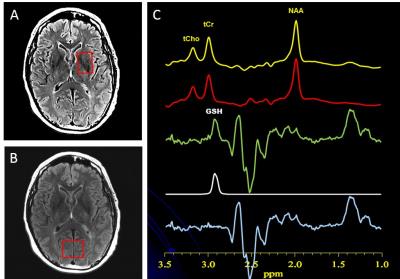 |
Striatal Glutathione Deficit in Parkinson’s Disease Measured In Vivo with J-edited 1H MRS Directly Implicates Oxidative Stress in Disorder Pathophysiology
Dikoma Shungu, Xiangling Mao, Nora Weiduschat, Aneliya Hanineva , Yize Zhao, Halinder Mangat, Guoxin Kang, James Carter, Natalie Hellmers, Claire Henchcliffe
Postmortem studies of Parkinson’s disease (PD) brain have consistently reported deficits of nigrostriatal glutathione (GSH) – the most abundant antioxidant in living tissue – of up to 40% compared to normal brain, strongly implicating oxidative stress in the pathophysiology of PD. However, direct evidence corroborating a striatal GSH deficit in PD brain in vivo is currently lacking. Using J-edited 1H MRS, this study measured striatal GSH in vivo in patients with PD and in matched control subjects, and found not only a 15% deficit of striatal GSH in PD that corroborated postmortem data, but also evidence of nigrostriatal neurodegeneration in the disorder.
|
17:03
|
0614.
 |
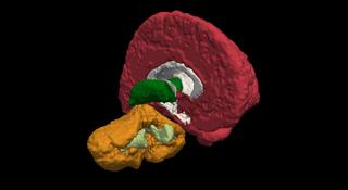
Subtypes Evaluation of Motor Dysfunction in Parkinson’s Disease using Neuromelanin-sensitive Magnetic Resonance Imaging
Tao Gong, Yuanyuan Xiang, Guangbin Wang, Richard Edden
The aim of this study was to evaluate the differences in NM-MRI between PD motor subtypes. We compared the signal intensity contrast ratios in medial and lateral regions of the SNc using NM-MRI in TD, PIGD and controls. The results demonstrated more severe signal attenuation in the medial part of SNc in PIGD patients compared with TD group. And the medial part of SNc showed high power to discriminate the PD motor subtypes. NM-MRI affords us a valuable examination method to discriminate the PD motor subtypes, providing a new evidence for the neuropathological basis of differences between the two subtypes.
|
17:15
|
0615.
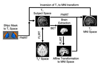 |
Reproducible detection of nigral iron deposition in Parkinson’s disease: Validation in two cohorts
Jason Langley, Naying He, Daniel Huddleston, Fuhua Yan, Xiaoping Hu
A characteristic of Parkinson's disease is neuronal loss in substantia nigra pars compacta (SNpc).In healthy subjects, SNpc contains a dense distribution of neuromelanin containing dopaminergic neurons and significant degeneration in SNpc has occurred at the time Parkinsonian symptom onset. Furthermore, extensive evidence suggests that iron deposition is related to neuronal loss and reduction of neuromelanin in SNpc. In this abstract, we examine the reproducibility of iron deposition in SNpc.
|
17:27
|
0616.
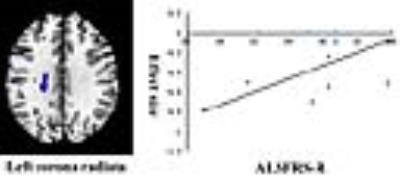 |
Altered white matter microarchitecture in amyotrophic lateral sclerosis:a voxel-based meta-analysis of diffusion tensor imaging
Guangxiang Chen, Feifei Zhang, Song Wang, Xiaoqi Huang, Qiyong Gong
The results of recent diffusion tensor imaging (DTI) studies on amyotrophic lateral sclerosis (ALS) have been inconclusive and controversial. We performed a voxel-based meta-analysis to identify statistical consensus between published DTI studies for altered white matter (WM) microarchitecture in ALS. Our findings provides a thorough profile of WM microarchitecture alterations in ALS and further evidence that the neuronal degeneration is not limited to the corticospinal tract but also includes the extra-motor areas, supporting the view of ALS being a multisystem degenerative disorder involving WM.
|
17:39
|
0617.
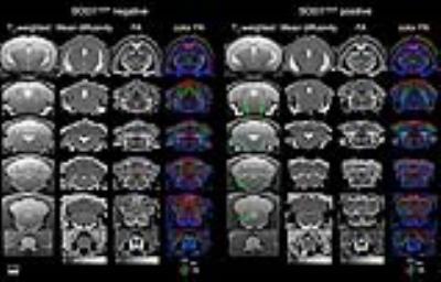 |
Diffusion and T2 characterizations in hindbrain and spinal cord in SOD1 mouse model of ALS
Luke Xie, Robert Brendza, Maj Hedehus, Sara Dominguez, Oded Foreman, Arundhati Ghosh, William Meilandt, Gai Ayalon, Richard Carano
Amyotrophic lateral sclerosis (ALS) is a devastating neurological disease characterized by motor neuron loss and eventual paralysis and respiratory failure. The SOD1 transgenic mouse model exhibits many aspects of human ALS and is useful for evaluating treatment strategies. MRI of the nervous system can provide the critical insights to motor neuron and upper body functional deterioration in ALS. In this study, we applied T2 and DTI to assess gray and white matter degeneration in the brain and cervical spine in a SOD1 mouse model.
|
17:51
|
0618.
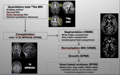 |
Sodium accumulation in primary motor areas, an early feature of amyotrophic lateral sclerosis patients
Aude-Marie Grapperon, Annie Verschueren, Adil Maarouf, Lauriane Pini, Sylviane Confort-Gouny, Jean-Philippe Ranjeva, Maxime Guye, Shahram Attarian, Wafaa Zaaraoui
Amyotrophic lateral sclerosis (ALS) is a rapidly fatal neurodegenerative disease characterized by upper (in brain) and lower (in spine) motor neuron degeneration. As conventional MRI fails to show brain motor neurons impairment in ALS, advanced techniques are needed to improve the diagnosis and to monitor the progression of the disease. In this study, brain 23Na MRI was applied in 15 ALS patients and 31 controls. A common pattern of sodium accumulation was found in patients in the primary motor areas while no atrophy was detected. The occurrence of sodium accumulation without atrophy probably reflects early neuronal injury in ALS.
|
18:03
|
0619.
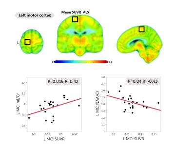 |
Glial activation measured by [11C]-PBR28 PET correlates with 1H-MRS brain metabolites in amyotrophic lateral sclerosis
Eva-Maria Ratai, Mohamad Alshikho, Nicole Zürcher, Marco Loggia, Paul Cernasov , Jennifer Fish, Raghav Seth, Sabrina Paganoni, Bruce Rosen, Jacob Hooker, Nazem Atassi
The purpose of our study was to evaluate the relationship between glial activation assessed by [11C]-PBR28 positron emission tomography, and neuronal integrity and gliosis/neuroinflammation measured by magnetic resonance spectroscopy in people with amyotrophic lateral sclerosis (ALS). Glial activation measured by increased [11C]-PBR28 uptake correlated with increased levels of myo-Inositol/Creatine, a spectroscopic marker of gliosis/neuroinflammation in the brain stem and motor cortices. Furthermore, increased [11C]-PBR28 uptake correlated with neuronal damage measured by decreased N-acetylaspartate/Creatine levels. To our knowledge, this is the first study to evaluate the relationship between glial activation, measured by [11C]-PBR28 PET, and brain metabolites assessed by MRS.
|
|