Wednesday, 26 April 2017
| Room 312 |
08:15 - 10:15 |
Moderators: Dimo Ivanov, Jennifer McNab |
Slack Channel: #s_neuro
Session Number: O07
08:15
 |
0695.
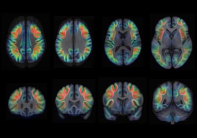 |
Interhemispheric differences of the U-shape fibre system in the human brain 
Francisco De Santiago Requejo, Pedro Luque-Laguna, Ahmad Beyh, Steven Williams, Marco Catani, Flavio Dell'Acqua
The white matter of the human brain is mostly composed of myelinated axonal bundles connecting different brain regions. Within these tracts, U-shape fibres are short association connections that link adjacent gyri. Although they were first described by Meynert in the 19th century most of the mapping in the human brain has remained incomplete (Catani et al., 2012).
|
08:27
|
0696.
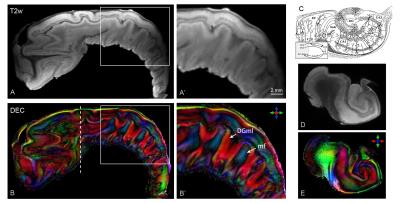 |
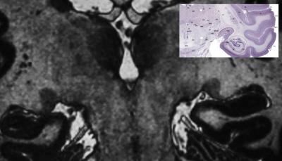
Laminar microstructure and subfield connectivity of the human hippocampus revealed with ultra-high field diffusion MRI at 11.7T
Manisha Aggarwal, Priya Sathyanarayan, David Nauen
In this work, we demonstrate high-field (11.7T) 3D diffusion MRI (dMRI) of the fixed human hippocampal formation to investigate its internal organization. The results of this study show delineation of fine hippocampal microstructure and inner connectivity of the human hippocampal subfields based on HARDI. Further, we demonstrate 3D reconstruction of intra-hippocampal subfield connectivity, e.g. the trisynaptic hippocampal circuit, using fODF-based probabilistic tractography with combined high spatial and angular resolution, which has important future applications for mapping of pathology-induced changes in the hippocampus in Alzheimer’s disease and related neurological disorders.
|
08:39
|
0697.
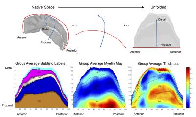 |
Unfolded hippocampal coordinate system for quantitative mapping and subfield segmentation
Jordan DeKraker, Kayla Ferko, Jonathan Lau, Stefan Köhler, Ali Khan
This work presents a novel computational technique for unfolding the hippocampus, providing a smooth and consistent mapping from native 3D space to a common coordinate system that is intrinsically defined by hippocampal anatomy. This coordinate system allows for laminar-based sampling of quantitative volumetric data and a means to pool data across subjects without additional registration or warping. We demonstrate the value of this technique with data from a set of healthy young participants scanned at 7T, taking advantage of high-resolution isotropic imaging for visualizing intra-hippocampal features, and employ a single surgical patient case with histology for validation.
|
08:51
|
0698.
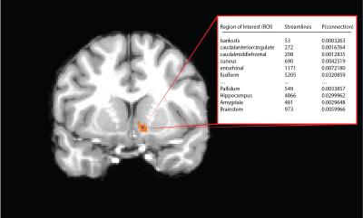 |
Segmentation of the Human Nucleus Accumbens Using High-Resolution Diffusion Tractography
Samuel Cartmell, Qiyuan Tian, Grant Yang, Christoph Leuze, Jennifer McNab, Casey Halpern
Tractography-based parcellation of the Nucleus Accumbens (NAc) was performed using high-resolution diffusion data from the Human Connectome Project. Analysis of clustering indicated the NAc was best separated into 2 subregions, consistent with anatomical and histological studies of animals. Output of the procedure gave qualitatively similar results across subjects, producing clusters that tended to occupy the ventromedial and dorsolateral portions of the NAc. The ventromedial subregion demonstrated increased task fMRI activation and displayed preferential connections to known biased projection areas such as medial orbitofrontal cortex. Finally, qualitatively similar results were obtained by performing the clustering procedure on a separate, lower-quality dataset.
|
09:03
|
0699.
 |
Optimization of Lateral Geniculate Nucleus Volume Determination at 3T and 7T
Njoud Aldusary, Lars Michels, Birgit Keisker, Michael Wyss, Karen Huebel, Arwa Baeshen, David Brunner, Klaas Pruessmann, Klara Landau, Spyridon Kollias, Marco Piccirelli
The clinical 2D PD scans and the research very high resolution 3D T1 scans used to determine the lateral geniculate nucleus (LGN) have high contrast respectively high resolution, but too low resolution respectively too long acquisition time. Nevertheless, volumetric and contrast quantification of the LGN and of other brain tissues might increase in clinical relevance. Therefore, we optimized the contrast of 3D T1 MPRAGE to obtain the best contrast with sufficient resolution to quantify volume and contrast of the LGN.
|
09:15
|
0700.
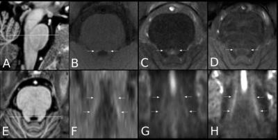 |
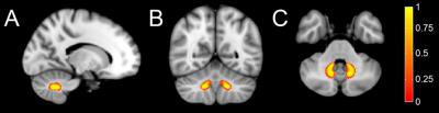
In vivo imaging of human Locus coeruleus at 3 and 7 Tesla
Nikos Priovoulos, Heidi IL Jacobs, Dimo Ivanov, Kamil Uludag, Frans Verhey, Benedikt A Poser
Structural and functional alterations of the Locus coeruleus (LC) are implicated in various neurological diseases, including Alzheimer’s. Current standard spin-echo based approaches to imaging the LC suffer from several limitations at 3T, including long acquisition time and highly anisotropic resolution. In this study, we have evaluated in healthy human subjects different MRI contrasts both at 3 and 7 Tesla to image LC. We show that visualization of the locus coeruleus is best achieved with magnetization transfer contrast at 7T superior to standard T1- and T2-weighted methods.
|
09:27
|
0701.
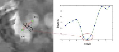 |
A seed point discontinuity-based level set method for accurate substantia nigra and red nucleus segmetation in QSM images
Tian Guo, Binshi Bo, Xinxin Zhao, Xu Yan, Yang Song, Caixia Fu, Dongya Huang, Hedi An, Nan Shen, Yi Wang, Jianqi Li, Guang Yang
Accurate segmentation of substantia nigra (SN) and red nucleus (RN) in quantitative susceptibility mapping (QSM) images has great clinical value in quantifying iron deposition and measuring disease severity. We propose a new segmentation algorithm which uses the discontinuity of seed points in different tissues as prior knowledge. Seed points in SN or RN can be obtained from standard atlas or specified manually. This prior was then incorporated into level set method to segment SN and RN. Experiments on in-vivo MR images showed that the proposed method achieved more accurate segmentation results than the atlas-based method and classic level-set method.
|
09:39
|
0702.
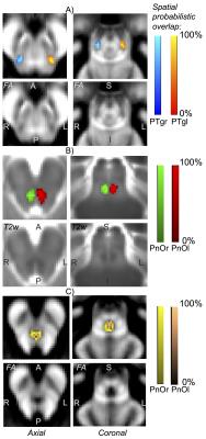 |
In vivo 7 Tesla probabilistic neuroimaging structural atlas of human pedunculotegmental, oral pontine reticular and paramedian-raphe nuclei
Marta Bianciardi, Christian Strong, Nicola Toschi, Brian Edlow, Bruce Fischl, Emery Brown, Bruce Rosen, Lawrence Wald
Mesopontine-tegmental nuclei such as the pedunculotegmental, oral-pontine-reticular and paramedian-raphe nuclei modulate arousal and motor functions. Dysfunction of these nuclei is implicated in the pathogenesis of disorders of consciousness, sleep disorders, and neurodegenerative diseases. However, a stereotaxic probabilistic atlas of these nuclei in humans does not exist. We used segmentation of 1.1 mm-isotropic 7 Tesla diffusion-fractional-anisotropy and T2-weighted images to generate and validate an in vivo probabilistic neuroimaging structural atlas of these nuclei in MNI space. We constructed this atlas to aid the localization of these nuclei in conventional images in future research and clinical studies of arousal and motor functions.
|
09:51
|
0703.
 |
An Improved Probabilistic Atlas of the Dentate Nucleus Derived with QSM
Naying He, Jason Langley, Daniel Huddleston, Huawei Ling, Hongmin Xu, Chunlei Liu, Yong Zhang, Fuhua Yan, Xiaoping Hu
Prior dentate nucleus (DN) atlases were derived using T1-, T2*- or susceptibility-weighted imaging. Accurate delineation of the DN boundary is difficult in these images. We present an atlas derived from quantitative susceptibility maps, which exhibit better contrast between DN and surrounding cerebellum tissue. An improved delineation of DN boundaries was achieved, resulting in increased maximum overlap in our DN atlas as compared to previously published results. We anticipate the atlas to be used in a variety of imaging applications ranging from functional imaging of DN to measuring DN iron deposition arising from disease.
|
10:03
|
0704.
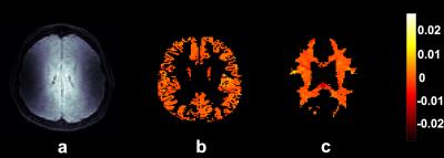 |
Measurements of cardiac related pulsatile volumetric strain in grey and white matter brain tissue with high resolution DENSE at 7T
Ayodeji Adams, Peter Luijten, Jaco Zwanenburg
The aim of this study was to investigate the cardiac induced volumetric strain differences between grey and white matter at 7T with DENSE in young, healthy volunteers, which may provide information on properties of the microvasculature. Increasing volumetric strain was seen in the systolic phase of the heart. The average strain in the grey matter was larger compared to white matter, but this was not significant. Measuring whole brain volumetric strain is feasible with DENSE at 7T, and has the potential to assess microvascular function (passive swelling over the cardiac cycle) in various brain areas.
|
|