Wednesday, 26 April 2017
| Room 313A |
13:45 - 15:45 |
Moderators: Petra Huppi, Jeff Neil |
Slack Channel: #s_neuro
Session Number: O12
13:45
|
0806.
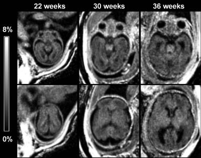 |
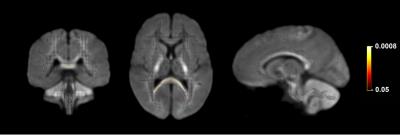
Quantitative assessment of the fetal brain myelination in vivo using fast macromolecular proton fraction mapping 
Alexandra Korostyshevskaya, Irina Prihod'ko, Andrey Savelov, Vasily Yarnykh
Macromolecular proton fraction (MPF) is a biophysical parameter describing cross-relaxation and closely correlated with myelin content in neural tissues. This study presents the first evaluation of the fast MPF mapping method in prenatal clinical neuroimaging and suggests that MPF in the fetal brain structures is sensitive to the earliest stages of myelin development.
|
13:57
|
0807.
 |
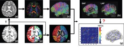
Anatomically constrained tractography and structural connectome of the neonatal brain
Manuel Blesa, Ahmed Serag, Devasuda Anblagan, Emma Telford, Sarah Sparrow, Scott Semple, Mark Bastin, James Boardman
The Anatomically-Constrained Tractography framework (ACT) is an advanced method to create tractography that relies on very accurate segmentation of the brain. To evaluate its role in tractography of the developing brain, we optimised the pipeline for neonatal data and compared against other methods (FA and WM seeding, with deterministic and probabilistic algorithms) used for the computation of neonatal structural connectivity. The results suggest that ACT with seeding at the GM / WM interface enhances anatomic accuracy of neonatal tractography, compared with other methods, and it has a significant impact on network measures.
|
14:09
 |
0808.
 |
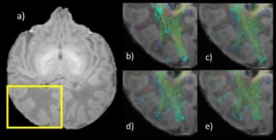
The short-range association fibers underlie brain network reconfiguration in typically and atypically developing children
Minhui Ouyang, Jennifer Muller, Hua Cheng, Yun Peng, J. Edgar, John Detre, Timothy Roberts, Hao Huang
Short-range association fibers (SAF) or U-fibers, connect adjacent gyri and constitute the majority of brain white matter. During development, SAF undergo dramatic changes in conjunction with brain network reconfiguration. How SAF reshape the brain network configuration during typical and atypical development is unknown. In this study, SAF was quantified with an index defined as normalized short-range association fibers (NSAF). We found that NSAF decreases were associated with increases in brain network efficiency in the typical developing brain from 2-7 years. Similar association were not observed in children with autism.
|
14:21
|
0809.
 |
Multimodal thalamocortical connectivity patterns in early brain development - permission withheld
Silvina Ferradal, Borjan Gagoski, Camilo Jaimes Cobos, Francesca Yi, Clarissa Carruthers, Catherine Vu, Ryan Larsen, Brad Sutton, P. Ellen Grant, Lilla Zollei
Understanding normal thalamocortical organization in early brain development has significant clinical relevance as it could provide early indicators of neurodevelopmental disorders which could originate from alterations in functional and structural brain maturation. Here we show structural and functional thalamocortical connectivity patterns derived from twenty healthy term infants scanned within the first weeks of life. Our results show that while there is a general good spatial agreement between both modalities, there are certain regions that exhibit different thalamocortical patterns. These discrepancies are possibly mediated by maturational processes such as axonal myelination and synaptogenesis.
|
14:33
|
0810.
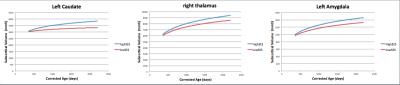 |
Socioeconomic Status Influences Early Longitudinal Cortical and Subcortical Development
Justin Remer, Douglas Dean III, Sean Deoni
Brain development may be influenced by socioeconomic status (SES), a marker of a family’s income and parental education. SES differences may result in exposure to extra stress during critical periods of neurodevelopment. We performed the first longitudinal analysis of differential brain development in 70 healthy infants and young children (1 year to 6 years of age) stratified by familial SES using high resolution T1 MRI. We demonstrated that trajectories of subcortical and cortical maturation are significantly different between infants and children from low and high SES families over the first 6 years of life.
|
14:45
|
0811.
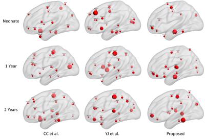 |
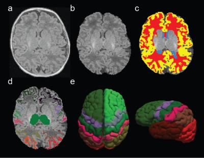
Novel Functional Brain Development Patterns during Infancy Revealed through the Application of Functionally Derived Brain Parcellations
Feng Shi, Andrew Salzwedel, Weili Lin, John Gilmore, Wei Gao
Signal heterogeneity within the predefined regions of interest (ROIs) may confound the functional connectivity estimation. In this study, we generate brain parcellations for neonate, 1-year, and 2-year-old infants, respectively, and use them to reveal potential novel functional developmental patterns. Our results show the progression of local functional specialization during early brain development. Moreover, different patterns of hub distributions are observed using different functional parcellation schemes suggesting the importance of selecting appropriate functionally-derived brain parcellations in characterizing infant whole brain connectivity pattern.
|
14:57
|
0812.
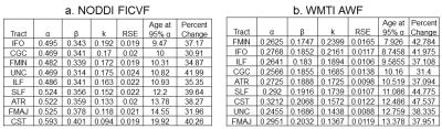 |
Comparison of NODDI and WMTI microstructural parameters in typical development
Kirsten Lynch, Farshid Sepehrband, Arthur Toga, Kristi Clark
Childhood and adolescence is an extended period of postnatal maturation characterized by dynamic changes in white matter microstructure. Multi-shell diffusion MRI (dMRI) models, such as neurite orientation dispersion and density imaging (NODDI) and white matter tract integrity (WMTI) provide an invaluable measure for the study of child development with tissue compartment estimates. NODDI and WMTI are based on similar frameworks, however they differ in several model assumptions. This study provides a comparison of NODDI and WMTI intra-axonal volume fraction model fittings in a cohort of children ages 0 -18 years in order to determine which model best reflects neurodevelopmental features.
|
15:09
|
0813.
 |
Pubertal contributions to white matter apparent fibre density in late childhood: a fixel-based analysis
Sila Genc, Marc Seal, Thijs Dhollander, Charles Malpas, Philip Hazell, Timothy Silk
Recent evidence supports the contribution of pubertal stage to local and global grey and white matter remodelling. Using fixel-based analyses, we show that pubertal children have greater apparent fibre density in the splenium of the corpus callosum compared with age-matched pre-pubertal children. This finding suggests that pubertal onset itself, rather than chronological age, drives the remodelling of white matter microstructure – which is an important consideration for assessing biological age. This is particularly important for studying paediatric and adolescent populations, as pubertal stage may be an important factor to consider in addition to chronological age.
|
15:21
|
0814.
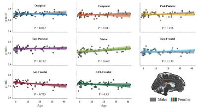 |
In vivo measurement of g-ratio in the Corpus Callosum using the macromolecular tissue volume: evaluating changes as a function of Callosal subregions, age and sex.
Shai Berman, Jason Yeatman, Aviv Mezer
Recent developments in quantitative and diffusion MRI, have made it possible to estimate the axonal g-ratio in human white-matter in-vivo. g-ratio is the ratio between the inner and outer radii of the myelin sheath wrapped around the axon. We suggest a simplified measurement of g-ratio incorporating proton density mapping, and implement it in the Corpus-Callosum of 100 subjects (ages 8-80). We find the g-ratio values agree with previously results. Furthermore, g-ratio values are stable over the lifespan and between the sexes. These results converge with theoretical evidence suggesting g-ratio has an optimal value for white-matter function.
|
15:33
|
0815.
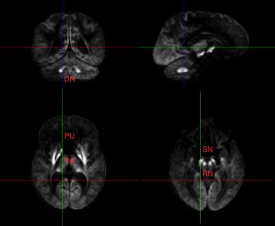 |
Greater Relaxivity in Brain Regions Indicates Tissue Iron Deposition in Adolescence to Adulthood
Eric Peterson, Dongjin Kwon, Beatriz Luna, Bart Larsen, Devin Prouty, Edith Sullivan, Adolf Pfefferbaum
This study investigates non-heme iron deposition in the adolescent brain in specific iron-susceptible regions as a function of age, sex, body mass index, supratentorial brain volume, handedness, scanning site, and race. A large cohort of 531 healthy adolescents, ages 12 to 22 years, were scanned at five sites on GE and Siemens systems using standard DTI and fMRI pulse sequence. This study demonstrates that in bilateral pallidum, putamen, dentate nucleus, red nucleus, and substantia nigra, both T2 and T2* show age-related declines. These results suggest ferratin-encapsulated iron deposition in specific brain regions is associated with normal adolescent brain development.
|
|