Wednesday, 26 April 2017
| Room 313A |
08:15 - 10:15 |
Moderators: Janine Lupo |
Slack Channel: #s_neuro
Session Number: O13
08:15
|
0705.
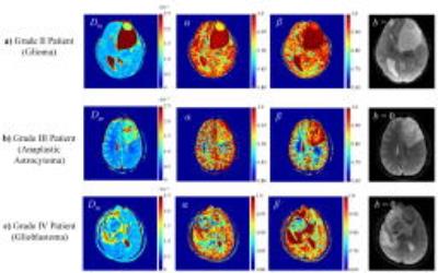 |
Towards Glioma Grading Using Non-Gaussian Diffusion Imaging with a Continuous-time Random Walk Model and A Quantile Histogram Analysis 
Muge Karaman, Jiaxuan Zhang, Zheng Zhong, Kejia Cai, Wenzhen Zhu, Xiaohong Zhou
Gliomas are the most common primary tumors of the central nervous system. As surgical biopsy may not be always feasible, an accurate noninvasive glioma grading is highly desirable for planning treatments. Recently, a number of non-Gaussian diffusion models were developed to characterize the anomalous diffusion behavior of the complex biological tissue. Among these, the continuous-time random walk (CTRW) model showed a great potential to probe the tissue heterogeneity and complexity that is elevated with tumor progression. In this study, we show that the CTRW parameters are capable of differentiating glioma grades, beyond simply separating low-grade and high-grade as in many diffusion MR studies on gliomas.
|
08:27
|
0706.
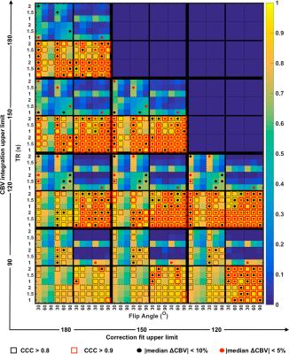 |
Optimization of acquisition and analysis methods for clinical dynamic susceptibility contrast (DSC) MRI using a validated digital reference object
Natenael B Semmineh, Ashley M Stokes, Laura C Bell, Jerrold L Boxerman, C Chad Quarles, Natenael Semmineh
Brain tumor DSC-MRI studies can be confounded by T1 and T2* effects that occur when the contrast agent extravasates. Traditionally a combination of contrast agent pre-loading and leakage correction techniques are used to minimize T1 leakage effects, but currently there is no consensus on the most robust dosing scheme. Using a validated DSC-MRI digital reference object we characterize the influence of pre-load dosing schemes, acquisition pulse parameters, and leakage correction methods on CBV accuracy. Our goal is to leverage this computational approach to identify the optimal combination of parameters for brain tumor CBV mapping.
|
08:39
|
0707.
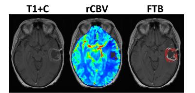 |
MRI-perfusion derived fractional tumor burden (FTB) is predictive of overall and progression free survival in newly diagnosed glioblastoma following concomitant chemoradiotherapy
Melissa Prah, Jennifer Connelly, Kathleen Schmainda
The phenomenon of pseudoprogression (PsP) on standard imaging can make response assessment difficult in patients with glioblastoma who have undergone standard chemoradiation treatment (CRT). PsP mimics tumor progression on standard imaging, yet is thought to represent a positive biological response to treatment. Recent efforts to define rCBV thresholds to distinguish tumor from treatment effect has enabled the creation of fractional tumor burden (FTB) maps. FTB maps quantify the percent of tumor within an enhancing lesion. This study shows that, within 4-months post-CRT, FTB is a better indicator of PFS and OS than median rCBV or methylation status alone.
|
08:51
|
0708.
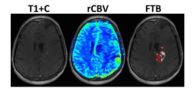 |
MRI-perfusion derived Fractional Tumor Burden (FTB) stratifies survival in recurrent glioblastoma following treatment with bevacizumab
Melissa Prah, Jennifer Connelly, Scott Rand, Kathleen Schmainda
Imaging response, in patients with recurrent glioblastoma (rGBM) who are treated with bevacizumab (which decreases vascular permeability), is often difficult to assess since decreased contrast agent uptake might falsely underestimate lesion size or biologic activity. Alternatively, relative cerebral blood volume (rCBV) has shown promise to identify true responders. Fractional tumor burden (FTB) maps, which are derived from rCBV, allow spatially quantifiable characterization of rGBM within lesion enhancement, and therefore may provide additional value to post-treatment response-assessment. This work demonstrates that patients with less than 75% FTB following treatment with bevacizumab have a clear progression-free and overall survival benefit.
|
09:03
|
0709.
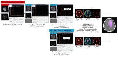 |
Improvement in Diagnostic Accuracy and Reliability of Pharmacokinetic Parameters from DCE MR Imaging by using Arterial Input Function Obtained from DSC MR imaging: Differentiation of High Grade Glioma from Low Grade Glioma. - video not available
Sung-Hye You, Seung Hong Choi
The aim of this study was to compare two AIFs derived from DCE (AIFDCE) and DSC MR imaging (AIFDSC) in terms of the diagnostic accuracy and reliability of pharmacokinetic parameters from DCE MRI for differentiation of high grade from low grade glioma. This retrospective study included 70 patients with pathologically confirmed gliomas. In all of the patients, we performed preoperative DSC and DCE MRI, and two AIFs (AIFDSC and AIFDCE) were obtained from each image. Pharmacokinetic parameters (Ktrans, Vp, and Ve) were processed. DCE MRI parameters obtained using AIFDSC showed better accuracy and reliability than those derived from AIFDCE.
|
09:15
|
0710.
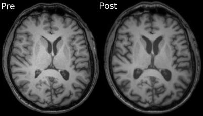 |
Brain volume loss in glioblastoma patients following photon and proton radiochemotherapy
Jan Petr, Frank Hofheinz, Andreas Gommlich, Felix Raschke, Esther Troost, Bettina Beuthien-Baumann, Annekatrin Seidlitz, Ivan Platzek, Michael Baumann, Mechthild Krause, Jörg van den Hoff
Gray matter (GM) atrophy in healthy brain tissue following radiochemotherapy was shown in brain-tumor patients in several studies. Here, we aimed to study GM and white matter (WM) changes in glioblastoma patients undergoing photon (n=43) and proton (n=12) radiochemotherapy. In photon-therapy patients, a statistically significant decrease of both GM (~2%) and WM (1.3-2.3%) volume was found with a positive influence of the RT-dose on the GM volume loss. In proton-therapy patients, no significant changes in GM and WM volumes were observed after therapy. This indicates that the proton-therapy has the potential to reduce structural GM changes in healthy tissue.
|
09:27
|
0711.
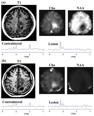 |
Volumetric 3D analysis of high grade glioma at pre- and post-radiation therapy by magnetic resonance echo-planar spectroscopic imaging
Yanqin Lin, Doris Lin, Karim Snoussi, Anouk Marsman, Andrew Maudsley, Sulaiman Sheriff, Katie Link, Peter Barker, Lawrence Kleinberg
31 patients with high grade glioma were recruited at both pre- and post-radiation therapy. 3D EPSI was employed on a Siemens 1.5 T scanner. NAA and Cho in lesion are normalized to Cr at contralateral part. At pre-radiation, Cho/Cr at lesion is significantly higher than that in contralateral part (p = 0.003). From pre- to post-radiation, Cho/Cr at lesion is significantly decreased (p < 0.007). Elevated Cho level can be an indicator for residual glioma tumor after surgery. Cho/Cr ratio can be used to monitor the treatment response. EPSI technique can be a promising way to monitor treatment response.
|
09:39
|
0712.
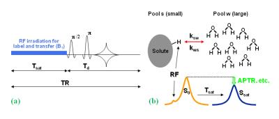 |
Differentiating Recurrent Glioma from Treatment Effects Using Amide Proton Transfer-Weighted MRI
Shanshan Jiang, Yi Zhang, Hye-Young Heo, Peter Van Zijl, Jinyuan Zhou
We assess the feasibility and the value of amide proton transfer-weighted (APTw) MRI in identifying viable malignant glioma. 22 patients with suspected recurrent glioma following chemoradiation were scanned at 3T. A total of 64 stereotactic biopsy specimens were obtained from gadolinium-enhancing regions of interest with varying APTw signals, 47 of which were histopathologically assigned as recurrent tumor and 17 treatment effects. APTw MRI revealed different signal intensities in the biopsied sites showing recurrent tumor (hyperintense signal) or treatment effects (iso-intense to minimally hyperintense signal). APTw signal intensity identified recurrent tumor with 97.9% sensitivity and 88.2% specificity.
|
09:51
|
0713.
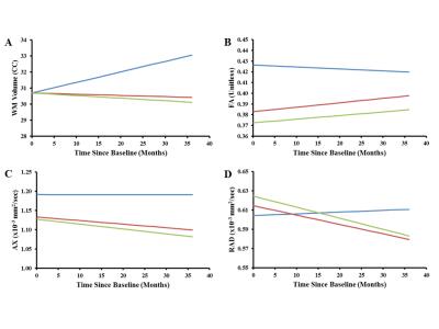 |
Disrupted Integrity of Frontal White Matter and Neurocognitive Correlates in Patients Treated for Pediatric Medulloblastoma
John Glass, Robert Ogg, Jung Hyun, Julie Harreld, Jane Schreiber, Yimei Li, Amar Gajjar, Wilburn Reddick
This study assessed the longitudinal white matter (WM) microstructure of 146 patients and 72 normal healthy age-similar controls. WM volume, fractional anisotropy (FA), and radial (RAD) and axial (AX) diffusivity trajectories were examined and correlated with neurocognitive performance at 36 months. After surgery but before any additional therapy, frontal WM volume in patients was similar to controls but FA was significantly reduced and was significantly correlated with neurocognitive performance three years later. Over the next three years, WM volume significantly decreased in patients and was significantly correlated with decreased Working Memory.
|
10:03
 |
0714.
 |
Improved prediction of meningioma-brain adhesion with normalized octahedral shear strain using slip interface imaging based on MR-elastography
Ziying Yin, Joshua Hughes, Joshua Trzasko, Kevin Glaser, Armando Manduca, Jamie Van Gompel, Michael Link, Anthony Romano, Richard Ehman, John Huston III
Knowledge of meningioma-brain adhesion can be important to surgical outcome but has been reliably assessed only during surgery. Slip interface imaging (SII), a recently developed MR-elastography based technique, is capable of determining the degree of meningioma-brain adhesion preoperatively. In SII, a non-adherent meningioma demonstrates a hyper-intense octahedral shear strain (OSS) contour along the tumor-brain interface. In 25 meningiomas, an algorithm improved by normalizing OSS to the combined wave amplitude provided a more accurate prediction in the setting of peritumoral edema. Normalized OSS increased SII accuracy from 72% to 92%, and the kappa coefficient increased from 0.37 (fair) to 0.86 (good).
|
|