Monday, 24 April 2017
| Room 313BC |
13:45 - 15:45 |
Moderators: Konstantinos Arfanakis, Elizabeth Meyerand |
Slack Channel: #s_neuro
Session Number: O15
13:45
|
0182.
 |
Quality Assurance of Neuro-MRI within Multicenter Clinical Trials 
Preethi Subramanian, David Poon, Shivangi Vora, Sydney Cearlock, Xiangyu Yang, Jun Zhang, Michael Knopp
Volumetric analysis and quantification especially of dynamic MR sequences is increasingly invaluable for assessing treatment response and time to progression in the majority of clinical trials focusing on neuro-oncology. These innovative therapeutic trials rely heavily on consistent image acquisitions even in multi-center Phase 2/3 trials. We have developed a highly structured DICOM tag based, parameter driven, semi-automated QC approach that readily enables visualization of acquisition inconsistencies using a heat-mapping spectrum. As the imaging core laboratory for several NCI-NCTN clinical trials, we expanded the QC methodology to also enable constructive feedback education / training to help improve the quality of Neuro-MRI submissions.
|
13:57
|
0183.
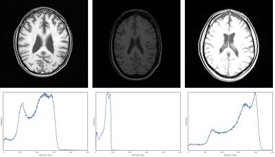 |
Brain MR image intensity normalization in the presence of pathology
Hugo Kuijf, Mariëlle Jansen, Mirjam Geerlings, Max Viergever
Brain MR image intensities do not have a fixed tissue-specific value. Especially in longitudinal studies, where a subjects’ anatomy might change, pathology can arise, scanner software and hardware may be replaced, the resulting image intensities can differ widely. This thwarts subsequent post-processing or image analysis. Various image intensity normalization techniques exist, but are often evaluated on healthy subjects. In this work, we evaluate six normalization techniques on 25 image-pairs (five year interval) of subjects with brain pathology. Traditional methods (e.g. Gaussian and Z-Score) are clearly affected by the presence of pathology and perform less than more recent techniques.
|
14:09
|
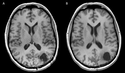
0184.
 |
Novel Design of a 3D-Printed Anthropomorphic Brain Phantom for Segmentation Validation in Magnetic Resonance Imaging - permission withheld
Anna Altermatt, Francesco Santini, Xeni Deligianni, Stefano Magon, Philippe Cattin, Jens Wuerfel, Laura Gaetano
Precise brain phantoms are important for evaluating the quality of segmentation tools for brain MRI. Here we suggested the construction of a 3D physical brain phantom as gold standard to validate the performance of those tools. Folding patterns of grey and white matter compartments were replicated using 3D-printed models from a real structural brain scan. T1 and T2 intensities of these brain regions in a 3 Tesla MRI were mimicked by a 0.6% agar mixture containing the appropriate concentrations of the paramagnetic compounds Ferumoxide and Manganese chloride. With its 3D-printed brain-like design, the phantom showed to be a promising alternative to existing methods for MRI segmentation validation.
|
14:21
|
0185.
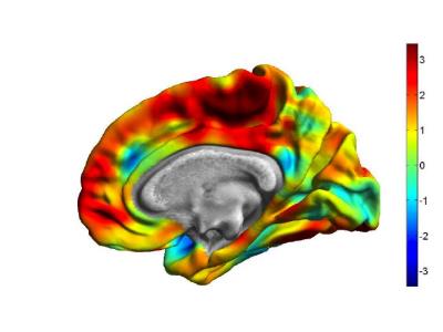 |
Traffic-Related Air Pollution Associated with Reduced Cortical Thickness and Altered White Matter Organization in a Longitudinally Studied, Pediatric Cohort
Kim Cecil, Travis Beckwith, Mekibib Altaye, Rachel Severs, Christopher Wolfe, Zana Percy, Thomas Maloney, Kimberly Yolton, Grace LeMasters, Patrick Ryan
Traffic-related air pollution (TRAP) is strongly associated with adverse cardiopulmonary health effects. Evidence suggests the developing brain may also be a target organ for particulate matter due to translocation either from the respiratory system or through the olfactory nerve. Using a pediatric cohort, we tested the hypothesis that exposure to TRAP during critical windows of brain development is significantly associated with changes in brain structure and organization. Children with high exposure levels at time of birth were associated with reductions in brain volume, cortical thickness, and diffusion abnormalities in white matter at 12 years compared with children at low exposure.
|
14:33
|
0186.
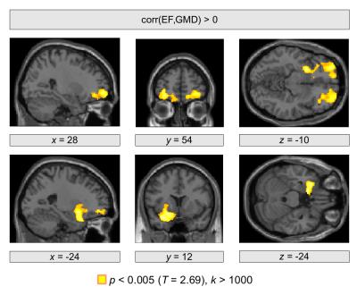 |
Investigating structural brain change with heart failure using voxel-based morphometry
Karsten Mueller, Friederike Thiel, Andrej Teren, Frank Beutner, Stefan Frisch, Joachim Thiery, Harald Möller, Arno Villringer, Matthias Schroeter
Heart failure is a multifactorial disease including a reduced pump efficiency leading to an insufficient oxygen supply for all body organs. However, the consequence of heart failure to brain structure is an important issue that needs further investigation. We used structural MRI with voxel-based morphometry to investigate a relationship between gray matter density and heart failure using ejection fraction and N-terminal prohormone of brain natriuretic peptide as markers for disease severity. These markers were found to be associated with decreased gray matter density in orbitofrontal and hippocampal brain regions indicating local gray matter abnormalities in these regions with heart failure.
|
14:45
 |
0187.
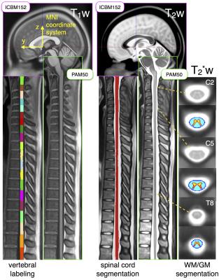 |
PAM50: Multimodal template of the brainstem and spinal cord compatible with the ICBM152 space
Benjamin De Leener, Vladimir Fonov, D. Louis Collins, Virginie Callot, Nikola Stikov, Julien Cohen-Adad
Template-based analysis of multi-parametric MRI data of the spinal cord sets the foundation for multi-center studies with minimum bias, thereby helping the discovery of new biomarkers of spinal-related diseases. In this study, we introduce a spinal cord MRI template, the PAM50, which is anatomically compatible with the ICBM152 brain template and uses the same coordinate system. The fusion of the PAM50 and ICBM152 templates facilitates group studies and multi-center studies of combined brain and spinal cord MRI and also allows the use of existing atlases of the brainstem compatible with the ICBM template.
|
14:57
|
0188.
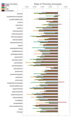 |
Consistency of Inter-Database Cortical Thinning with Age
M. Ethan MacDonald, Rebecca Williams, Nils Forkert, Avery Berman, Cheryl McCreary, Richard Frayne, Bruce Pike
This work investigates cerebral cortical thinning as a function of age, and how this relationship varies between four healthy subject databases, with a consolidated 1,382 subjects. Cortical thickness measurements of each subject were computed for 68 regions. Linear regression was used to determine the thinning rate for each region in each database as well as for the consolidated database. ANCOVA tests were run to test the effect of database. Correlation matrices were used to test the intra-relationship of locations between databases. Statistically significant correlations were found with age and differences were found between databases in all regions.
|
15:09
|
0189.
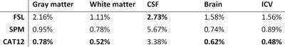 |
A test-retest analysis of brain volume measurement techniques
Hugo Kuijf, Geert Jan Biessels, Max Viergever, Jaco Zwanenburg
Brain volume measurements should both be accurate and precise. Accuracy of brain segmentation techniques is well studied. With the availability of test-retest datasets, precision (low coefficient of variation (COV)) can be investigated. In this work, we studied the COV of the FSL, SPM, and CAT12 software packages on 120 3T brain MR images of three subjects (40 images each) and compare it to previous results of FreeSurfer on this dataset. CAT12 performs best on total gray matter, white matter, and brain volume; whereas FSL has the lowest COV for CSF. COV values should be considered when studying brain volume change.
|
15:21
|
0190.
 |
Altered grey matter volume in patients with type 2 diabetes mellitus - video not available
Jia Liu, Taiyuan Liu, Wenhui Wang, Lun Ma, Xiaoyue Ma, Shaojie Shi, Meiyun Wang
Our meta-analysis indicates that patients with T2DM have significantly and robustly reduced grey matter, mainly in the cortical-striatal-limbic networks. The meta-regression results suggest that T2DM patients with longer illness duration may have smaller grey matter volume in the right MTG. Our finding supports the notion that T2DM could lead to subtle diabetic brain structural changes, which may be correlated with cognitive impairment inT2DM patients.
|
15:33
 |
0191.
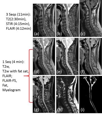 |
Volumetric T2-weighted and FLAIR Imaging of Spine with Uniform Fat Suppression in a Single Acquisition
Xinzeng Wang, Joshua Greer, Marco Pinho, Robert Lenkinski, Ananth Madhuranthakam
2D T2-weighted turbo spin-echo (T2w-TSE), fluid attenuated inversion recovery (FLAIR) with and without fat suppression are widely used in the clinical brain and spine protocols to improve diagnosis. However, FLAIR suffers from low SNR and long scan times. In this work, we developed a dual-acquisition 3D TSE sequence combined with dual-echo Dixon based approach to generate T2-weighted and FLAIR images of the spine with and without fat suppression in a single acquisition using the similar acquisition time as 2D FLAIR. Uniform fat/water separation was achieved using a shared-field-map and complex subtraction was used to generate FLAIR-like images without artifacts.
|
|