Wednesday, 26 April 2017
| Room 311 |
13:45 - 15:45 |
Moderators: Gareth Barker, Dan Wu |
Slack Channel: #s_neuro
Session Number: O16
13:45
 |
0786.
 |
Diffusion MRI of the entire postmortem human spinal cord at microscopic resolution 
Evan Calabrese, Gary Cofer, Nandan Lad, G. Allan Johnson
Diffusion MR imaging of the human spinal cord has become increasingly important in both clinical diagnostics, and research science.1 As MRI methods improve, there is a need to understand the limits of diffusion MRI in the human spinal cord. Here, we present a microscopic resolution diffusion MRI dataset of the entire postmortem human spinal cord, generated from a multi-segment acquisition, using an automated image-processing pipeline. These data provide unique insights for spinal cord research, future diagnostic imaging applications, and for postmortem pathologic evaluation of spinal cord specimens.
|
13:57
|
0787.
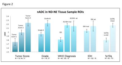 |
Diffusion imaging in grade II and III gliomas depends upon new vs recurrent status and enhancing vs nonenhancing status.
Tracy Luks, Tracy McKnight, Evan Neill, Llewellyn Jalbert, Arie Perry, Soonmee Cha, Joanna Phillips, Annette Molinaro, Susan Chang, Sarah Nelson
The relationship of diffusion imaging parameters with prognostic histological and molecular factors for patients with grade II and III gliomas is unclear, particularly for tumors that are non-enhancing on post-Gadolinium images. We investigated the relationship of ADC and FA values with histological tumor score, tumor grade, and molecular characteristics for non-enhancing (NE) vs contrast-enhancing (CE) and newly-diagnosed (ND) vs recurrent (REC) disease. In NE patients, histopathological and molecular characteristics associated with poorer clinical outcome were found to have higher ADC and lower FA. In CE patients, some characteristics associated with poorer outcome hadlower ADC and higher FA.
|
14:09
|
0788.
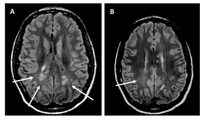 |
Prevalence of diffusely abnormal white matter in individuals with clinically isolated syndromes suggestive of multiple sclerosis
Cornelia Laule, Jimmy Lee, Guojun Zhao, Rick White, Irene Vavasour, Andrew Riddehough, Anthony Traboulsee, Luanne Metz, David Li
Diffusely abnormal white matter (DAWM) is present in individuals with clinically isolated syndromes (CIS) suggestive of multiple sclerosis (MS) at a similar frequency as seen in definite MS. CIS subjects with DAWM showed reduced brain volume and greater lesion load, both of which are known to correlate with clinical disability and progression. DAWM may have prognostic importance in CIS so examining its impact on conversion to MS, future disability and progression is warranted.
|
14:21
|
0789.
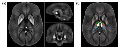 |
Decreasing magnetic susceptibility (QSM) of thalamic nuclei in Multiple Sclerosis (MS) – the thalamus as a target of projected inflammation?
Ferdinand Schweser, Ana Martins, Fuchun Lin, Jesper Hagemeier, Jannis Hanspach, Bianca Weinstock-Guttman, Nicola Bertolino, Dhaval Shah, Niels Bergsland, Michael Dwyer, Robert Zivadinov
This work studied intra-thalamic magnetic susceptibility changes in 120 patients with clinically isolated syndrome (CIS), relapsing-remitting MS (RRMS), and secondary progressive MS (SPMS). We detected decreased magnetic susceptibility in several nuclear groups of the thalamus in MS patients compared to controls, indicative of decreased iron concentration.
|
14:33
|
0790.
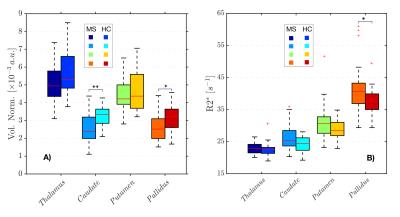 |
Iron loss occurs in the deep gray matter of multiple sclerosis patients
Enedino Hernández-Torres, Vanessa Wiggermann, David Li, Lindsay Machan, A Sadovnick, Anthony Traboulsee, Simon Hametner, Alexander Rauscher
In this work, a new approach for looking at the “iron deposition” in deep gray matter is presented. We investigated iron deposition in the deep gray matter indirectly by measuring R2*. In addition, we assessed the normalized volume of the structures of the DGM. We found a stronger association between increases in R2* and volume reductions of the same DGM structures in the MS group compared with the control group. Finally, we corrected the R2* measurements by the volume of the structures (R2*m). The R2*m values were reduced in the MS group suggesting that iron accumulation is not a common feature of MS but on the contrary a redistribution/reduction of the iron takes place, which may be masked by structural atrophy.
|
14:45
|
0791.
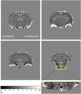 |
Phase imaging of axonal integrity of cranial corticospinal tract in experimental spinal cord injury at 9.4T
Genxia He, Junchao Qian
Spinal cord injury (SCI) leads to neuronal cell death, axonal damage and demyelination. Brain undergo anatomical changes following SCI. Recently MR phase imaging has shown promising application in visualizing demyelination. In this study we explored the possibility to investigate integrity of cranial corticospinal tract in SCI using phase imaging. Diffusion tensor imaging was also used to verify the axonal integrity. The results showed phase contrast decreased along with axial diffusivity did not significantly change in contralateral pyramid two weeks post-injury compared to pre-injury levels. Thus, phase imaging is a potential endogenous biomarker for brain axonal integrity after SCI.
|
14:57
|
0792.
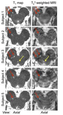 |
7 Tesla imaging of microstructural brainstem changes in REM sleep behavior disorder
Marta Bianciardi, Laura Lewis, Lawrence Wald, Bruce Rosen, Aleksandar Videnovic
REM-sleep-behavior-disorder (RBD) is characterized by the absence of muscle-atonia during REM-sleep and is thought to be related to a dysfunction of brainstem-nuclei (Bn) of the arousal/motor networks. Yet, a precise identification of the Bn involved in vivo is still missing, thus limiting our understanding of this disease. Through multi-contrast high-spatial-resolution 7Tesla-MRI and a recently developed stereotaxic-Bn-atlas, we consistently detected across RBD-patients microstructural-changes in a subregion of the substantia nigra, consistent with pars reticulata, and in a peri-nigral area. Interestingly, these changes were compatible with the presence of lacunar infarcts, finding that differs from recent reports of nigral iron-accumulation in RBD.
|
15:09
|
0793.
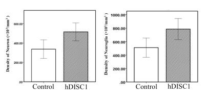 |
Detecting DISC1 Related Microstructural Abnormalities using Non-Gaussian Diffusion (DKI & NODDI) and QSM: with Histological Validation
Nan-Jie Gong, Russell Dibb, Kyle Decker, Mikhail Pletnikov, Eric Benner, Chunlei Liu
Metrics provided by DKI could be used to detect microstructural changes. However, they bear no explicit neurobiological interpretations. In contrast, the biophysical model of NODDI could provide metrics sensitive to density of neuron. The magnetic susceptibility derived from QSM method can sever as a sensitive biomarker for quantifying density of cells including both neurons and neuroglia.
|
15:21
|
0794.
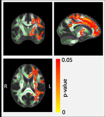 |
A quantitative MRI study of APOE-dependent microstructural differences in young healthy volunteers using NODDI, qMT and g-ratio
Nicholas Dowell, Simon Evans, Sarah King, Jennifer Rusted
We present the first NODDI, qMT and g-ratio study of microstructural differences between carriers and non-carriers of the APOE-e4 gene. This gene is a risk factor for the development of Alheimer's Disease later in life. Our work shows that the more specific microstructural measures offered by the NODDI technique has revealed an increase in the non-tissue (Viso) component in the brain among APOE-e4 carriers. Importantly, the other NODDI parameters and g-ratio show no difference suggesting that although the tissue component among APOE-e4 carriers is reduced, there is no detectable difference to the underlying microstructure. This important finding has not been shown before, and supports the large body of evidence that shows young healthy APOE-e4 carriers are not disadvantaged across a number of cognitive domains and (in some tasks) out-perform their non-e4 peers.
|
15:33
|
0795.
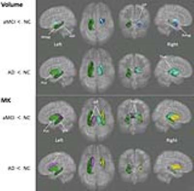 |
Spatial and Temporal Relationship between Microstructural and Morphological Abnormalities of Alzheimer’s disease: Evidence in Cortical and Deep Gray Matter
Nan-Jie Gong, Chun-Chung Chan, Lam-Ming Leung, Chun-Sing Wong, Russell Dibb, Chunlei Liu
Non-Gaussian diffusion metrics such as MK from DKI can complement conventional MD and FA for detecting microstructural changes, especially in deep gray matter. This can potentially improve the efficacy of diffusion metrics for serving as diagnostic imaging biomarkers. We also provided evidence supporting the proposed notion that microstructural changes in cortical and deep gray matter predate macrostructural changes such as volume and cortical thickness.
|
|