Tuesday, 25 April 2017
| Room 313BC |
13:45 - 15:45 |
Moderators: Florian Knoll, Xiaohong Joe Zhou |
Slack Channel: #s_neuro
Session Number: O17
13:45
|
0496.
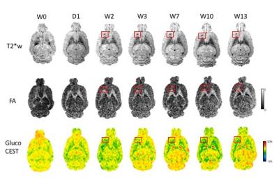 |
LONG TERM EFFECTS OF PULSED FOCUSED ULTRASOUND AND MICROBUBBLES DETECTED BY MULTIVARIATE IMAGING MODALITIES 
Zsofia Kovacs, Tsang-Wei Tu, Georgios Papadakis, William Reid, Dima Hammoud, Joseph Frank
Blood brain barrier (BBB) opening by MR-guided pulsed Focused Ultrasound (pFUS) and microbubbles (MB) is a non-invasive treatment of various central nervous system diseases. However, the potential adverse effects of repeated pFUS+MB exposure have not been thoroughly elucidated and may limit clinical translation. To date MRI scans of repeated BBB opening by pFUS+MB have been achieved without hemorrhage, edema and behavioral changes in non-human primates (Arvanitis, et al. 2015; Downs, et al. 2015). By incorporating detailed multivariate imaging modalities we characterized the long term effects of single or repeated pFUS+MB in the rat brain.
|
13:57
|
0497.
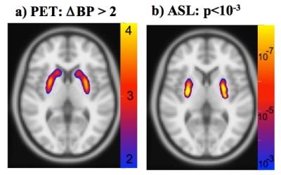 |
Function versus occupancy in the human brain: PET/fMRI during infusion of D2 antagonist
Tracy Barbour, Christin Sander, Daphne Holt, Joseph Mandeville
Simultaneous PET and fMRI were employed in healthy human subjects to investigate the dose-dependent relationship between drug occupancies of a D2-receptor antagonist and induced CBF responses measured by arterial spin labeling. Results indicate a super-linear relationship between CBF and occupancy, with a larger CBF response in putamen than in caudate at matched occupancies. These results inform dopaminergic neurophysiology, and the method may provide general utility for probing dopaminergic function in human subject cohorts.
|
14:09
 |
0498.
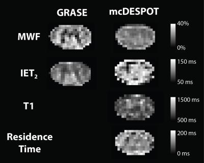 |
Myelin Water Imaging Using T2-Relaxation in the Spinal Cord; Comparison of Multi-echo GRASE and mcDESPOT
Emil Ljungberg, Irene Vavasour, Alexander Rauscher, Anthony Traboulsee, Alex MacKay, Shannon Kolind
There are currently several techniques for in vivo myelin water imaging using T2 relaxation. In this study we compare two clinically feasible myelin water imaging protocols for the cervical spinal cord: mcDESPOT and multi-echo GRASE. Myelin estimates from GRASE were consistently higher than mcDESPOT in white matter, but both techniques were able to differentiate between white and gray matter. T1 estimates from mcDESPOT also showed clear differences between white and gray matter. By combining GRASE and mcDESPOT, with total acquisition time less than 15 min, we can build a better picture of the tissue microstructure of the spinal cord.
|
14:21
|
0499.
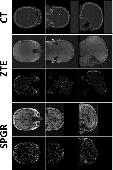 |
Improved Localisation of DBS Electrodes using Pseudo-Positive Contrast from a Zero-Echo-Time Acquisition
Rolf Schulte, Jeffrey Ashe, Sohan Ranjan, Julia Prusik, Julie Pilitsis, Stanley Hayes, Anne Menini, Florian Wiesinger, Ileana Hancu
The goal of this work is to reduce artefact extent and improve localisation of deep-brain stimulation (DBS) electrodes with MRI. A Zero Echo-Time (ZTE) sequence was used for data acquisition in a phantom and a patient; the minimal signal voids around electrodes, the ZTE’s natural proton-density weighting and low SAR/B1rms make it ideal for imaging implants. A pseudo-positive contrast image was first generated by inverting ZTE image intensity values, fitting and subtracting the background signal; a centre of mass calculation and singular value decomposition were then employed for electrode detection. Similar-sized artefacts as in CT images and improved precision over standard T1-weighted imaging were demonstrated.
|
14:33
|
0500.
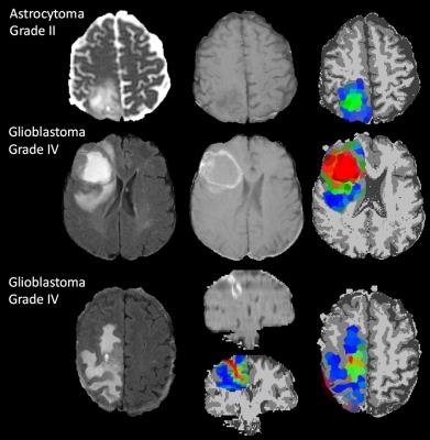 |
Tissue type mapping of gliomas using multimodal MRI
Felix Raschke, Thomas Barrick, Guang Yang, Timothy Jones, Xujiong Ye, Franklyn Howe
1H MRSI can assess glioma infiltration margins and malignant invasion but technical limitations prevent widespread use. In this study we used 2D 1H MRSI to determine voxels of specific tumour tissue type from which we extracted multimodal MRI (M-MRI) image characteristics. Subsequently, we applied superpixel segmentation and Bayesian statistical analysis to M-MRI alone to derive nosologic tumor images of these same tissue types with whole brain coverage. We obtained 100% classification accuracy for overall glioma grade, and an average 0.77 Dice overlap coefficient with the manual segmentation volume. Such methodology could aid prognostic assessment, surgical treatment and radiotherapy dose planning.
|
14:45
|
0501.
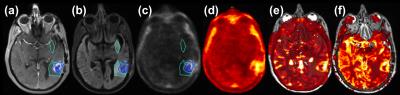 |
Multimodal Imaging of Vascularity and Drug Delivery in GBM Patients Treated with Anti-angiogenesis Inhibitor
Yi-Fen Yen, Jayashree Kalpathy-Cramer, Ciprian Catana, Xiao Da, Yangming Ou, Andrew Beers, Jacob Hooker, Bruce Rosen, Tracy Batchelor, Elizabeth Gerstner
Anti-angiogenic agents can decrease vessel permeability and perfusion, raising concerns that these agents may also decrease blood brain barrier penetration of concomitantly administered chemotherapy. Using MR-PET multimodal imaging, we found radiolabeled temozolomide correlated with permeability and perfusion in patients with recurrent GBM treated with bevacizumab. MR assessment of vascularity may be a surrogate marker for concomitant drug delivery.
|
14:57
|
0502.
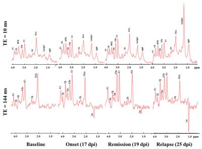 |
Haemodynamic and metabolic MR biomarkers of disease progression in an animal model of relapsing-remitting multiple sclerosis - permission withheld
Mohamed Tachrount, Andrew Davies, Flavia Rosianu, Roshni Desai, David Thomas, Kenneth Smith, Xavier Golay
Multiple sclerosis (MS) is a chronic inflammatory demyelinating and neurodegenerative disorder of unknown cause affecting the central nervous system. We have assessed haemodynamic and metabolic alterations within spinal cord during disease progression in an animal model (experimental autoimmune encephalomyelitis (EAE) by combining both arterial spin labelling (ASL) and MR Spectroscopy. We demonstrate for the first time that the neurological deficits are strongly correlated with impaired blood flow and reveal both reversible and irreversible simultaneous metabolic alterations.
|
15:09
|
0503.
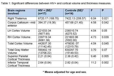 |
Multimodal MR investigation of brain and behavioral changes in patients with HIV-infections
Michael Thomas, Rajakumar Nagarajan, Eric Daar, Santosh Yadav, Charles Hinkin, Manoj Sarma, Zohaib Iqbal, Sathya Arumugam, Mario Guerrero , Mohammad Haris, Ebrahim Haroon
Regional brain volumes and cortical thickness using 3D T1-weighted MP-RAGE and neurometabolites quantitated using 5D EP-JRESI MRSI were obtained from a group of HIV+ (n=16) and HIV-subjects (n=15). Compared to HIV- subjects, following findings were observed in HIV+: i) decreases in the volume of right thalamus, mid anterior corpus callosal region and cortex (right, left, combined), and ii) decreases in cortical thickness of superior parietal and inferior temporal regions. The cortical thickness and volumetric changes were predicted by (increased choline, decreased NAA and Glx). Right basal ganglia glutamate/glutamine ratios and HIV+ status together significantly predicted psychomotor slowing during neurocognitive testing.
|
15:21
|
0504.
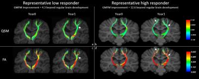 |
Combined tract-based analysis of diffusion fractional anisotropy and quantitative susceptibility mapping: a joint assessment of axonal and myelin microstructural changes in children with cerebral palsy
Lijia Zhang, Lyon Chen, Susan Ellor, Jessica Sun, Joanne Kurtzberg, Allen Song
In this report we developed a tract-based diffusion anisotropy and magnetic susceptibility analysis approach to jointly evaluate the potential mechanisms for axonal growth and myelin repair in children undergoing autologous cord blood stem cell therapy. Advancing from our prior findings that baseline brain connectivity is correlated with CP disease severity and that brain connectivity increase is correlated with functional motor improvement in CP patients, we provide further evidence that the increased brain connectivity may be the result of increased myelination of the affected neural pathways, in addition to the possibility of axonal regeneration.
|
15:33
|
0505.
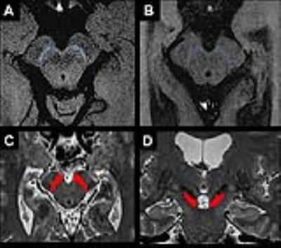 |
Multimodal MRI biomarker study of substantia nigra damage in idiopathic REM sleep behavior disorder
Nadya Pyatigorskaya, Rahul Gaurav, Dario Arnaldi, Smaranda Leu-Semenescu, Lydia Yahia-Cherif, Romain Valabregue, Marie Vidailhet, Stephane Lehericy
We quantified substantia nigra (SN) damage in idiopathic REM sleep behavior disorder (iRBD) patients using multimodal MRI biomarkers and determined biomarker efficacy. Nineteen patients with iRBD and 18 healthy volunteers underwent 3-Tesla MRI, including diffusion tensor imaging, neuromelanin (NM)-sensitive imaging and T2* mapping. The volume and normalized signal intensity in NM-sensitive images, R2* and diffusion tensor measures were quantified in the SN. Patients with iRBD showed reduced NM-sensitive volume and signal intensity and reduced fractional anisotropy versus controls in the SN. Combination of the three biomarkers had excellent diagnostic accuracy. These measures may represent valuable biomarkers for prodromal Parkinson’s disease.
|
|