Monday, 24 April 2017
| Room 312 |
08:15 - 10:15 |
Moderators: Jihye Jang, Graham Wright |
Slack Channel: #s_cv
Session Number: O18
08:15
|
0037.
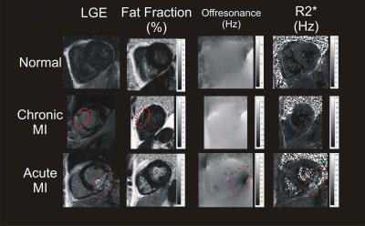 |
Joint Native Myocardial Fat Fraction, Off-Resonance and R2*/T2* Mapping in Ischemic Cardiomyopathies 
James Goldfarb, Usama Hasan, Jie Cao
Myocardial fat content, R2*/T2* and off-resonance frequency can be measured with high resolution using a native MR water-fat separation imaging technique applied to multiple gradient echo images. Significant differences in myocardial fat fraction were found consistent with fatty metaplasia in a subset of chronic myocardial infarction patients. Off-resonance and T2* changes consistent with intramyocardial hemorrhage were observed in a subset of acute myocardial infarction patients.
|
08:27
 |
0038.
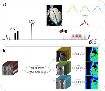 |
Simultaneous Multi-Slice Imaging For Whole Heart Myocardial T1 Mapping In A Single Breath-Hold
Sebastian Weingärtner, Steen Moeller, Kâmil Ugurbil, Chetan Shenoy, Mehmet Akçakaya
Myocardial T1-mapping bears promise for evaluation of numerous cardiomyopathies, but requires multiple breath-hold scans with conventional techniques. In this study we explored the acceleration potential of multi-band imaging for myocardial SAPPHIRE T1-mapping with 3-slice coverage in a single breath-hold. Three linear methods for slice and in-plane unaliasing were evaluated. Phantom studies confirmed the accuracy of the proposed T1-mapping method, and in-vivo evaluation has shown reliable image quality with 3-fold acceleration at the cost of 1.3 to 1.7-fold increased in-vivo variability. Smaller loss in-vivo precision was achieved using regularized methods, for the trade-off against increased inter-slice leakage.
|
08:39
|
0039.
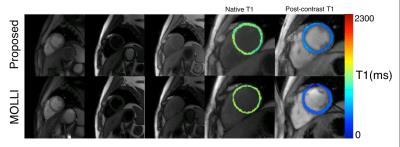 |
Free-Breathing, Non-ECG-Gated, Continuous Myocardial T1 Mapping and ECV Quantification with Multitasking
Jaime Shaw, Anthony Christodoulou, Behzad Sharif, Debiao Li
Currently used T1 mapping techniques utilize both ECG gating and breath-holds/navigators with low imaging efficiency and/or respiratory motion artifacts. We removed the need for ECG gating and respiratory monitoring with Cardiac MR Multitasking, a continuous acquisition technique using a low-rank tensor (LRT) imaging framework. The aim of this work is to validate a free-breathing, non-ECG-gated native T1 mapping and ECV quantification method in healthy subjects against standard MOLLI T1 mapping.
|
08:51
|
0040.
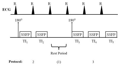 |
Optimized Single Pre/Post Contrast Protocol for MOLLI T1 Mapping with Inversion Group (IG) Fitting
Marshall Sussman, Luigia D'Errico, Bernd Wintersperger
A major focus in cardiac research is the assessment of myocardial pathology using quantitative T1 mapping. A number of sequences are being investigated for this task. One candidate is MOLLI. It provides superior precision to other cardiac T1mapping techniques. However, its precision is dependent on heart rate and range of T1 values present. Current attempts at optimizing precision are somewhat impractical, as they utilize separate MOLLI protocols for different heart rates, and for pre/post contrast imaging. This study identifies a single MOLLI protocol optimal for precision over a broad range of heart rates and pre/post contrast T1 values.
|
09:03
|
0041.
 |
Early Gradual Assessment of Tissue Injury and Functional Outcome after Myocardial Infarction by Cardiovascular Magnetic Resonance T1 Mapping
Sebastian Haberkorn, Christoph Jacoby, Jürgen Schrader, Malte Kelm, Uli Flögel
The value of CMR to distinguish between the severity of ischemic injuries after myocardial (MI) has yet to be established. Here, we quantified local tissue injuries and their correlation with functional outcome in two different experimental models of MI including native and post-contrast T1 maps, T2 maps and LGE. Only native T1 maps enabled in the acute phase after MI the detection of substantial differences in myocardial tissue texture between the two models, where neither of the other measures was indicative. In conclusion, native T1 mapping enables a gradual assessment of myocardial injury and holds predictive potential for the functional outcome.
|
09:15
|
0042.
 |
Multiparametric Characterization of Myocardial Tissue by Contrast-Enhanced Cardiac Magnetic Resonance Imaging in Subjects with Prediabetes, Diabetes and Controls from a Western General Population – Results of the KORA-MRI-Study
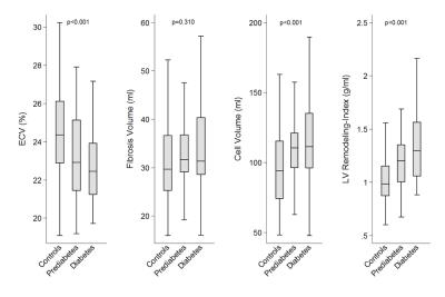
Corinna Storz, Holger Hetterich, Roberto Lorbeer, Sigrid Auweter, Wolfgang Rathmann, Christopher Schlett, Annette Peters, Konstantin Nikolaou, Fabian Bamberg, Jeanette Schulz-Menger
Cardiac magnetic resonance imaging (CMR) allows for detailed characterization of the myocardium, which may be beneficial in assessing cardiomyopathy in the setting of hyperglycemic states. We performed a comprehensive CMR protocol in subjects with prediabetes, diabetes and controls and preserved left ventricular (LV) ejection fraction (EF) in a western population-based sample. Subjects with prediabetes and diabetes had an increased LV-remodeling-index as well as higher estimates of cell volume compared with controls, while extracellular volume, as a parameter of diffuse myocardial fibrosis (MF), was decreased. This may highlight the role for hypertrophy in the pathogenesis of diabetic cardiomyopathy in this western population.
|
09:27
 |
0043.
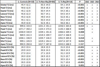 |
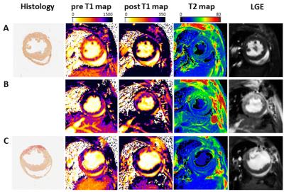
Comprehensive Cardiac Structure-Function MRI in Heart Transplant Recipients: Influence of Acute Cardiac Allograft Rejection
Ryan Dolan, Amir Rahsepar, Julie Blaisdell, Kenichiro Suwa, Allen Anderson, Kambiz Ghafourian, Esther Vorovich, Jonathan Rich, Jane Wilcox, Clyde Yancy, Jeremy Collins, James Carr, Michael Markl
Cardiac MRI provides a comprehensive structure-function evaluation of the heart with increasingly strong evidence of its ability to detect acute cardiac allograft rejection following heart transplant. In this large cohort of transplant recipients, quantitative T2 and ECV were significantly elevated during episodes of biopsy-proven rejection compared to baseline.
|
09:39
|
0044.
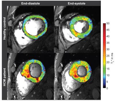 |
Myocardial T2* Changes Periodically across the Cardiac Cycle and is Prolonged in Hypertrophic Cardiomyopathy: A 7.0 Tesla MRI Patient Study
Till Huelnhagen, Fabian Hezel, Teresa Serradas Duarte, Min-Chi Ku, Bert Flemming, Erdmann Seeliger, Marcel Prothmann, Jeanette Schulz-Menger, Thoralf Niendorf
Ultrahigh field MR (UHF-MR) enables temporally resolved myocardial T2* mapping which benefits probing the myocardium at different physiological states. Myocardial BOLD contrast or T2* are commonly regarded as surrogates for myocardial tissue oxygenation, but the factors influencing T2* are manifold including cardiac macromorphology. Meaningful interpretation of myocardial T2* could be beneficial for understanding cardiac (patho)physiology in vivo, but requires careful identification of influential factors and their contributions to T2*. To this end, this study examines the relationship between myocardial T2* and myocardial wall thickness and investigates it’s capability to distinguish between healthy myocardium and myocardium affected by hypertrophic cardiomyopathy (HCM).
|
09:51
|
0045.
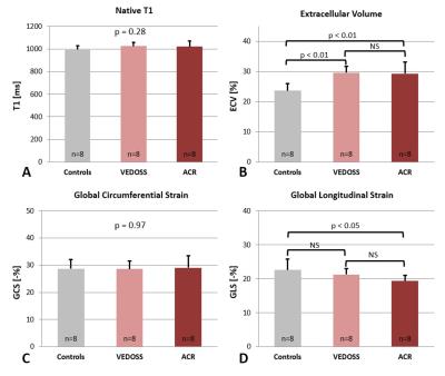 |
Myocardial extracellular volume expansion precedes functional myocardial alterations during the evolution of systemic sclerosis - permission withheld
Alexander Gotschy, Constantin von Deuster, Christian Stoeck, Valeriy Vishnevskiy, Lukas Wissmann, Kerem Tezcan, Markus Niemann, Suzanna Jordan, Britta Maurer, Sebastian Kozerke, Oliver Distler, Robert Manka
Myocardial involvement is common in patients with systemic sclerosis and is known to cause myocardial fibrosis and subtle ventricular dysfunction. Both can be characterized by novel CMR methods like native T1-mapping, ECV quantification or LV deformation imaging. However, the temporal onset of myocardial fibrosis and functional impairment during the progression of the disease is still unknown. Therefore, we investigated the presence of subclinical functional and fibrotic myocardial involvement in patients with very early diagnosis of systemic sclerosis and found that the expansion of ECV can be detected before LV functional impairment, assessed by CMR feature tracking, can be observed.
|
10:03
|
0046.
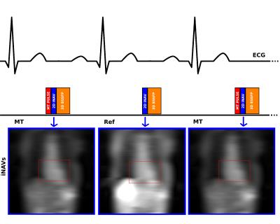 |
Non-contrast free breathing and motion corrected 3D whole heart quantitative magnetization transfer imaging for assessment of myocardial fibrosis - permission withheld
Karina Lopez, Radhouene Neji, Rahul Mukherjee, John Whitaker, Reza Razavi, Camila Muñoz, Claudia Prieto, Sebastien Roujol, Rene Botnar
We have developed a contrast-free free-breathing motion corrected (100% scan efficiency) 3D whole heart imaging technique for measurement of myocardial magnetization transfer ratio (MTR) maps. The sequence is based on the interleaved acquisition of MT weighted and non-MT weighted datasets and beat-to-beat rigid motion correction using 2D image navigators. Initial results in 4 healthy volunteers have shown good image quality, enabling the visualisation of the coronary arteries and MTR maps of healthy myocardium (MTR=41±7.2%). This approach promises higher sensitivity for measuring changes in macromolecule content associated with myocardial fibrosis than previous studies, justifying further investigation in a patient cohort.
|
|