Oral
Body: Breast, Chest, Abdomen, Pelvis
Thursday, 27 April 2017
| Room 320 |
08:15 - 10:15 |
Moderators: Chris Flask, Houchun Hu |
Slack Channel: #s_body
Session Number: O25
08:15
|
1067.
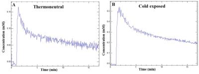 |
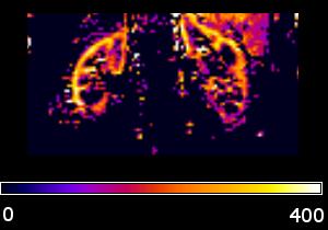
Evaluation of the Vascular Perfusion in Activated Brown Adipose Tissue by Dynamic Contrast Enhanced MR Imaging 
Jadegoud Yaligar, Sanjay Kumar Verma , Venkatesh Gopalan , Anantharaj Rengaraj, Tian Xianfeng, S. Sendhil Velan
Vasculature plays an important role in white and brown adipose tissue (WAT and BAT) metabolism. In expanding WAT, abnormal vasculature may lead to energy (fat) deposition whereas in activated BAT it may potentially facilitate the energy consumption by oxidizing the fat. Understanding the vascular network and blood perfusion properties of the activated BAT is important for triglyceride clearance, increased blood flow and oxygen. In this feasibility study we have investigated the vascular properties and blood perfusion rate constant of the activated BAT by quantitative dynamic contrast enhanced MR imaging in a rodent model.
|
08:27
|
1068.
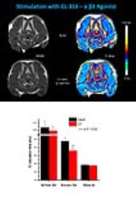 |
Functional Assessment of Brown Adipose Tissue by T2 Mapping
Ulrich Flögel, Christoph Jacoby, Jens Fischer, Maria Grandoch
Within the last decade, the scientific interest in brown adipose tissue (BAT) was greatly invigorated by the discovery of functional BAT in adult humans. Importantly, reduced BAT activity has recently been associated with predisposition to obesity and abnormal glucose homeostasis. The current gold standard for functional BAT imaging is CT-guided 18FDG-PET. Here, we describe a robust T2 mapping method to assess the metabolic activity of BAT independent of substrate selection and without the need of harmful radiation or contrast agents. Compared to previous T2*-based techniques the present approach is less prone to susceptibility and physiological artifacts especially at high fields.
|
08:39
|
1069.
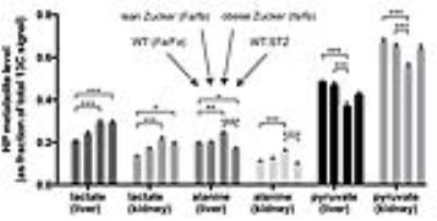 |
Hyperpolarized 13C-pyruvate MRI of the liver and kidneys in rodent models of type 1 and type 2 diabetes
Cornelius von Morze, Prasanna Allu, Gene-Yuan Chang, Irene Marco-Rius, Eugene Milshteyn, Catherine Gleason, Daniel Vigneron, David Pearce
The purpose of this study was to characterize changes in hyperpolarized 13C-pyruvate spectra in the liver and kidneys of two contrasting models of diabetes, obese Zucker diabetic fatty (ZDF) rats and streptozotocin(STZ)-treated (insulin-deficient) wild type Zucker rats. The results were interpreted in combination with transcriptional analysis of freeze-clamped tissue samples from these animals. Hyperpolarized lactate levels were elevated in both models while hyperpolarized alanine signals clearly diverged, decreasing in the type 1 model but increasing in type 2. Overall, the results suggest a complex interplay of transcriptional and substrate-level effects in determining the metabolic phenotype in diabetes.
|
08:51
 |
1070.
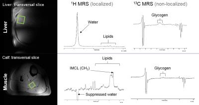 |
Multinuclear in vivo MR spectroscopy at 7T reveals differences in liver and muscle metabolism in NAFLD patients and healthy controls
Martin Gajdošík, Sabina Smajiš, Christian Kienbacher, Lorenz Pfleger, Anton Luger, Siegfried Trattnig, Michael Trauner, Michael Krebs, Martin Krššák
Recent developments at 7T allowed for qualitative assessment of the hepatic lipids, faster measurement of glycogen and ATP turnover. Improved spectral resolution also enabled better separation and quantitation of hepatic metabolites. The aim of the study was to employ the advanced methods of multinuclear in vivo MR spectroscopy at 7T in the liver and muscles in non-alcoholic fatty liver disease (NAFLD) patients and healthy controls. Static and dynamic multinuclear MRS data, supported by mixed meal test and euglycemic-hyperinsulinemic clamp study, showed metabolic changes and disturbed interorgan crosstalk in NAFLD patients.
|
09:03
|
1071.
 |
Renal Arterial Spin Labeling in Diabetes Mellitus
José Mora-Gutierrez, Nuria García-Fernández, María Slon, Danny Wang, Alberto Benito, José Páramo, María Fernández-Seara
Diabetic nephropathy (DN) is a microvascular complication of diabetes mellitus (DM) and a leading cause of chronic kidney disease (CKD). However evidence of renal damage is not detected until the advanced disease stages, using current clinical diagnostic tools. The goal of this study was to investigate renal hemodynamic changes in diabetic patients using ASL and evaluate whether the technique is sensitive enough to detect renal dysfunction early in the disease course, which could have relevant clinical and therapeutic implications. The results demonstrated detection of hemodynamic changes in kidney microvasculature in diabetic patients. Moreover, ASL was able to detect small changes in kidney perfusion across different stages of CKD in the diabetic population.
|
09:15
|
1072.
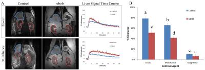 |
Dynamic Contrast-Enhanced Magnetic Resonance Imaging: A Pre-Clinical Approach to Detect and Monitor Diabetes
Dorela Shuboni-Mulligan, Christiane Mallett, Maciek Parys, Barbara Blanco-Fernandez, Erik Shapiro
The link between diabetes and dysregulation of OATP transporters provides an avenue for imaging the disease with MRI. The aim of this work was to use DCE-MRI to non-invasively measure decreases of OATPs during the onset of diabetes. The data presented here verifies that both Gd-EOB-DTPA and Gd-BOPTA enhanced MRI reveals underlying alterations in OATPs that are present in the liver and kidney as a result of diabetes. Preliminary data further suggests that there could be a link between changes in organ enhancement and disease onset. Thus, there is the potential to monitor diabetes and perhaps even diagnose pre-diabetes.
|
09:27
|
1073.
 |
Preliminary Analysis of Longitudinal Functional MRI Data in Diabetic Nephropathy and Healthy Controls - permission withheld
Lu-Ping Li, Jon Thacker, Wei Li, Huan Tan, Chi Wang, Orly Kohn, Stuart Sprague, Pottumarthi Prasad
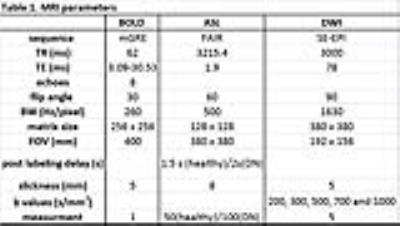
BOLD, Diffusion and ASL MRI data were acquired in diabetic nephropathy (DN) patients with stage-3 chronic kidney disease and healthy subjects. Consistent with the chronic hypoxia model for progression of DN, kidneys in subjects with DN had significantly lower blood flow and medullary R2* compared to controls. Intra-subject coefficient of variation (CV) for R2* and ADC was less than 10% in the control group when comparing baseline and 18 month data, suggests R2* and ADC measurements were reproducible over 18 month. CV was higher for blood flow.
|
09:39
|
1074.
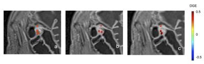 |
Parametric mapping with spatial registration of abdominal MRI is feasible and may inform upon the spatial distribution of fibrosis in intestinal lesions of patients with Crohn’s Disease
Elizabeth Li, Jordi Rimola, MD PhD, Timothy Lu, MD, Alexandre Coimbra, PhD, Alex De Crespigny, PhD
Parametric mapping may provide estimates of the degree and spatial distribution of fibrosis in Crohn’s Disease (CD) patients but is subject to respiratory and peristaltic motion. Validation of out-of-the-box registration strategies and their impact on quality and robustness of parametric maps-delayed gain of enhancement (DGE) and magnetization transfer ratio (MTR) were evaluated.
|
09:51
 |
1075.
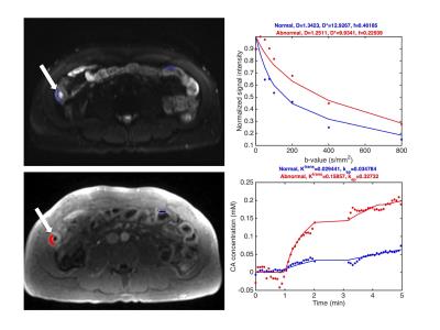 |
Quantitative IVIM-DWI and DCE-MRI for assessment of bowel inflammation in Crohn’s disease
Stefanie Hectors, Sonja Gordic, Octavia Bane, Joana Torres, Judy Cho, Jean-Frederic Colombel, Bachir Taouli
The aims of this study were to quantify intravoxel incoherent motion DWI (IVIM-DWI) and DCE-MRI metrics and to assess the correlation between IVIM-DWI and DCE-MRI metrics in abnormal and normal appearing bowel segments in Crohn’s disease (CD) patients. ADC was significantly reduced in abnormal vs. normal bowel wall segments. A significant negative correlation between Ktrans (measured with DCE-MRI) and perfusion fraction f (measured with IVIM-DWI) was observed. Our preliminary results suggest that IVIM-DWI could potentially be used for simultaneous assessment of perfusion and diffusion in the bowel wall of CD patients, which needs to be verified in a larger cohort of patients.
|
10:03
|
1076.
 |
Inflammatory activity of Crohn's disease: Evaluation by MR T2*mapping without intravenous enhancement - permission withheld
Si yun Huang, Xue hua Li, Zhong wei Zhang, Jin jiang Lin, Li Huang, Zhuang nian Fang, Meng chen Zhang, Meng jie Jiang, Hua song Cai, Margaret H. Pui, Shi ting Feng, Can hui Sun, Zi ping Li
Crohn's disease (CD) is a chronic relapsing inflammatory bowel disease leading to structurally irreversible bowel damage. The incidence of CD had been increasing in the past century epidemiologically . Accurate evaluation of CD activity is crucial for new therapeutic goals of mucosal healing, preventing bowel damage, limiting disability, and improving quality of life . Although colonoscopy remains the gold standard for assessing CD activity, it is invasive, limited to assessment of the small bowel, and not suitable for continuing monitoring of CD activity . Thus, seeking for alternative noninvasive and accurate approaches to assess CD activity is necessary.
|
|