Oral
Body: Breast, Chest, Abdomen, Pelvis
Wednesday, 26 April 2017
| Room 316A |
13:45 - 15:45 |
Moderators: David Bluemke, Jens Vogel-Claussen |
Slack Channel: #s_body
Session Number: O26
13:45
|
0826.
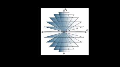 |
Free-Breathing Pediatric Chest MRI: Utility of Self-Navigated Golden-Angle Ordered Conical Ultrashort Echo Time (UTE) Acquisition 
Evan Zucker, Joseph Cheng, Anshul Haldipur, Michael Carl, Shreyas Vasanawala
To assess feasibility of conical k-space trajectory free-breathing UTE chest MRI versus 4D flow and effects of 50% data subsampling and soft-gated motion correction, 32 consecutive children were recruited. Images scored by two blinded radiologists showed good to excellent delineation of all evaluated structures. UTE surpassed 4D flow for lungs and airways and was equivalent for pulmonary arteries. 50% subsampling mildly reduced but maintained diagnostic image quality, favoring its shorter scan time. Soft-gating slightly improved pulmonary artery delineation for one reader but overall degraded images, possibly due to noise from data subsampling, and suggesting motion-robustness of the conical golden-ordered trajectory.
|
13:57
|
0827.
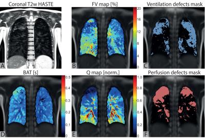 |
Functional lung MRI using matrix pencil decomposition and N2 multiple-breath washout measurements in cystic fibrosis
Grzegorz Bauman, Sylvia Nyilas, Orso Pusterla, Tanja Haas, Michael Ith, Bernd Jung, Carmen Casaulta, Gregor Sommer, Enno Stranzinger, Urs Frey, Philipp Latzin, Oliver Bieri
This study examines a correlation between the functional lung MRI using matrix pencil decomposition and lung function tests in patients with cystic fibrosis. A strong correlation between the global ventilation inhomogeneity index (LCI) from multiple breath washout and ventilation/perfusion impairment in the lung determined by functional MRI is observed. The results of our study support the potential of functional MRI as a diagnostic tool in monitoring disease progression in cystic fibrosis.
|
14:09
 |
0828.
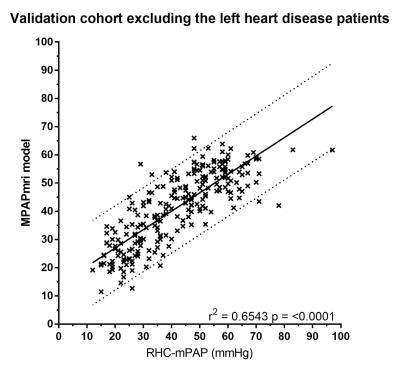 |
Cardiopulmonary MRI as a diagnostic tool in Pulmonary Hypertension
Christopher Johns, David Kiely, David Capener, Charlotte Hammerton, Neil Hamilton, Robin Condliffe, Charlie Eliott, Athanasios Charalampopoulos, Jim Wild, Andy Swift
Pulmonary hypertension has a poor prognosis. Invasive right heart catheter measured mean pulmonary artery pressure (RHC-MPAP) is the gold standard for clinical diagnosis. Here we present a parametric model derived from cardio-pulmonary MRI for the prediction of pulmonary hypertension with a strong correlation with RHC-MPAP and a high diagnostic accuracy. In certain patients, right heart catheterisation may be avoided due to high specificity of this cardio-pulmonary MR model.
|
14:21
|
0829.
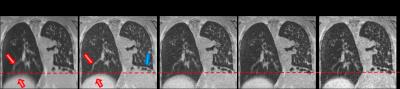 |
Assessment of cystic fibrosis disease using UTE imaging with XD-GRASP reconstruction: a comparison with CT
Jean Delacoste, Catherine Beigelman, Li Feng, Jerome Yerly, Davide Piccini, Daniel Sodickson, Ricardo Otazo, Matthias Stuber, Alain Sauty
Motion resolved reconstructions, using compressed sensing, of 3D ultra short echo time (UTE) acquisitions in cystic fibrosis patients were performed. The definition of the lung-liver interface was quantified and found to be significantly higher than that in motion corrupted reconstructions of the whole datasets. The Helbich-Bhalla score for cystic fibrosis was determined using both computed tomography (CT) and MRI data. Correlation between scores obtained with both modalities was good (ρ=0.77) but consistency was moderate (ICC=0.62). This was due to average MRI scores being lower by 15%, most likely because mosaic perfusion could not be assessed with MRI.
|
14:33
|
0830.
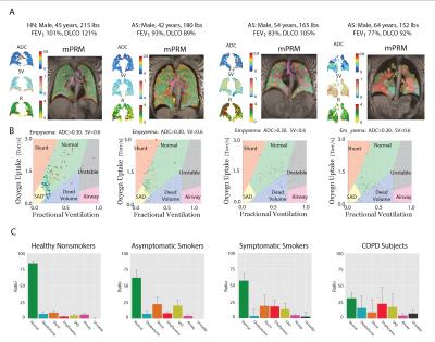 |
Multi-parametric Response Map Using Hyperpolarized Gas MR Imaging to Retain Regionality: Making Hyperpolarized Gas Imaging Great Again
Hooman Hamedani, Yi Xin, Stephen Kadlecek, Ian Duncan, Sarmad Siddiqui, Mehrdad Pourfathi, Nicholas Drachman, Kai Ruppert, Joe Naji, Maurizio Cereda, Rahim Rizi
By establishing multi-parametric response map (mPRM) of Gas MRI, we suggested a multifaceted regional combination of the imaged mPRM that best explain lung function deterioration while had a meaningful representation of each subject lung function condition.
|
14:45
|
0831.
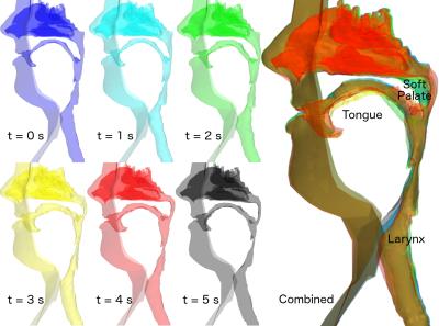 |
Using dynamic MRI to create moving boundary conditions for CFD: determining the causes of upper airway motion in sleep apnea
Alister Bates, Andreas Schuh, Brynne Williams, Matthew Lanier, Keith McConnell, Wolfgang Loew, Robert Fleck, Jason Woods, Charles Dumoulin, Raouf Amin
The upper airway consists of complex mobile structures such as the tongue, soft palate and larynx that make predicting surgical outcomes in obstructive sleep apnea difficult. Dynamic computational fluid simulations provide a method to assess the causes of airway deformation, but require information on the airway shape and motion. Combining MR imaging from three sequences provides this information in high spatiotemporal resolution. Simulations allow characterization of airway wall motion as either in the same or opposite direction as the force applied by the intraluminal airflow, which can be used to better understand the subject-specific mechanics of sleep apnea.
|
14:57
|
0832.
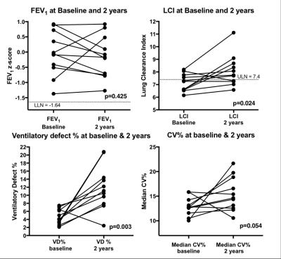 |
LONGITUDINAL MONITORING OF DISEASE PROGRESSION IN CHILDREN WITH MILD CYSTIC FIBROSIS USING HYPERPOLARISED GAS MRI AND LUNG CLEARANCE INDEX
Laurie Smith, Paul Hughes, Felix Horn, Helen Marshall, Graham Norquay, Guilhem Collier, David Hughes, Chris Taylor, Noreen West, Ina Aldag, Alex Horsley, Jim Wild
Hyperpolarised gas ventilation MRI is a sensitive method for evaluating disease progression in subjects with cystic fibrosis and normal spirometry. Ventilation defect % (VD%) increased in 10/11 subjects studied with a mean change of 201%. The MRI coefficience of variance (CV) of signal intensity was similarly sensitive to change. 10/11 subjects had increased lung clearance index (LCI) at 2-years but no subject had abnormal spirometry at either visit. The % change in LCI demonstrated strong correlations with the % change in CV outcomes. VD% and CV reflect different but complimentary aspects of lung disease that appear to track disease progression.
|
15:09
|
0833.
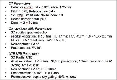 |
3D Radial UTE MRI outperforms 3D Cartesian conventional echo time MRI for evaluation of cystic fibrosis lung disease
Scott Nagle, Christopher Francois, Madeline Poranski, Laura Bell, Kevin Johnson, Sean Fain
In this prospective cross-sectional study of 30 cystic fibrosis (CF) subjects, 3D radial ultrashort echo time (UTE) MRI significantly outperformed 3D Cartesian conventional echo time MRI when compared with reference standard computed tomography (CT), especially with respect to the depiction of air trapping. Short-term 1-2 week repeatability was comparable with CT. Since air trapping is considered one of the earliest signs of CF lung disease, and potentially reversible, the use of UTE MRI could significantly improve the utility of MRI as a biomarker for treatment effect in mild CF lung disease.
|
15:21
|
0834.
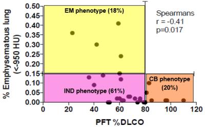 |
A new COPD phenotype characterized by hyperpolarized xenon-129 MRI
Kun Qing, Sahar Mansoor, John Mugler, III, Talissa Altes, Nicholas Tustison, Kai Ruppert, Jaime Mata, G.Wilson Miller, Iulian Ruset, F.William Hersman, Joanne Cassani, Yun Shim
Existing literature describes two distinctive phenotypes of chronic obstructive pulmonary disease (COPD): airway-predominant chronic bronchitis and alveolar-predominant emphysema. In this study, based on results from pulmonary function tests and computed tomography, we found a new mixed phenotype of COPD. This mixed phenotype showed minimal emphysematous tissue destruction, but low diffusion lung capacity (DLCO). Subsequent hyperpolarized xenon-129 MRI results indicated that gas exchange to the pulmonary blood in lungs for this mixed phenotype was significantly impaired as compared to controls and the classic COPD phenotypes.
|
15:33
|
0835.
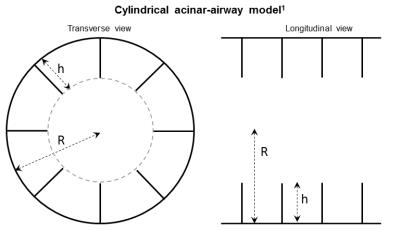 |
Hyperpolarized 3He gas MRI in infant lungs: investigating alveolar-airspace size with restricted gas diffusion
Nara Higano, Robert Thomen, James Quirk, Kenneth Parks, Heidie Huyck, Andrew Hahn, Sean Fain, Michael Baker, Gloria Pryhuber, Jason Woods
Acinar development in infant humans has not been extensively studied. Hyperpolarized gas diffusion MRI has been shown to relate directly to alveolar-airspace size in adults, pediatrics, canines, and mice. Using ex-vivo lungs from 7 healthy and 1 diseased infant humans, we investigated the relationship between 3He apparent diffusion coefficient (ADC) via mono-exponential decay, alveolar-duct radius via a restricted diffusion model originally developed for mice, and radius via histological measurement. While the mouse model is invalid in the infant diffusion regime, ADC measurements reflect changes in alveolar-airspace size. This method shows promise for longitudinal in-vivo acinar-airway monitoring in neonatal patients.
|
|