Oral
Body: Breast, Chest, Abdomen, Pelvis
Tuesday, 25 April 2017
| Room 310 |
08:15 - 10:15 |
Moderators: Sachin Jambawalikar, Eric Sigmund |
Slack Channel: #s_body
Session Number: O29
08:15
 |
0362.
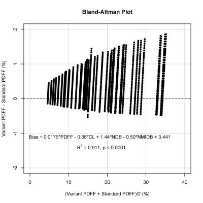 |
Robust agreement between MRI and MRS hepatic proton density fat fraction despite biologically plausible variability in fat spectra in patients with nonalcoholic steatohepatitis 
Cheng Hong, Adrija Mamidipalli, Jonathan Hooker, Gavin Hamilton, Tanya Wolfson, Soudabeh Fazeli Dehkordy, Scott Reeder, Rohit Loomba, Claude Sirlin
MRI- and MRS-based proton density fat fraction (PDFF) techniques require accurate modeling of the multi-peak spectrum of triglycerides (TG) in order to achieve accurate hepatic fat quantification. However, variations in TG spectrum may lead to quantification variability. We performed a secondary analysis of adults with biopsy-confirmed nonalcoholic steatohepatitis undergoing confounder-corrected chemical-shift-encoded 3T MRI and MRS, and calculated variant PDFF values using a range of biologically plausible spectral models. Within the range of fat fractions seen in the liver, PDFF estimation using MRI and MRS was robust to variability in the TG spectrum. Greater bias was seen when the baseline fat fraction was higher, but remained low.
|
08:27
|
0363.
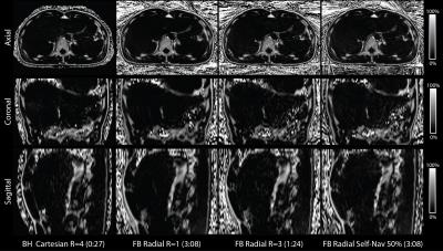 |
Free-breathing Fat Quantification in the Liver Using a Multiecho 3D Stack-of-Radial Technique: Investigation of Motion Compensation and Quantification Accuracy
Tess Armstrong, Thomas Martin, Alto Stemmer, Xinzhou Li, Yutaka Natsuaki, Kyunghyun Sung, Holden Wu
Multiecho Cartesian MRI methods can non-invasively quantify liver fat, but are susceptible to motion artifacts and limited by breath-hold (BH) imaging. We have developed a new free-breathing (FB) liver fat quantification technique using 3D stack-of-radial imaging (Radial). In this work, we further investigate motion compensation and quantification accuracy for FB Radial. In n=11 healthy volunteers, FB Radial fat quantification demonstrated significant correlation (ρ > 0.9876) and low mean difference (< -1.19%) compared to BH Cartesian and BH single-voxel spectroscopy. FB Radial can potentially achieve accurate whole-liver fat quantification with either a fast 1-2 minute scan or a 3-minute self-navigated scan.
|
08:39
|
0364.
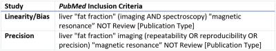 |
Linearity, Bias, and Precision of Proton-Density Fat Fraction for Liver Fat Quantification: A Meta-Analysis
Ali Pirasteh, Mustafa Bashir, Scott Reeder, Claude Sirlin, An Tang, Guido Kukuk, Jens-Peter Kuhn, Holger Hetterich, Ji Soo Song, Takeshi Yokoo
Proton-density fat fraction (PDFF) is a quantitative imaging biomarker (QIB) of hepatic triglyceride concentration and steatosis. Liver PDFF can be measured noninvasively using magnetic resonance imaging (MRI) or spectroscopy (MRS). Various MRI-based PDFF methods have been validated in single-center studies at 1.5T or 3T field strength using a specific reconstruction algorithm on a single vendor platform. However, its technical performance as a QIB is unknown in a multi-center, multi-vendor setting. In this meta-analysis of previously published data from multiple studies, we demonstrated excellent linearity, negligible bias, and high repeatability/reproducibility of MRI-PDFF across different field strengths, vendors, and reconstruction algorithms.
|
08:51
|
0365.
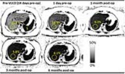 |
Monitoring Resolution of Fatty Liver Disease with MRI following Bariatric Surgery: A Prospective, Multi-center Study
B. Dustin Pooler, Curtis Wiens, Alan McMillan, Nathan Artz, Alexandra Schlein, Yesenia Covarrubias, Jonathan Hooker, Jeffrey Schwimmer, Luke Funk, Guilherme Campos, Jacob Greenberg, Garth Jacobsen, Santiago Horgan, Claude Sirlin, Scott Reeder
The temporal resolution of fatty liver disease following bariatric surgery is poorly understood. We used a validated chemical shift encoded MRI (CSE-MRI) method to measure liver proton density fat fraction (PDFF) as a biomarker of liver fat. We followed a cohort of 50 obese adults undergoing bariatric surgery with pre-operative very low calorie diet (VLCD) and conclude that average liver PDFF normalizes to <5% by 6 months following bariatric surgery. Normalization of liver fat is seen in 79% of patients who lower body mass index (BMI) by ≥10 mg/k2 and 83% of patients who lose ≥30 kg.
|
09:03
|
0366.
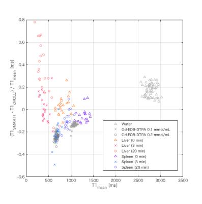 |
A paradoxical systemic bias in Gd-EOB-DTPA-enhanced T1 relaxometry of the liver: a comparison of SMART1Map and MOLLI.
Akira Yamada, Sachie Fujita, Yoshihiro Kitoh, Yasuo Adachi, Hayato Hayashibara, Aya Shiobara, Atsushi Nozaki, Yuji Iwadate, Glenn Slavin, Yasunari Fujinaga, Masumi Kadoya
SMART1Map (saturation method using adaptive recovery times for cardiac T1 mapping) is a new single-point T1 mapping technique that directly measures true T1 unlike look-locker approaches. The feasibility of Gd-EOB-DTPA-enhanced T1 relaxometry of the liver using SMART1Map was evaluated comparing with modified look-locker inversion recovery (MOLLI). A significant paradoxical systemic bias was observed between and within SMART1Map and MOLLI in Gd-EOB-DTPA-administrated liver, although SMART1Map may be more reproducible than MOLLI in the rest of conditions. Careful consideration should be given to the effect of the paradoxical systemic bias in the evaluation of liver function using Gd-EOB-DTPA-enhanced T1 relaxometry.
|
09:15
 |
0367.
 |
Determining the T1 of the water in the liver by modelling the effects of fat, iron and off-resonance frequencies on MOLLI T1 measurements
Ferenc Mozes, Elizabeth Tunnicliffe, Thomas Marjot, Christina Levick, Michael Pavlides, Matthew Robson
The frequency dependence of balanced steady-state free precession signals causes significant alterations in modified Look-Locker inversion recovery T1 measurements of livers with fat accumulation, leading to either under- or over-estimation of liver T1 values. This is further to the already-known influence of iron. The present study shows a possibility to correct for these effects, yielding a T1 measurement that represents the T1 of the water component independent of the fat and is tested both in phantoms and human participants.
|
09:27
|
0368.
 |
Bayesian prediction for insufficient liver enhancement in gadoxetic acid-enhanced hepatobiliary phase imaging
Yuki Mori, Utaroh Motosugi, Tatsuya Shimizu, Shintaro Ichikawa, Hiroshi Onishi
Insufficient liver enhancement due to decreased liver function is a major limitation in gadoxetic acid-enhanced hepatobiliary phase imaging (HBP). Recent research shows that insufficient liver enhancement is associated with liver function tests including total bilirubin level, Child-Pugh classifications, indocyanine green tests, and liver stiffness measured by MR elastography. However, none of these tests have been practically used for determining the patients with insufficient liver enhancement before MR imaging. We used univariate tests and logistic regression to determine predictive factors and performed cross validation to reveal utility of Bayesian method for predicting patients with insufficient liver enhancement in gadoxetic acid-enhanced HBP.
|
09:39
|
0369.
 |
Flexible and Efficient 2D Radial TSE T2 Mapping with Tiered Echo Sharing and with “Pseudo” Golden Angle Ratio Reordering - permission withheld
Yutaka Natsuaki, Mahesh Keerthisavan, Ali Bilgin, Bradley Bolster, Kevin Johnson, Xiaoming Bi, Gerhard Laub, Maria Altbach
There has been recent increased interest in quantitative T2 mapping for accurate diagnosis of many pathological disorders. 2D radial TSE with tiered echo sharing and bit-reverse view ordering acquires TE data for T2 mapping in an efficient and motion robust fashion, but imposes limits on the choice of Echo Train Length (ETL). The current work introduces a novel view ordering algorithm with “pseudo” Golden Angle ratio (pGA) that removes restrictions in the ETL. With this algorithm, the scan time of 2D radial TSE is reduced (by18% in this study) without a compromise in image quality or in T2 mapping accuracy.
|
09:51
|
0370.
 |
Assessment of Nonalcoholic Fatty Liver Disease (NAFLD) Activity Score (NAS) with MR Elastography (MRE)
Meng Yin, Alina Allen, Kevin Glaser, Sudhakar Venkatesh, Taofic Mounajjed, Vijay Shah, Richard Ehman
To investigate the utility of a hepatic imaging protocol “hepatogram”, which includes multi-parametric MR Elastography (MRE) and fat fraction assessment, in predicting nonalcoholic fatty liver disease (NAFLD) activity score (NAS: 0-8). In both preclinical and clinical subjects with histology-proven NAFLD, generalized linear models of liver stiffness, damping ratio and fat fraction successfully distinguished each NAS score with excellent accuracy (AUROC>0.89 for all). Misclassifications in distinguishing steatohepatitis (NAS≥3) from NAFLD (NAS<3) was only 2/64 mice and 3/51 human subjects. Our findings indicate the hepatogram imaging protocol can predict NAS score and may be useful to monitor NAFLD disease progression and regression.
|
10:03
|
0371.
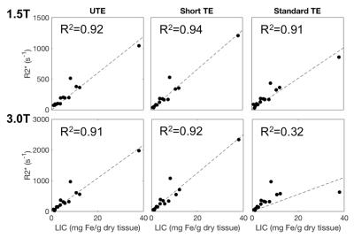 |
Accuracy and Reproducibility of Iron Quantification using Ultra-Short TE Imaging at 1.5T and 3.0T
Curtis Wiens, Ante Zhu, Kevin Johnson, Scott Reeder, Diego Hernando
This work examined the accuracy and reproducibility of ultra-short TE (UTE) R2* mapping in patients with liver iron overload. Fifteen subjects with known or suspected liver iron overload were scanned at 1.5T and 3.0T using a radial UTE, two Cartesian multi-echo, gradient-echo acquisitions, and an R2-based (FerriScan) reference acquisition. UTE R2* measurements demonstrated excellent reproducibility across field strengths (with expected linear increase with field strength) and high correlation with liver iron concentration. Cartesian approaches offered excellent reproducibility for R2*<1000s-1. However R2*>1000s-1, neither Cartesian approach were reproducible across field strength, suggesting that the range of R2* had been surpassed.
|
|