Oral
Body: Breast, Chest, Abdomen, Pelvis
Monday, 24 April 2017
| Room 320 |
08:15 - 10:15 |
Moderators: Gabriele Masselli, Evis Sala |
Slack Channel: #s_body
Session Number: O35
08:15
|
0097.
 |
Amide proton transfer magnetic resonance imaging of uterine endometrial cancer: Association with histologic grade 
Yukihisa Takayama, Akihiro Nishie, Osamu Togao, Yoshiki Asayama, Kousei Ishigami, Yasuhiro Ushijima, Daisuke Okamoto, Nobuhiro Fujita, Kenzo Sonoda, Jochen Keupp, Hiroshi Honda
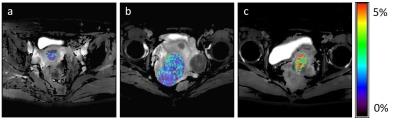
The histologic grade of endometrioid adenocarcinoma (EMCA) is one of the important factors in choosing a treatment plan. Currently, needle biopsy or surgical resection is necessary to diagnose the histological grade; a less invasive procedure is strongly desired. In this study, we evaluated the utility of amide proton transfer (APT) imaging in estimating the histologic grade of EMCA. APT signal intensities (SIs) of EMCA increased in accordance with the progression of histologic grade. APT SIs of high-grade EMCA were significantly higher than those of low-grade EMCA. APT imaging has potential as a biomarker for histologic grade of EMCA.
|
08:27
|
0098.
 |
The prognostic power of early and late phase DCE-MRI parameters in locally advanced cervix cancer.
Kjersti Lund, Trude Simonsen, Gunnar Kristensen, Einar Rofstad
DCE-MRI can provide prognostic information on locally advanced cervix carcinomas. Most studies have emphasis on the early phase of the Signal Intensity Time Curve (SITC). The purpose of this study was to explore the prognostic value of the late phase of the SITC and to reveal any added value to that of parameters from the early phase. Both the early phase parameter LETV and the late phase parameter TVIS was associated with overall survival. The association was independent of clinical factors like tumor volume, FIGO stage and lymph node status. TVIS did not provide any added prognostic value to LETV.
|
08:39
|
0099.
 |
Histogram analysis of intravoxel incoherent motion MRI in predicting chemoradiotherapy response in cervical cancer
Jose Perucho, Elaine Lee, Wing Chi Chan, Nanjie Gong, Queenie Chan
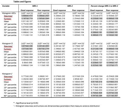
Histogram analysis of intravoxel incoherent motion (IVIM) diffusion-weighted MRI (DWI) could be a promising quantitative approach in predicting tumour response to chemoradiotherapy (CRT) in cervical cancer. We retrospectively studied twenty-five patients with cervical cancer who had paired IVIM MRI examinations before and at week-4 of treatment. We observed that histogram skewness of true diffusion coefficient (D) prior to treatment and that a large increase in the 90th percentile of D following CRT were predictive of better CRT response.
|
08:51
|
0100.
 |
A One-Step Biomarker Quantification Methodology for DCE-MRI of Complex Ovarian Masses: Capturing Kinetic Pattern from Early to Late Enhancement - video not available
Anahita Fathi Kazerooni, Mahnaz Nabil, Hamidreza Haghighat Khah, Hamidreza Saligheh Rad
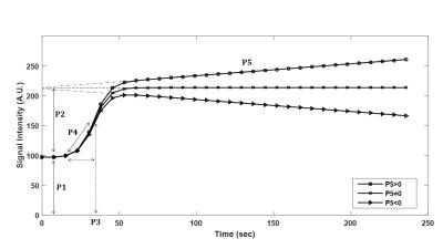
Accurate characterization of sonographically-indeterminate ovarian masses before surgery is crucial for proper disease management. While DCE-MRI has emerged as a problem-solving technique, accurate parameter estimations from semi-quantitative or PK analysis are dependent on multiple steps, including proper protocol design, motion reduction, selection of physiology-based PK model and AIF, which discourages development and reliability of computer-aided diagnostic procedures. Here, we aimed to develop a one-step pre-processing and quantification classification scheme based on a five-parameter Sigmoid model, capturing early- to late-enhancement kinetics, including washout as a previously overlooked parameter for ovarian masses, to generate accurate differentiation of complex ovarian masses.
|
09:03
|
0101.
 |
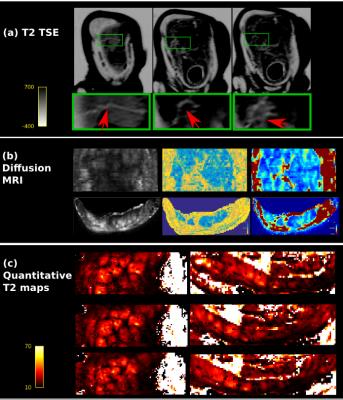
Ex vivo MRI evaluation of vulvar cancer to predict resection margins in fresh wide local excision specimens: a pilot study
Jan Heidkamp, Petra Zusterzeel, Andor Veltien, Arie Maat, Ilse Van Engen-Van Grunsven, Jurgen Fütterer
Currently there’s no accurate and topical peroperative information available on the margin status of wide local resection specimens containing vulvar cancer. In this pilot study we performed a qualitative image evaluation of ex vivo 7T MR images acquired of fresh specimens of the vulva containing vulvar cancer using different MRI sequences. High resolution T2 weighted images obtained the highest score for image quality, visibility of the tumor, and visibility of the transition between the epidermis and the resection surface.
|
09:15
|
0102.
 |
Assessment of uterine artery hemodynamics in normal pregnancy with 4D Flow MRI
Eileen Hwuang, Marta Vidorreta, Nadav Schwartz, John Detre, Daniel Licht, Walter Witschey
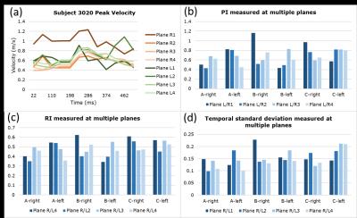
In vivo imaging of uterine artery blood flow during remodeling is potentially valuable in assessing placental function during pregnancy. We present 4D flow MRI of the uterine arteries, demonstrating inter- and intrasubject heterogeneity in vessel anatomy and hemodynamics. This high spatial resolution, multi-location approach potentially addresses the limitations of Doppler ultrasound in quantifying pulsatility and resistance indices as clinical biomarkers of placental health.
|
09:27
 |
0103.
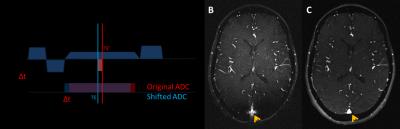 |
In-utero non-contrast MR angiography of the fetal vasculature using a double-echo radial sampling scheme
Uday Krishnamurthy, Brijesh Yadav, Pavan Jella, Swati Mody, Edgar Hernandez-Andrade, FeiFei Qu, Anabela Trifan, Ewart Haacke, Sonia Hassan, Roberto Romero, Jaladhar Neelavalli
To show that the isotropic gradient delay issue can be addressed by a simple shift of the readout-window . We also report the use of a fully flow-compensated, readout-shifted 2D radial gradient echo sequence to perform non-contrast MRA of the human fetus in-utero
|
09:39
|
0104.
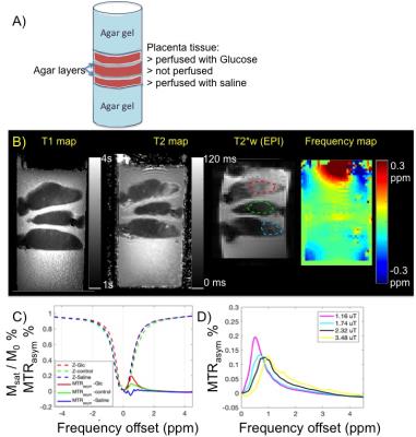 |
Feasibility of glucose CEST in the human placenta
Jie Luo, Yang Ji, Esra Abaci Turk, Iris Zhou, Drucilla Roberts, Patricia Grant, Phillip Sun
Placental glucose transfer is essential to sustain fetal development, yet there has been no report attempting to measure glucose transport across the human placenta with MRI-based approaches. Emerging glucose chemical exchange saturation transfer (glucoCEST) imaging is uniquely sensitive to glucose, which has been explored in tumor imaging. Herein, we have demonstrated glucoCEST MRI is a valid tool to monitor glucose perfusion in ex vivo human placenta, laying the groundwork for in vivo glucoCEST in human placenta.
|
09:51
|
0105.
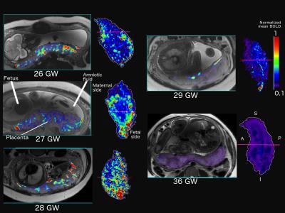 |
Parametric Mapping of Oxygen Activity in Human Placenta across Gestation using in utero BOLD imaging
Vidya Rajagopalan, Vince Schmithorst, Julie Coloigner, Jessica Wisnowski, Matthew Borzage, Hollie Lai, Skorn Ponrartana, Ashok Panigrahy, Stefan Bluml
We present here, for the first time, parametric maps of oxygen activity in normal human placenta using in utero functional MR imaging. Our method highlights anatomical and gestational age dependent patterns in placental activity. These maps can be used to gain insight into normative placental function and identifying insufficient or abnormal placental functioning at various points in gestation.
|
10:03
|
0106.
 |
An exploration of quantitative physiological multi-modal in-vivo imaging of the human placenta
Jana Hutter, Paddy Slator, Jonathan O'Muircheartaigh, Rui Azeredo Gomes Teixeira, Anthony Price, Ana Gomes, Laura McCabe, Sophie Arulkumaran, Mary Rutherford, Joseph Hajnal
The crucial role of the placenta in successful pregnancies is the transfer of oxygen within functional units – cotyledons. However, current screening falls short of visualizing this in-vivo. This study explores a multi-model in-vivo MRI acquisition able to visualize and depict a range of spatial and temporal processes and the underlying micro-structure. Diffusion characteristics such as Mean Diffusivity and fractional anisotropy, quantitative T2* maps, temporal characteristics and the depiction of vasculature allow insights and can be applied to a range of research questions.
|
|