Wednesday, 26 April 2017
| Room 320 |
13:45 - 15:45 |
Moderators: Richard Hodgson, Mary Kate Manhard |
Slack Channel: #s_msk
Session Number: O37
13:45
|
0846.
 |
A preliminary application of Porosity Index measured by UTE MRI sequence in the femoral neck 
Min Chen, Huishu Yuan, Lizhi Xie
The current study aims to assess the feasibility of porosity index (PI) measurements derived from ultra-short echo time (UTE) MRI technology in the femoral neck and to further investigate its latent associations with age, gender, body mass index (BMI) and tibial PI. It was concluded that cortical PI measured by UTE MRI sequence can be applied in femoral neck and cannot be replaced by tibial measurement. Femoral neck and tibial PI were observed to correlate with age and BMI , which worth further study.
|
13:57
 |
0847.
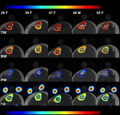 |
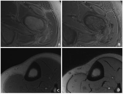
Feasibility of bone mineral and water quantification in vivo by solid-state 31P and 1H MRI at 3T
Xia Zhao, Hee Kwon Song, Alan Seifert, Cheng Li, Felix Wehrli
Surrogates for bone matrix density, pore volume fraction and mineral density can be studied with solid-state MRI. Here, we developed an in vivo MRI protocol to simultaneously quantify bone mineral 31P and bound and pore water on a 3 Tesla clinical MRI system with a dual-frequency extremity coil and 3D 1H UTE and 31P PETRA-ZTE pulse sequences. Measurements in the mid-tibia of 10 subjects yielded 7.06±1.53mol/L 31P, and 13.99±1.26 and 10.39±0.80mol/L H2O for total and bound water, respectively, in good agreement with prior ex vivo data. The work suggests that both organic and inorganic phases of cortical bone can be quantitatively evaluated in vivo with a single, integrated protocol.
|
14:09
|
0848.
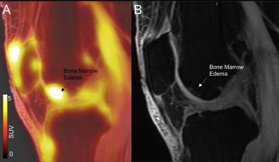 |
Study Of The Associations Between [18F]-NaF PET Bone Remodeling, MRI Trabecular Bone Structure and Patient Reported Outcomes in subjects with Osteoarthritis
Valentina Pedoia, Rohit Curucundhi, Dragana Savic, Misung Han, Youngho Seo, Matthew Bucknor, Benjamin Franc, Sharmila Majumdar
In Osteoarthritis (OA) degeneration of articular cartilage, are accompanied by changes in subchondral and trabecular bone. In this study we analyzed bone structure, cartilage degeneration and bone remodeling, quantified using simultaneous PET/MRI system in fourteen subjects with radiographic or symptomatic OA. The aim of this study was to evaluate these imaging biomarkers in association with patient reported outcomes for evaluating this technique as a tool for assessing OA. Our results showed associations between Standardized Uptake Values (SUV) and both level of pain measured by KOOS and cartilage degeneration measured using T1ρ and T2 relaxation times.
|
14:21
|
0849.
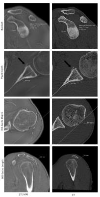 |
Zero Echo Time MRI: Osseous Shoulder Imaging
Ryan Breighner, Yoshimi Endo, Gabrielle Konin, Lawrence Gulotta, Matthew Koff, Hollis Potter
Routine MRI fails to provide direct visualization of bone due to the short tissue relaxation times and limited signal intensity. This study investigates the use of proton density zero echo time (ZTE) MRI for bone in the shoulder. Shoulder CT and ZTE images were acquired for 31 patients. Five measures of osseous defect and lesion sizes were compared between the two modalities. ‘Fair’ to ‘excellent’ intraobserver agreement was observed between CT and ZTE MRI. Zero Echo Time MRI may obviate the need for additional CT evaluation in some cases.
|
14:33
|
0850.
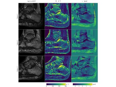 |
Simultaneous R2* and Quantitative Susceptibility Mapping of Trabecularized Yellow Bone Marrow: Initial Results in the Calcaneus
Maximilian Diefenbach, Jakob Meineke, Peter Foehr, Stefan Ruschke, Thomas Baum, Jan Kirschke, Andreas Hock, Hendrik Kooijman, Ernst Rummeny, Dimitrios Karampinos
R2* mapping has been previously used to measure trabecular bone density by quantifying the magnetic field inhomogeneity effects induced by the susceptibility difference between trabecular bone and marrow. Quantitative susceptibility mapping (QSM) has been emerging in other body parts for measuring the field-independent magnetic susceptibility. Trabecular bone is in many locations embedded in yellow fatty marrow. Therefore, trabecular bone QSM requires to account for the presence of fat. The purpose of the present work is to develop a methodology for simultaneous R2* mapping and QSM of trabecularized yellow bone marrow and apply the technique in the calcaneus of healthy volunteers.
|
14:45
|
0851.
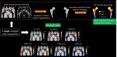 |
Proximal Femur Marrow Adipose Tissue Assessment Using 3T Multi-Parametric Chemical Shift Encoded MRI: Preliminary Results in Osteoporotic Patients - permission withheld
Dimitri MARTEL, Gregory CHANG, Mary BRUNO, Benjamin LEPORQ
Recent assessment of osteoporosis by monovoxel magnetic resonance spectroscopy states interesting feature about subregional femoral marrow fat composition. in this study , we evaluate differences in bone marrow fat of the proximal femurs of controls and patients with OP using a novel quantitative multi-parametric MRI approach that provides information about fat content, fatty acid composition, transverse relaxation and internal magnetic susceptibility.
|
14:57
|
0852.
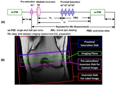 |
Epiphyseal Bone Marrow Perfusion Imaging in the Distal Femur Using Arterial Spin Labeling: Feasibility and Challenges
Xiufeng Li, Casey Johnson, Jutta Ellermann
Epiphyseal bone marrow perfusion of the distal femur is a valuable biomarker for knee diseases or injuries, such as anterior cruciate ligament tears and post-traumatic osteoarthritis. Bone marrow perfusion imaging is also of interest in the management of developmental knee diseases, such as osteochondritis dissecans, where the capacity to heal may be related to sufficient perfusion to the site of injury. ASL is well suited for monitoring of knee disease progression, assessment of therapy response, and use in pediatric populations. This study is to evaluate the feasibility and challenges of epiphyseal bone marrow ASL imaging in the distal femur.
|
15:09
|
0853.
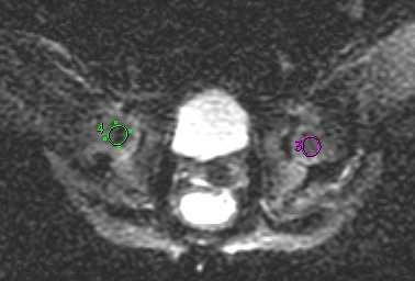 |
MRI quantification of diffusion and perfusion in epiphysis of femoral heads after close reduction of children with DDH by intravoxel incoherent movement
Xiang Hong Meng, Zhi Wang, Dan Dan Zheng, Jian Ping Yang, Zhong Li Zhang
Developmental dysplasia of the hip (DDH) is a common disease of the development of hips. Some children with DDH need close reduction followed by immobilization in spica casting, but excessive hip abduction may lead to avascular necrosis of the epiphysis of femoral head. Therefore, this study would like to use IVIM method to help the pediatric orthopaedic doctors to know whether the blood supply of epiphysis of femoral heads is insufficiency or not in patients with DDH after close reduction.
|
15:21
|
0854.
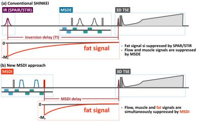 |
Motion-Sensitized Driven-Inversion (MSDI) for improvement of diffusion-prepared MR neurography (SHINKEI) in the brachial plexus
Masami Yoneyama, Iain Ball, Yasuhiro Goto, Hitoshi Tadenuma, Kayoko Abe, Makoto Obara, Tetsuo Ogino, Tomoyuki Okuaki, Michinobu Nagao, Marc Van Cauteren
Brachial plexus is anatomically complex and may be involved in a variety of pathologies that leads to significant morbidity. MR neurography, based on diffusion-prepared MR neurography (SHINKEI), plays a major role in the diagnostic work-up of plexus pathologies. To solve the problems that caused by the current fat suppression techniques (SPAIR and STIR) with SHINKEI, we developed motion-sensitized driven inversion (MSDI). MSDI could simultaneously suppress fat, flow and muscle signals using only one pre-pulse module. SHINKEI with MSDI provides uniform fat suppression in the brachial plexus, as in STIR, with no significant decrease in SNR compared to SPAIR.
|
15:33
|
0855.
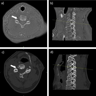 |
Diagnostic Accuracy of Zero Echo Time Magnetic Resonance Imaging for Grading of Cervical Spine Neural Foraminal Stenosis
Darryl Sneag, Parina Shah, Ryan Breighner, Yoshimi Endo, Erin Argentieri, Matthew Koff
A challenge for orthopaedic radiologists is utilizing MRI to assess neuroforaminal (NF) stenosis of the cervical spine as cortical bone does not display with sufficient signal intensity. This study utilized zero echo time (ZTE) imaging to visualize the cervical spine and compare evaluation of NF stenosis to corresponding CT imaging. Substantial agreement was found between ZTE and CT (κ=0.71). ZTE tended to underestimate stenotic grade in 25% of foramina, with a majority (66%) of differences within one grading level. Further development of ZTE and image processing may minimize the need for cervical spine CT in assessing NF stenosis.
|
|