Thursday, 27 April 2017
| Room 313A |
13:00 - 15:00 |
Moderators: Elisabeth Garwood, Valentina Taviani |
Slack Channel: #s_msk
Session Number: O39
13:00
 |
1142.
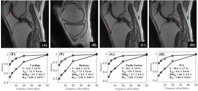 |
Three-dimensional Ultrashort Echo Time Cones Imaging with Magnetization Transfer Modeling (3D UTE-Cones-MT): Simulation, Specimen and Volunteer Studies of the Knee Joint 
Yajun Ma, Eric Chang, Michael Carl, Jiang Du
A major limitation associated with conventional clinical MRI sequences is the magic angle effect . The conventional T2 and T1rho measures may increase more than 100% when the fibers are oriented from 0º to the magic angle (~55º) relative to the B0 field, far more than that associated with osteoarthritis (OA). Magnetization transfer (MT) imaging has shown less sensitivity to the magic angle effect, and can indirectly evaluate macromolecules which have extremely short T2 (~10 us) and invisible with all MRI sequences. However, conventional MT techniques cannot be applied to short T2 tissues such as menisci, ligaments, tendons and bone. Ultrashort echo time (UTE) sequences with TEs 100-1000 times shorter than those of clinical sequences have been developed to image these short T2 tissues. In this study, we aimed to develop 3D UTE with Cones sampling and MT (3D UTE-Cones-MT) imaging and signal modeling to quantify water and macromolecules in both short and long T2 tissues in the knee joint at 3T.
|
13:12
|
1143.
 |
Two compartmental diffusion model of skeletal muscle: application to aging and chronic limb suspension induced DTI changes in the medial gastrocnemius - permission withheld
Usha Sinha, Vadim Malis, Shantanu Sinha
Diffusion tensor imaging (DTI) is a powerful technique that allows one to probe tissue at the microstructural level. Though microarchitecture determines the DTI indices, a diffusion model is required to make inferences about the microstructure. We applied a two compartmental diffusion model for muscle to explain differences in the DTI indices with age and with atrophy induced by limb suspension. The model qualitatively explains the changes in DTI seen in limb suspension that is linked to decrease in muscle fiber diameter and in intracellular volume fraction. Extensions to the model are required to explain the age related changes in DTI.
|
13:24
|
1144.
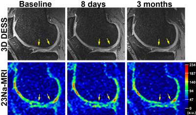 |
In Vivo Follow-up of Low-Grade Femoral Cartilage Defects using Sodium MRI at 7T
Stefan Zbyn, Vladimir Mlynarik, Vladimir Juras, Markus Schreiner, Pavol Szomolanyi, Didier Laurent, Celeste Scotti, Harry Haber, Joerg Goldhahn, Ewa Kubiak, Oliver Bieri, Stefan Marlovits, Miika Nieminen, Siegfried Trattnig
Sodium (23Na) MRI was employed for the evaluation of patients with ICRS Grade I-II cartilage defects at 7T. 23Na data from defect, weight-bearing, and non-weight-bearing region of femoral cartilage were obtained at baseline, 8-days, 3-months and 6-months follow-up. Significantly lower 23Na values were found in defect than in weight-bearing and non-weight-bearing regions at all time-points. While 23Na values in weight-bearing and in non-weight-bearing regions were stable over time, a significant decrease was found in the defects. 23Na-MRI allows noninvasive follow-up of changes in the cartilage GAG content and thus might be particularly useful for the evaluation of cartilage regenerating therapies.
|
13:36
|
1145.
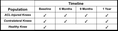 |
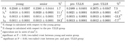
Cluster Analysis of Cartilage T2 and T1rho Relaxation Times: Can the Contralateral Knee be used as a Control in the ACL-injured population?
Uchechukwuka Monu, Emily McWalter, Caroline Jordan, Brian Hargreaves, Garry Gold
In an ACL-injured population, longitudinal studies that use advanced MRI techniques such as T2 and T1rho mapping to assess cartilage health, typically compare ACL-injured knees with a separate healthy group or the contralateral knees. It is still unclear whether the contralateral knees can be used as a control group. Using a cluster analysis-based technique, we identify in the contralateral knees, significant increase in T1rho relaxation times over 1-year that is comparable to the increase in the ACL-injured knees. These focal cluster areas may represent degenerative changes and demonstrate that the contralateral knees may not be good controls.
|
13:48
|
1146.
 |
Novel NMR biomarkers characterizing human knee synovial fluid after ACL-injuries: correlation with immunoassay and longitudinal cartilage MR T1? and T2 imaging
Kaipin Xu, Keiko Amano, Matthew Tanaka, Subramaniam Sukumar, John Kurhanewicz, Virginia Kraus, Benjamin Ma, Xiaojuan Li
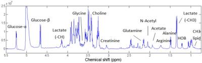
Subjects with acute anterior cruciate ligament (ACL) injury have a high risk of developing post-traumatic osteoarthritis (PTOA) even after ACL reconstruction. To identify novel NMR biomarkers of synovial fluid that may predict cartilage degeneration after acute injury and to develop potential preventative strategies for PTOA, human knee synovial fluid harvested from 25 anterior cruciate ligament (ACL) injured subjects were studied using high resolution magic angle spinning (HR-MAS) NMR spectroscopy and correlated to immunoassay and longitudinal cartilage MR T1ρ and T2 imaging.
|
14:00
|
1147.
 |
Biomechanical properties of bovine knee cartilage under compressive loading: A study at high field MRI (9.4T) using T1, T2 and T1rho relaxometry combined with DENSE-FID.
Willy Gsell, Willy Zevenbergen, Tom Dresselaers, Deva Chan, Corey Neu, Uwe Himmelreich, Ilse Jonkers
The aim of this study was to correlate the cartilage displacement pattern during cartilage on cartilage contact through displacement encoding imaging during compressive loading with the T1, T2 and T1rho changes, indices of changes in collagen matrix, water and proteoglycan contents. We demonstrated that local mechanical changes in the cartilage during loading were correlated to global molecular changes assessed through T1, T2 and T1rho. The localized cartilage deformation and strain fields suggest a differential response to loading of the different regions of the cartilage which could help in further optimizing cell based therapy for osteoarthritis.
|
14:12
|
1148.
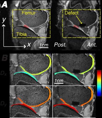 |
Intratissue Strains Increase in a Full Thickness and Critical Sized Ovine Cartilage Defect Model
Deva Chan, Luyao Cai, Kent Butz, Eric Nauman, Darryl Dickerson, Ilse Jonkers, Corey Neu
Functional imaging of intratissue strain in articular cartilage provides an opportunity to probe the healthy or diseased state of the tissue. We utilized displacement encoded MRI to document the increase in deformation following creation of a critical sized femoral defect in a large animal model ex vivo. Strain heterogeneity indicated that the implant replacement is critical to long-term repair success and restoration of physiological cartilage strain and mechanical function. This study represents a crucial step toward the evaluation of biomechanical imaging biomarkers to evaluate tissue damage, repair, and regeneration in the intact joint of a clinically-relevant and translational animal model.
|
14:24
|
1149.
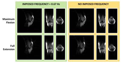 |
Dynamic knee imaging using 4D self-gated MRI with compressed sensing reconstruction
Valentina Mazzoli, Jasper Schoormans, Martijn Froeling, Andre Sprengers, Klaas Nicolay, Bram Coolen, Nico Verdonschot, Aart Nederveen, Gustav Strijkers
Knee abnormalities and pain are sometimes elucidated during motion, therefore the ability to obtain 4D images of the moving knee could add diagnostic value to the conventional static MRI scans. In this work we present a method to obtain 4D imaging of the human knee during motion, without the use of an external gating system.
|
14:36
|
1150.
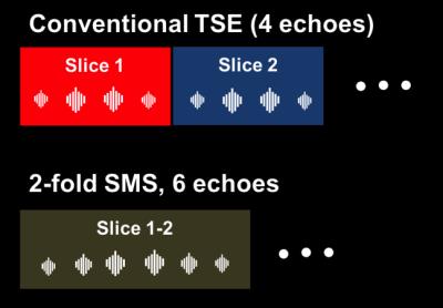 |
Rapid knee MRI using TSE sequences accelerated with a combination of simultaneous multislice, multicoil compressed sensing and elongated echo trains
Akio Yoshimoto, Steven Baete, Mary Bruno, Mitchell Kline, Elisabeth Garwood, Fernando Boada, Ricardo Otazo, Michael Recht
Turbo spin echo imaging is accelerated with a novel combination of simultaneous multislice, multicoil compressed sensing and elongated echo times to reduce the scan time of routine knee examinations to 7 minutes. The accelerated protocol is validated against the conventional technique in a cohort of 10 patients based on the opinion of expert radiologists. Diagnostic accuracy was found to be similar despite the reduction in scan time. This might be useful to expand the utilization of MRI and to increase patient throughput.
|
14:48
|
1151.
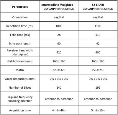 |
3D MRI of Knee in Pediatric Patients with CAIPIRINHA SPACE: Diagnostic Performance Assessment with Arthroscopic Correlation - permission withheld
Jan Fritz, Shivani Ahlawat, Gaurav Thawait, Esther Raithel, Wesley Gilson, Rushyuan Lee
3D CAIPIRINHA SPACE permits the acquisition of 4-fold accelerated, high quality data sets and has been shown to be feasible for efficient 3D MRI of the knee; however, the clinical application has not been demonstrated. We report the performance of 3D CAIPIRINHA SPACE MRI for the diagnosis of internal derangement of the knee in children and adolescents using arthroscopy correlation as the standard of reference. 3D CAIPIRINHA SPACE enables clinically feasible isotopic 3D MRI of the knee in children and adolescents with an acquisition time of 10 min and high accuracy for the diagnosis of meniscal, ligamentous and cartilage abnormalities.
|
|