Oral
General Cancer Imaging
Monday, 24 April 2017
| Room 313BC |
16:15 - 18:15 |
Moderators: Kristine Glunde, Eugene Kim |
Slack Channel: #s_cancer
Session Number: O40
16:15
|
0307.
 |
Imaging of the Tumor Type-specific Microenvironment in Preclinical Cancer Models of Varying Malignancy 
Ellen Ackerstaff, Natalia Kruchevsky, Ekaterina Moroz, H. Carl LeKaye, Kristen L. Zakian, SoHyun Han, HyungJoon Cho, Radka S. Stoyanova, Nirilanto Ramamonjisoa, Inna S. Serganova, Ronald G. Blasberg, Jason A. Koutcher
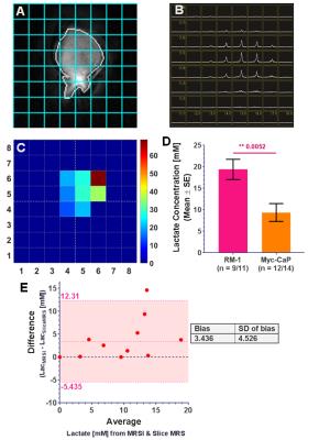
An abnormal tumor microenvironment characterized by hypoxia, low extracellular pH (pHe), vascular abnormalities, and high tumor lactate has been associated with aggressive, treatment-resistant tumors. Using tumor models of different origin and malignancy, and focusing on prostate cancer, we investigated the relationship of lactate metabolism and vascularity, and, in selected models, localized pHe. We found differences in whole-tumor lactate concentrations between tumor models and successfully mapped lactate concentrations. Vascular blood flow and permeability varied significantly between tumor models in well-vascularized areas, while being similar across all models in hypoxic areas, emphasizing a need for spatial characterization of the tumor microenvironment.
|
16:27
 |
0308.
 |
Brain Tumors Disrupt the Resting-State Connectome
Darian Hadjiabadi, Leland Pung, Jiangyang Zhang, BD Ward, Woo-Taek Lim, Meghana Kalavar, Nitish Thakor, Bharat Biswal, Arvind Pathak
Resting-state functional MRI (rsfMRI) has become indispensable for mapping the changes in ‘connectivity’ between brain regions in a range of diseases including brain tumors. However, the complex interplay between abnormal brain tumor vasculature, tumor blood flow, and cancer cell-induced neurovascular uncoupling can confound the interpretation of resting-state connectivity in patients. Therefore, in this preclinical study we quantified brain tumor-induced changes on resting-state connectivity relative to that in healthy brains, followed by histological validation. RsfMRI revealed that brain tumors alter the resting-state connectome, and histology confirmed that this was largely due to cancer cell-induced disruption of the neurovascular unit.
|
16:39
|
0309.
 |
Multiparametric MR for assessment of tissue characteristics of small intestine neuroendocrine tumour evaluated by histological correlations
Mikael Montelius, Oscar Gustafsson, Johan Spetz, Ola Nilsson, Eva Forssell-Aronsson, Maria Ljungberg
This study investigates the relations between MR derived, quantitative parameters reflecting perfusion, diffusion and relaxation, and histological indices reflecting apoptosis, proliferation, vascularity and fibrosis. We show that important biological characteristics of tumour tissue can be probed by multiparametric MRI.
|
16:51
|
0310.
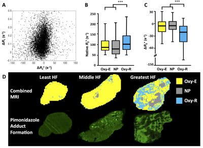 |
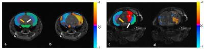
Understanding the relationship between R2* and R1 MRI biomarkers of hypoxia: insights from 786-0 renal cancer xenografts and patients with renal carcinoma
Ross Little, Yann Jamin, Jessica Boult, Josephine Naish, Yvonne Watson, Susan Cheung, Huiqi Lu, Damien McHugh, Geoff Parker, Joely Irlam, Catherine West, John Waterton, Simon Robinson, James O'Connor
Quantification of tumour R2* and oxygen-induced ΔR2* and ΔR1 are being investigated as potential biomarkers of tumour hypoxia, but their relationship is complex and not well understood. Here, we used a validated R1 biomarker (oxygen refractory fraction, termed “Oxy-R”) to segment tumours into hypoxic and non-hypoxic sub-regions. This revealed a clear relationship between hypoxic status and native R2* and hyperoxia-induced ΔR2*. Preclinical findings were replicated in clinical data from patients with renal carcinoma. These data highlight the importance of heterogeneity-based analysis of tumours and provide further validation of Oxy-R as a biomarker of tumour hypoxia.
|
17:03
|
0311.
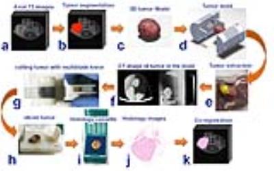 |
Co-registration of multi-parametric MRI and histology to study breast cancer Habitats in a preclinical model.
William Dominguez-Viqueira, Bruna Jardim-Perassi, Mikalai Budzevich, Epifanio Ruiz, Suning Huang, Pedro Enriquez-Navas, Jan Poleszczuk, Debora Zuccari, Robert Gillies, Gary Martinez
Different tumor micro-environments or habitats are discernible by MRI. In order study these; a co-registration framework was developed using 3D-printed tumor-molds created from in-vivo MRI images of five mice with implanted breast cancer tumors. The results of automated 3D-alignment of MRI images and histology slices are promising and encourage further experiments using the presented workflow. Tumor habitats clustering from multi-parametric MRI images showed encouraging results with similarities to the hypoxic pattern observed by immunohistochemistry. This work will help understanding MRI habitats to monitor cancer evolution as a means to aid treatment decisions in the future.
|
17:15
 |
0312.
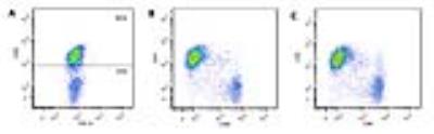 |
Fluorine-19 NMR cytometry to quantify human transgenic CAR T cell biodistribution in murine studies of glioblastoma immunotherapy
Fanny Chapelin, Hideho Okada, Eric Ahrens
Technologies to quantify the biodistribution of emerging immunotherapeutic cell therapies against cancer can accelerate the timeline to evaluate potential candidates. In this study, we describe the use of in ‘NMR cytometry’ to assay immunotherapeutic cell biodistribution in a mouse model of sub-cutaneous glioblastoma (U87) treated with chimeric antigen receptor (CAR) T-cells. We examine CAR T cell 19F labeling efficiency, phenotype and biodistribution with 19F NMR at day 2 and 7 post infusion to elucidate T-cell tumor homing, survival, and tissue distribution.
|
17:27
|
0313.
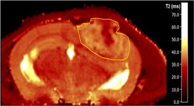 |
Correlated quantitative assessment of glioblastoma-angiogenesis by T2-mapping and in vivo multiphoton microscopy
Artur Hahn, Ke Zhang, Gergely Solecki, Michael Breckwoldt, Lukas Buschle, Sabine Heiland, Christian Ziener, Martin Bendszus, Frank Winkler, Felix Kurz
Microvasculatures in healthy cortical tissue, in untreated and in antiangiogenically treated glioblastoma multiforme are compared in a mouse model. From T2-maps, the information entropy is determined for each tissue type. In addition, capillaries are directly imaged through in vivo multiphoton microscopy to obtain sets of microvascular parameters. The T2-entropy is lowest in healthy tissue and significantly higher in glioblastoma, with a moderate decrease in treated tumors. Several vascular characteristics correlate with the T2-entropy. The correlations provide insight into the influence of microvasculature on MR-dephasing.
|
17:39
|
0314.
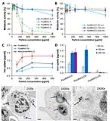 |
In vivo tracking of iron oxide labeled T-cells infiltrating preclinical tumor models - permission withheld
Johannes Riegler, Vincent Javinal, Maj Hedehus, Mike Reichelt, Meredith Sagolla, Jill Schartner, Franklin Peal, Richard Carano
The discovery of immune checkpoint pathways such as CTLA4 and PD1/PDL1, which control T-cell activation and activity, has fuelled interest in their modulation to achieve sustained anti-tumor immunity. This requires sufficient T-cell infiltration and activity in tumors. However, these processes are incompletely understood, in part due to the terminal nature of current analysis techniques. We therefore optimized labeling of activated T-cells with iron oxide nanoparticles, transferred labeled T-cells into tumor bearing hosts and performed serial MRI. Although, hypointense spots could be detected in the tumor rim following T-cell transfer, quantification is complicated by vascular abnormality induced susceptibility changes.
|
17:51
|
0315.
 |
Magnetisation Transfer MRI Facilitates Non-Invasive Identification of Fibrosis in Chemically-Induced Rat Mammary Carcinomas Imaged on a 1.5T Clinical Platform
Jessica Boult, Neil Jerome, Matthew Orton, James d'Arcy, Martin Leach, Dow-Mu Koh, David Collins, Simon Robinson
Intratumoural fibrosis is associated with poor prognosis in breast cancer patients. Non-invasive detection of such fibrosis may contribute to the provision of personalised treatment regimens. Multi-parametric MRI, using a clinical MRI scanner and incorporating endogenous contrast mechanisms, was performed on MNU-induced rat mammary carcinomas to identify parameters sensitive to the detection and quantification of fibrosis. Magnetisation transfer MRI derived parameters correlated with percentage picrosirius red staining, which detects collagen I/III, major components of fibrosis, in this heterogeneous tumour cohort. These results strongly support the inclusion of magnetisation transfer in clinical MR breast imaging protocols.
|
18:03
|
0316.
 |
MRI Tracking and Quantitative Analyzing Natural Killer Cell Infusion for Hepatocellular Carcinoma Treatment in a Rodent Model
Zhanliang Su, Xifu wang, Linfeng Zheng, Tianchu Lyu, Matteo Figini, Guohong Han, Daniel Procissi, Lei Qin, Bin Zhang, Jeremy Zhang, Wei Xing, Yihe Yang, Kejiang Wang, Shixin Wang, Vahid Yaghmai, Andrew Larson, Zhuoli Zhang
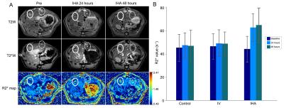
This paper presents the first evidence that transcatheter IHA NK cell local delivery for HCC adoptive transfer immunotherapy in a rat model. We use a clinically feasible method by combining FDA-approved drugs heparin, protamine and ferumoxytol to compound HPF nanocomplexes for magnetic NK cell labeling so that their biodistribution can be visualized and quantized in vivo with MRI.
We demonstrated: a) transcatheter IHA NK cell infusion improved their homing efficacy to target tumors; b) quantitative analysis result of serial MRI monitoring of NK cell migrate to target tumors could serve as a key early biomarker for predicting longitudinal response. |
|