Wednesday, 26 April 2017
| Room 316A |
08:15 - 10:15 |
Moderators: Kai-Hsiang Chuang, Ferdia Gallagher |
Slack Channel: #s_mrs_molecular
Session Number: O47
08:15
|
0725.
 |
Phase II Clinical Hyperpolarized 13C 3D-Dynamic Metabolic Imaging of Prostate Cancer using a B1-insensitive Variable Flip Angle Design 
Hsin-Yu Chen, Peder Larson, Jeremy Gordon, Robert Bok, Marcus Ferrone, Mark van Criekinge, Lucas Carvajal, Rahul Aggarwal, James Slater, Ilwoo Park, Eugene Milshteyn, Sarah Nelson, John Kurhanewicz, Daniel Vigneron
The 3D compressed-sensing echo-planar spectroscopic imaging (CS-EPSI) sequence has enabled 5-dimensional HP-13C imaging of human prostate cancer with full-prostate coverage and high spatial and temporal resolution. A new variable flip angle design substantially desensitizes the sequence to B1 variations for improved quantitative analysis of pyruvate-to-lactate rate constant (kPL) measurements. Pre-clinical animal and clinical patient studies demonstrate that the resulting sequence enabled improved quantitative accuracy while maintaining excellent metabolite SNR.
|
08:27
|
0726.
 |
Demonstrating the Randle Cycle In Vivo: Assessment of Physiological Alterations in Human Cardiac Metabolism Using Hyperpolarised 13C MR Spectroscopy
Damian Tyler, Oliver Rider, Michael Dodd, Angus Lau, Andrew Lewis, Jack Miller, Mark Peterzan, Claire Trumper, Stefan Neubauer
The recent introduction of dissolution Dynamic Nuclear Polarization (DNP) has opened up a new window on in vivo metabolism and in this work we present the first demonstration that dissolution-DNP can observe physiological modulation of metabolism in the healthy human heart. The transition from the fasted to the fed state is shown to lead to an increase in flux through the key regulatory enzyme, pyruvate dehydrogenase, due to a metabolic switch away from fatty acid oxidation towards glucose oxidation. Such studies will provide the basis for future clinical studies exploring the metabolic alterations that occur in the diseased heart.
|
08:39
|
0727.
 |
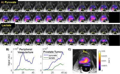
Utilizing hyperpolarized MRI in prostate cancer to assess metabolic dynamics and histopathologic grade
Kristin Granlund, Hebert Vargas, Serge Lyashchenko, Phillip DeNoble, Vincent Laudone, James Eastham, Ramon Sosa, Matthew Kennedy, Duane Nicholson, YanWei Guo, Albert Chen, James Tropp, Hedvig Hricak, Kayvan Keshari
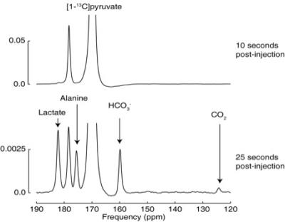
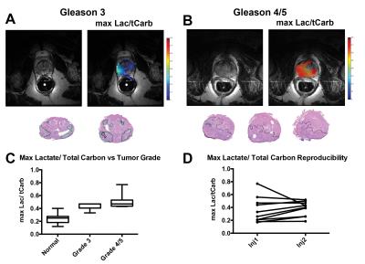
A hallmark of prostate cancer is the reprogramming of prostate cancer metabolism which has been exploited for both 31P and 1H MRSI. In recent work, we and others have shown that hyperpolarized substrates can be used in living systems to measure changes in metabolic dynamics. Many studies have focused on the use of HP pyruvate in preclinical models, though the characterization of prostate cancer in man using HP MRI has been limited. In this work, we demonstrate the use of HP pyruvate MRI in prostate cancer patients. We assess the metabolic dynamics to the prostate of pyruvate as well as its conversion to lactate. Moreover we extend this analysis to the comparison of HP lactate to prostate cancer grade showing that not only does this approach have the potential to measure differences in grade it also can provide reproducible metabolic signatures across patients.
|
08:51
|
0728.
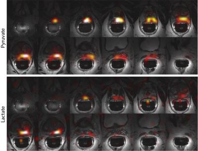 |
Human Hyperpolarized C-13 MRI Using a Novel Echo-Planar Imaging (EPI) Approach
Jeremy Gordon, Hsin-Yu Chen, Peder Larson, Ilwoo Park, Mark Van Criekinge, Eugene Milshteyn, Robert Bok, Rahul Aggarwal, Marcus Ferrone, James Slater, John Kurhanewicz, Daniel Vigneron
In this pilot study, we developed and applied a specialized symmetric EPI approach for the study of hyperpolarized [1-13C]pyruvate metabolism in the human prostate. We show that this approach can acquire ghost-free hyperpolarized [1-13C]pyruvate and [1-13C]lactate images with high spatiotemporal resolution in a clinical setting.
|
09:03
|
0729.
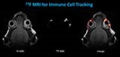 |
In Vivo Visualization of Orbital Immune Cell Infiltration During Early Development of Graves’ Disease by 19F MRI
Ulrich Flögel, Anja Eckstein, Utta Berchner-Pfannschmidt
Graves’ disease is an antibody-mediated autoimmune condition of the thyroid gland, which is frequently associated with Graves’ orbitopathy (GO), where inflammation of the orbit results in a detrimental remodeling of the orbital soft tissue. To investigate the molecular basis of GO and evaluate new therapeutic targets for treatment, we used a recently established mouse model of GO and analyzed the interplay of inflammation and adipogenesis using anatomical 1H MRI and 19F MRI for immune cell tracking. Hereby, the infiltration of immune cells could be identified as initial step leading to the remodeling of the orbital soft tissue in GO mice.
|
09:15
|
0730.
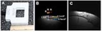 |
19F-perfluorocarbon-labeled human peripheral blood mononuclear cells can be detected in vivo using clinical MRI parameters in a therapeutic cell setting
Corby Fink, Jeffrey Gaudet, Matthew Fox, Shashank Bhatt, Sowmya Viswanathan, Michael Smith, Joseph Chin, Paula Foster, Gregory Dekaban
A major hurdle in advancing cell-based cancer vaccines is the inability to track where therapeutic cells migrate post-injection. We present the first study conducted in which primary Good Manufacturing Practice-grade 19Fluorine (19F) labeled all peripheral blood mononuclear cell lineages (PBMC) and 19F cellular MRI was used to track and quantify in vivo migration in a mouse model. Secondly, we present the highest sensitivity for 19F detection reported in the literature and the first time a 1.2cm deep injection of 19F-labeled PBMC using a small dual-tuned surface coil has been detected using a clinical scanner and MR protocol.
|
09:27
 |
0731.
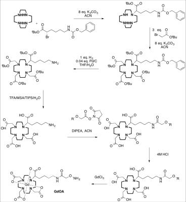 |
Molecular MR imaging of renal fibrogenesis in a mouse model - permission withheld
Philip Waghorn, Iris Chen, Nicholas Rotile, Chloe Jones, Diego Ferreira, Lan Wei, Bryan Fuchs, Peter Caravan
The fibrotic deposition and remodeling of extracellular matrix (ECM) proteins to form cross-linked collagen and elastin fibers is a characteristic feature of chronic kidney disease (CKD). Critical to fiber formation is the presence of allysine, which facilitates fibril cross-linking through condensation reactions with neighboring allysine and lysine residues on collagen and elastin. We have developed a novel Gd-based MR probe, GdOA, designed to target allysine for non-invasive molecular imaging of active fibrogenesis in kidney disease, allowing the onset of fibrogenesis to be detected and provide a quantitative measure of the efficacy of future anti-fibrotic targeted therapies.
|
09:39
|
0732.
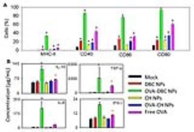 |
Chitin and dibutyrylchitin nanoparticles as imaging enabled vaccine nanocarriers - permission withheld
Barbara Blanco Fernandez, Yasser Aldhamen, Shatadru Chakravarty, Andrea Amalfitano, Erik Shapiro
We describe a strategy for fabricating chitin and dibutyrylchitin –an organo-soluble chitin derivative- nanoparticles (NPs) encapsulating a model antigen (ovalbumin) or iron oxide nanocrystals. The use of MRI enabled the determination of the cell migration of vaccinated cells, allowing the determination of the immunization efficiency in early stages. To form DBC, a reversible acylation of chitin was performed and NPs were fabricated using an emulsification/evaporation method, then chitin was regenerated by alkaline saponification. NPs had vaccine adjuvant properties and allowed the track of immune cells to lymph nodes, proving their utility as vaccine adjuvants and imaging agents.
|
09:51
|
0733.
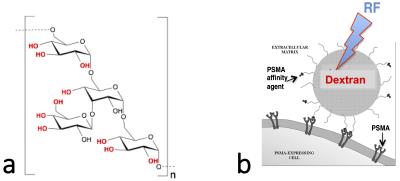 |
CEST MRI of PSMA expression using natural dextran-based contrast agents
Guanshu Liu, Sangeeta Ray Banerjee, Xing Yang, Nirbhay Yadav, Ala Lisok, Anna Jablonska, Jiadi Xu, Yuguo Li, Martin Pomper, Peter van Zijl
It is desirable to have non-invasive imaging methods able not only to render the expression and distribution of receptors, enzymes and other targets relevant to cancer, but also have a safety profile enabling use as a screening tool. We have developed an enzyme-targeted MR imaging approach that uses non-metallic, non-radioactive dextran as the imaging agent detected by CEST MRI. Our results indicate that imaging tumors expressing the prostate-specific membrane antigen (PSMA) could be accomplished using urea-conjugated dextran particles that could be detected in micromolar (per particle) concentration by MRI.
|
10:03
|
0734.
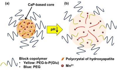 |
Detection of tumor hypoxic region using pH-activatable nanoparticles containing manganese contrast agent
Daisuke Kokuryo, Peng Mi, Horacio Cabral, Tsuneo Saga, Ichio Aoki, Nouhiro Nishiyama, Kazunori Kataoka
Assessing the environment inside a tumor would aid the development of an effective therapeutic strategy. Our group has recently developed a pH-activatable nanoparticle containing MR contrast agents, which is called MnCaP micelle. For an in vivo tumor model, a specific and strong enhancement of MR signal in the tumor was obtained after the administration. The enhanced area was agreed with the high lactate region found with 1H-MRS and which corresponds to the hypoxic region inside tumor. We conclude that MnCaP micelle detects hypoxic regions in the tumor clearly and therefore it has potential to provide important information for tumor therapy.
|
|