Thursday, 27 April 2017
| Room 311 |
15:30 - 17:30 |
Moderators: Karin Shmueli, Hongfu Sun |
Slack Channel: #s_contrastmechanisms
Session Number: O51
15:30
|
1207.
 |
Assessing the cellular distribution of iron in deep gray matter based on R2* and Quantitative Susceptibility Mapping (QSM) - Application to healthy controls and patients with Multiple Sclerosis (MS) 
Yanis Taege, Robert Zivadinov, Jannis Hanspach, Jesper Hagemeier, Michael Dwyer, Balint Sule, Nicola Bertolino, Dhaval Shah, Dejan Jakimovski, Niels Bergsland, Bianca Weinstock-Guttman, Ferdinand Schweser
This work introduces a clinically applicable technique to assess the cellular distribution of iron based on R2* and Quantitative Susceptibility Mapping (QSM). The method was applied to 68 MS patients and 29 controls, showing significant differences in the cellular iron distribution with age and disability.
|
15:42
 |
1208.
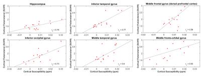 |
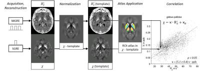
Correlations of beta-Amyloid and brain iron load (QSM): preliminary results of simultaneous assessment in a large sample
Jiri van Bergen, Xu Li, Frances-Catherine Quevenco, Sandra Leh, Anton Gietl, Valerie Treyer, Rafeal Meyer, Alfred Buck, Roger Nitsch, Peter van Zijl, Christoph Hock, Paul Unschuld
To extend findings on the use of QSM in Alzheimer’s Disease and possible direct interactions with Amyloid-β, this study is investigating a growing sample of elderly subjects using simultaneous assessment of Amyloid-PET for Aβ-load and QSM for estimation of iron load (indicated by susceptibility) using a combined PET-MRI instrument. Our preliminary data suggests a significant correlation between susceptibility and Aβ in subjects with high brain Aβ load or clinically diagnosed Mild Cognitive Impairment, in several cortical and sub-cortical regions. The sample is expected to grow considerably in the upcoming months.
|
15:54
|
1209.
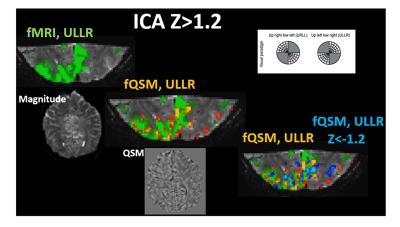 |
Discrimination of volumes with positive and negative functional QSM activation within fMRI-positive volumes in high-resolution, high-field task-based and resting-state data
Pinar Özbay, Lars Kasper, Klaas Pruessmann, Daniel Nanz
In this work, we wanted to explore the bidirectional activation in functional Quantitative Susceptibility Mapping (fQSM) via Independent Component Analysis (ICA) in various cases, hence we included i) visual paradigm, ii) motor task and iii) resting state experiments with high-resolution data acquired at 7-Tesla. We investigated the behavior in terms of activation patterns and temporal evaluations of functional-PHASE, fQSM and traditional fMRI. Furthermore, we compared regions of activation with phase-contrast-angiography data. In all scenarios, we have found out that the total (positive + negative) activated area in fQSM was well matching with positive activation in fMRI.
|
16:06
|
1210.
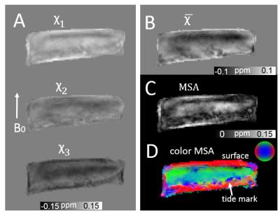 |
Magnetic Susceptibility Anisotropy of Collagen Fibrils in the Articular Cartilage
Hongjiang Wei, Kyle Decker , Yuyao Zhang, Chunlei Liu
Articular cartilage with depth dependent ultra-layer structure is constructed by collagen fibrils, which is shown by histology having three distinct layers: the fibrils are mostly parallel to the surface, randomly distributed and mostly oriented perpendicular to the surface in superficial zone, middle zone and deep zone, respectively. Quantitative susceptibility mapping is particularly sensitive to molecular content and cellular arrangement, thus is suitable to probe such highly organized microstructure and evaluate its magnetic susceptibility. Our study shows a clear B0-orientation-dependent susceptibility contrast in the articular cartilage and that the collagen fibril orientations can be well measured by susceptibility tensor imaging.
|
16:18
|
1211.
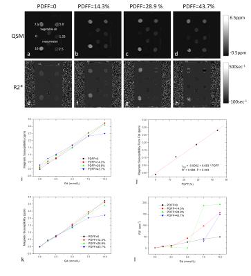 |
Quantitative Susceptibility Mapping (QSM) Overcomes R2* Confounding Factors for Measuring Liver Iron
Jianqi Li, Qi Song, Tian Liu, Zhuwei Zhang, Martin R Prince, Kelly Gillen, Xu Yan, Shu Cheng, Ting Hua, Xiance Zhao, Miao Zhang, Yu Zhao, Gaiying Li, Guangyu Tang, Guang Yang, Gary M Brittenham, Yi Wang
A major challenge in the R2 and R2* methods for mapping liver iron content is that they can be confounded by fat, fibrosis and other changes in cellularity that are known to contribute to R2 and R2*. In this paper, the fat contribution to liver susceptibility was estimated and removed from the measured liver susceptibility with validation on a gadolinium-fat-water phantom. In patients, fat-corrected QSM was found to be insensitive to liver diseases including fat and tumor, which had extensive effects on R2*. Therefore, QSM can overcome confounding factors in R2* for mapping liver iron content.
|
16:30
|
1212.
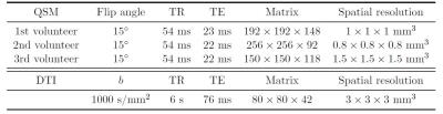 |
The effect of white matter fiber orientation in Quantitative Susceptibility Mapping at 7T
Marta Lancione, Mauro Costagli, Graziella Donatelli, Mirco Cosottini, Michela Tosetti
The tensorial nature of magnetic susceptibility affects frequency-shift images and quantitative maps (QSM), whose reliability should be questioned. Three healthy volunteers underwent 7T MRI exams including diffusion tensor imaging (DTI) and three QSM acquisitions, each with different orientation of the head. In order to assess the effect of susceptibility anisotropy, we sorted brain voxels depending on their fractional anisotropy (FA) and, by plotting their QSM values against the angle between the static field and the eigenvector corresponding to the largest eigenvalue in DTI, we observed an empirical threshold of FA that reflects the reliability of susceptibility measures.
|
16:42
|
1213.
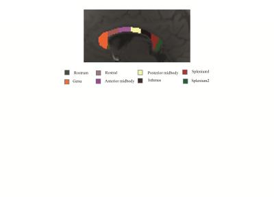 |
Signal compartments modelled from 7T multi-echo GE data showed variation across the corpus callosum
Kiran Thapaliya, Steffen Bollmann, Viktor Vegh, Markus Barth
Quantitative assessment of myelin water fraction using a multi-compartment model can be useful to improve our understanding of white matter diseases. Our work aims to explore tissue microstructure information contained in voxel signals by analysing voxel compartment volume fraction, frequency shift and $$$T_2^*$$$ from data acquired at 7T. We performed our analysis across from the rostrum to the splenium of corpus callosum. Parameterisation of tissue characteristics can potentially delineate structural and chemical changes in tissue with biologically meaningful information. This in turn provides a framework for new imaging biomarker development in neurodegenerative diseases and disorders, such as multiple sclerosis
|
16:54
|
1214.
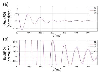 |
Nuclear susceptibility shift
Seung-Kyun Lee, Jinil Park, Jeongtaek Lee, Jang-Yeon Park
We have observed in a clinical scanner that the center frequency of water MRI changed with the 1H spin flip angle. The amount of the change could be explained by the tip-angle dependent nuclear spin paramagnetic susceptibility, much the same way as the usual, tissue electronic para- and dia-magnetic susceptibility induces B0 shift in MRI. The observed shift, corresponding to nuclear susceptibility of +0.004 ppm in water, may affect the ultimate accuracy of MR-based tissue magnetic susceptibility measurements if not properly accounted for.
|
17:06
|
1215.
 |
An interleaved sequence for simultaneous MRA, SWI and QSM
Yongsheng Chen, Saifeng Liu, Yan kang, E. Mark Haacke
MRA, SWI and QSM are important for identifying thrombus, hemorrhage, CMBs and assessing oxygen saturation and iron deposition in diseases such as stroke and traumatic brain injury (TBI). Practically, it is important to acquire these data with sufficient resolution, good SNR, co-registered and rapidly. Therefore, we developed a 3D interleaved GRE sequence that produces MRA, SWI, R2* and QSM for imaging arteries, veins and the basal ganglia in 4 minutes at 3T for the entire brain with a resolution of 0.67x1.33x2.0mm3. Five healthy volunteers’ data were acquired approved by the local IRB to demonstrate the utility of this approach.
|
17:18
|
1216.
 |
Quantitative Susceptibility Mapping from Unsuppressed Water Signals in 1H-MRSI Data
Xi Peng, Fan Lam, Bryan Clifford, Yudu Li, Zhi-Pei Liang
This work presents a new method to extract QSM from the unsuppressed water signals in 1H-MRSI data and enables simultaneous MRSI and QSM in a single acquisition. The proposed method builds on the recently proposed subspace imaging method called SPICE (SPectroscopicImaging by exploiting spatiospectral CorrElation), which allows MRSI acquisition without water suppression, thus encoding susceptibility induced phase variations in the water spectroscopic signals. Parallel imaging, subspace-based modeling and constrained reconstruction are integrated to generate QSM and high-resolution MRSI reconstruction from such data. In-vivo experiment results demonstrate the capability of proposed method in producing susceptibility map along with metabolite spatiospectral distributions from a single 6-minute scan.
|
|