Monday, 24 April 2017
| Room 314 |
13:45 - 15:45 |
Moderators: Kejia Cai, Mark Pagel |
Slack Channel: #s_contrastmechanisms
Session Number: O52
13:45
 |
0192.
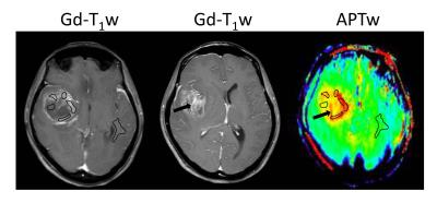 |
Amide proton transfer-weighted MRI signal as a surrogate biomarker to assess MGMT promoter methylation status in glioblastoma 
Shanshan Jiang, Xianlong Wang, Yu Wang, Hao Yu, Tianyu Zou, Yongxing Du, Charles Eberhart, Maria Adelita Vizcaino Villalobos, Yi Zhang, Hye-Young Heo, Peter Van Zijl, Zhibo Wen, Jinyuan Zhou
We explored the feasibility of using the APTW signal intensity as a surrogate biomarker to identify the methylation status of MGMT promoter in glioblastoma (GBM). Eighteen patients with newly diagnosed GBM were recruited and scanned. Results showed that the APTW signal intensities were significantly higher in the unmethylated MGMT promoter group than in the methylated MGMT promoter group. The area under the ROC curve (AUC) for APTW to differentiate these two GBM groups was 0.857. Preoperative APTW imaging may assist in predicting the MGMT promoter methylation status in patients with GBM.
|
13:57
|
0193.
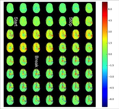 |
Dynamic Glucose Enhanced Imaging at 3T: First Human Data
Xiang Xu, Akansha Sehgal, Nirbhay Yadav, Linda Knutsson, John Laterra, Martin Pomper, Hailey Rosenthal, Peter van Zijl
Recently, it has been demonstrated that D-glucose has potential as an MRI contrast agent at 7T for imaging dynamic changes upon glucose infusion in brain tumors using chemical exchange saturation transfer (CEST) MRI. Here we show first data for the possibility of translating such technique to 3T using pseudo-continuous wave saturation and extend the method to acquire a 3D volume (10 slices) for better brain coverage. We present dynamic glucose-enhanced (DGE) data from healthy volunteers and a brain tumor patient with a low grade glioma showing the feasibility of glucose enhanced imaging at clinical field strength.
|
14:09
|
0194.
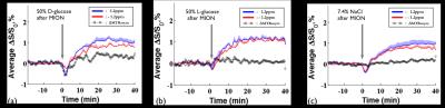 |
Effect of Osmolality on Dynamic Glucose Enhanced(DGE) MRI
Wonmin Choi, Julius Chung, Tao Jin, Seong-Gi Kim
Dynamic glucose enhanced(DGE) MRI has shown promise in glucose metabolism studies. In recent studies, a hypertonic dextrose solution was used for reliable detection of glucose in the brain. However, the effects of the hypertonic solution on DGE signal have not been verified yet. This study aimed to investigate the signal contributions from non-glucose related components. We used hypertonic D-, L-glucose, and NaCl solution to identify osmolality effects. Our data show an osmotic shift of water between the extravascular and intravascular space, induced by administering D-glucose(50%), can highly affect the DGE signal but negligible contributions were observed from the intravascular space.
|
14:21
|
0195.
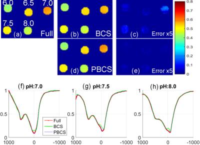 |
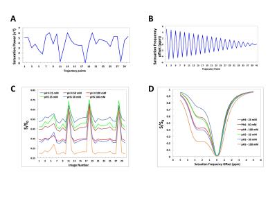
Accelerated CEST Imaging with Parallel Blind Compressed Sensing
Huajun She, Bian Li, Joshua Greer, Jochen Keupp, Ananth Madhuranthakam, Ivan Dimitrov, Robert Lenkinski, Elena Vinogradov
This work investigates accelerating CEST imaging using parallel blind compressed sensing (BCS). BCS method assumes a few functions are enough to represent the dynamic behavior. In CEST imaging, the Z-spectrum performs similar in the same compartment, which is suitable for BCS reconstruction. The traditional BCS method does not consider the coil sensitivity, which is complementary sparse information with spatial-temporal dictionary. The proposed method addresses the coil sensitivity information and the sparsity prior information in CEST and further improves the BCS method, demonstrating a better estimation of the CEST effect for both phantom and in vivo brain data.
|
14:33
|
0196.
 |
Quantitative Chemical Exchange Saturation Transfer (CEST) Imaging with Magnetic Resonance Fingerprinting (MRF)
Shuning Huang, Ouri Cohen, Michael McMahon, Young Kim, Matthew Rosen, Christian Farrar
CEST MRI suffers from several limitations including long image acquisition times and the qualitative nature of the CEST contrast. Clinical translation of CEST MRI would benefit greatly from the development of quantitative and rapid CEST methods. Here we build on the recently developed Magnetic Resonance Fingerprinting (MRF) technique and report the first use of a fast CEST fingerprinting method for generating quantitative exchange rate and exchangeable proton concentration maps of L-Arginine phantoms and a permanent MCAO rat stroke model.
|
14:45
|
0197.
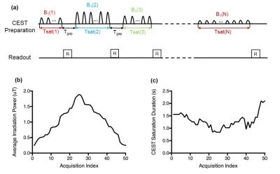 |
CEST Fingerprinting: A Novel Approach for Exchange Rate Quantification
Zhengwei Zhou, Pei Han, Debiao Li
In this work, we developed a CEST fingerprinting technique for exchange rate quantification. This method utilizes CEST saturation with varying B1 amplitudes and durations to create uniqueness of signal evolution for different exchange rates. The acquired signal was matched to a predefined dictionary. Preliminary studies were performed in phantoms to show the feasibility.
|
14:57
|
0198.
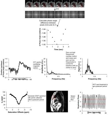 |
Measuring APT contrast in the lung using CEST FT-phase MRI and a retrospective gating technique
Kyle Jones, Carol Steum, Charles Hsu, Phillip Kuo, Mark Pagel, Edward Randtke
We have developed a CEST FT-phase MRI method that can measure endogenous Amide Proton Transfer (APT) contrast in lung tumors and other tissues that are affected by lung motion. The method monitors the breathing cycle based on the relative phase angle between adjacent pixels, and selects a subset of images during the quiescent period between breaths. The resulting MTRasym contrast of an oscillating egg white phantom, volunteers, and patients with lung tumors showed that CEST FT-phase MRI produced more precise quantitative assessments of APT.
|
15:09
|
0199.
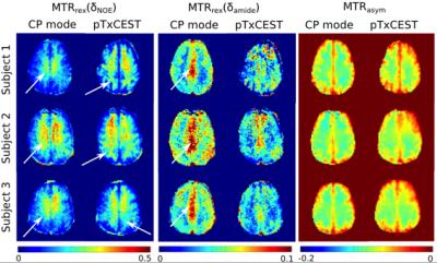 |
B1+ inhomogeneity mitigation in CEST using parallel transmission: pTxCEST
Nuno da Silva, Desmond Tse, Benedikt Poser, N Shah
To demonstrate the benefits from the increased spectral bandwidth at ultra-high field (UHF) by using parallel transmission (pTx) to mitigate flip-angle inhomogeneity in chemical exchange saturation transfer (CEST) imaging. A pTx basis pulse is homogenised by magnitude least-squares (MLS) optimisation and expanded to form a frequency-selective saturation pulse for CEST. The pTx saturation pulse was validated by both Bloch-McConnell simulation and in vivo imaging at 7T. Improved homogeneity in contrasts and relaxation-compensated CEST metrics were observed in our in vivo data when the pTx saturation pulse was used instead of the standard CP-mode Gaussian pulse.
|
15:21
|
0200.
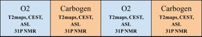 |
Understanding concomitant effects between CEST and ASL contrast.
Francisco Torrealdea, Marilena Rega, Mohamed Tachrount, Magdalena Sokolska, Xavier Golay
This study aims to assess the relationship between brain perfusion and CEST measurements. For this purpose, an oxygen-carbogen challenge experiment was designed in order to compare CEST measurements of the rat brain in low and high perfusion conditions. Comparison of the CEST with CBF measurement show strong correlation (p<0.005 with Spearman Rho=0.976). From the results of the study it is notable that blood perfusion is a strong modulator of the observed CEST signal in the rat brain.
|
15:33
|
0201.
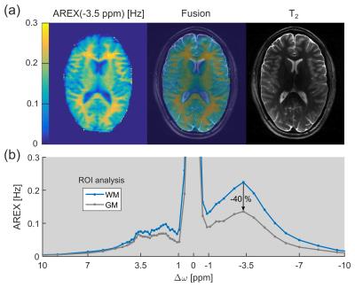 |
CEST Signals of Lipids
Steffen Goerke, Moritz Zaiss, Dario Longo, Francesca Garello, Enza Di Gregorio, Johannes Breitling, Mark Ladd, Peter Bachert
In this study, lipids were identified to be an important contributor to the upfield chemical exchange saturation transfer (CEST) signals of brain tissue. This finding can explain the pronounced CEST image contrast between gray and white matter observed in healthy volunteers.
|
|