Oral
Acquisition, Reconstruction & Analysis
Thursday, 27 April 2017
| Room 314 |
13:00 - 15:00 |
Moderators: Dimitrios Karampinos, Christopher Roy |
Slack Channel: #s_acq_recon_analysis
Session Number: O61
13:00
 |
1162.
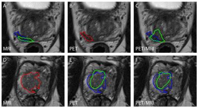 |
Multimodal multiparametric 18F-Fluciclovine PET/MRI improves computer-assisted detection of primary prostate cancer 
Mattijs Elschot, Kirsten Selnæs, Elise Sandsmark, Jose Teruel, Brage Krüger-Stokke, Øystein Størkersen, May-Britt Tessem, Siver Moestue, Helena Bertilsson, Tone Bathen
Computer-assisted algorithms have been proposed to support radiological reading of multiparametric MRI (mpMRI) images for the detection of primary prostate cancer, but have limited sensitivity. In this work, we investigated if standardized uptake values (SUV) from combined 18F-Fluciclovine PET/mpMRI can improve automated tumor detection. We found that, at the same number of false positives, a model based on combined PET/mpMRI correctly detected more tumors than models based on mpMRI only or PET only. These findings suggest that there is a potential role for multimodal multiparametric 18F-Fluciclovine PET/MRI for computer-assisted detection of primary prostate cancer.
|
13:12
|
1163.
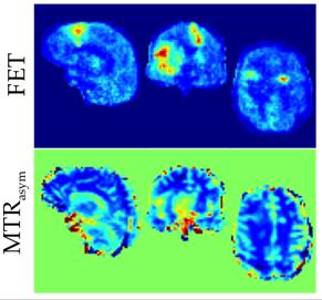 |
Hybrid MR-PET of Brain Tumours with amino acid-PET and Amide Proton Transfer MRI
Nuno da Silva, Philipp Lohmann, James Fairney, Arthur Magill, Ana-Maria Oros-Peusquens, Chang-Hoon Choi, Rüdiger Stirnberg , Xavier Golay, Karl-Josef Langen, N Shah
Information on amino acid metabolism has become an important component of the diagnostic and prognostic precision in the investigation of brain tumours. Both MRI and PET present methods to obtain amino acid weighted images, such as chemical exchange saturation transfer (CEST) or O-(2-18F- fluoroethyl)-L-tyrosine (FET), respectively. In this work, FET-PET and CEST imaging of brain tumours is investigated using hybrid MR-PET in 8 patients with brain tumours. The results suggest that the tumour-to-brain ratio of magnetization transfer ratio asymmetry (MTRasym) encodes different information than that from FET PET.
|
13:24
|
1164.
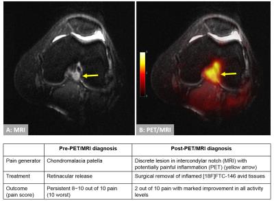 |
Management of complex regional pain syndrome (CRPS) with [18F]FTC-146 PET/MRI
Daehyun Yoon, Peter Cipriano, Trine Hjoernevik, Bin Shen, Dawn Holley, Harsh Gandhi, Vivianne Tawfik, Catherine Curtin, Ian Carroll, Christopher McCurdy, Brian Hargreaves, Frederick Chin, Sandip Biswal
Complex regional pain syndrome (CRPS) is a debilitating chronic pain condition affecting millions of patients worldwide. However, no specific diagnosis is currently available to accurately detect the pain generators in CRPS, and thus successful pain management for CRPS is very challenging. In this abstract, we propose a PET/MRI approach with a novel pain-specific PET tracer for identifying pain generators in CRPS. Our early experience with the proposed PET/MRI approach demonstrates that the image findings could alter the pain management for CRPS patients to achieve better pain-relief outcome.
|
13:36
 |
1165.
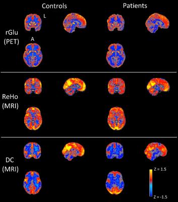 |
Using simultaneous PET-MRI to investigate the mechanisms of neurodegeneration in frontotemporal dementia
Esther Warnert, Udunna Anazodo, Kristy Coleman, Julia MacKinley, John Butler, Frank Prato, Elizabeth Finger, Keith St. Lawrence
Frontotemporal dementia (FTD) is a neurodegenerative disease characterized by progressive degeneration of brain function and structure. We present a proof-of-principle study in which simultaneous PET-MRI is used to inform on the pathophysiology of progressive neurodegeneration. By correlating glucose metabolism (PET) with functional connectivity (MRI), we found altered neuronal signaling in brain regions known to be critical in progression of FTD.
|
13:48
|
1166.
 |
The MRI signature of glial activation: a PET-MRI study using PBR28 radioligand and MR relaxometry
Guillaume Bonnier, Tom Hilbert, Daniel Albrecht, Marco Loggia, Cristina Granziera
Radioligands used in PET imaging and binding to the translocator protein (TSPO) have proved to be useful to study inflammatory brain processes. Relatedly, quantitative Magnetic Resonance Imaging provides metrics of tissue-level microstructure, which permit to study the consequences of brain inflammation. In this work, we have investigated the relationship between PBR28 radioligant uptake and T1 and T2 metric of brain tissue microstructure based on analysis of PET images, T1 and T2 relaxometry images of 5 HIV subjects.
|
14:00
|
1167.
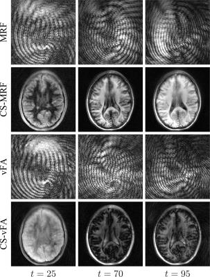 |
Accelerated parameter mapping with compressed sensing: an alternative to MR Fingerprinting
Pedro Gómez, Guido Buonincontri, Miguel Molina-Romero, Jonathan Sperl, Marion Menzel, Bjoern Menze
We introduce a method for MR parameter mapping based on three concepts: 1) an inversion recovery, variable flip angle acquisition strategy designed for speed, signal, and contrast; 2) a compressed sensing reconstruction which exploits spatiotemporal correlations through low rank regularization; and 3) a model-based optimization to simultaneously estimate proton density, T1, and T2 values from the acquired measurements. Compared to MR Fingerprinting, the proposed method achieves a five-fold acceleration in acquisition time, reconstructs an unaliased series of images, and does not rely on dictionary matching for parameter estimation.
|
14:12
|
1168.
 |
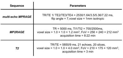
Ultra-high resolution in vivo multi-parameter mapping of the human brain
Kerrin Pine, Luke Edwards, Martina Callaghan, Pierre-Louis Bazin, Nikolaus Weiskopf
We present the first multi-parameter maps of R1, R2* and effective proton-density (PD*) acquired at 400 µm isotropic resolution at 7T. Prospective motion correction (PMC) by external optical tracking was used to reduce motion artifacts, as well as to avoid a co-registration step during calculation of the maps. The maps allow for characterizing of subtle subcortical and cortical features such as the line of Gennari in the visual cortex.
|
14:24
|
1169.
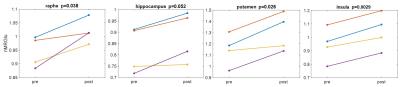 |
Changes in Hippocampal Subfield Volumes, Diffusivity and Brain Metabolism after Electroconvulsive Therapy: a Pilot PET/MR Study
Chuan Huang, Laura Kunkel, Adeeb Yacoub, Jie Ding, Christine DeLorenzo, Ramin Parsey
Major depression disorder is a highly prevalent illness with low treatment response rates. Fortunately, electroconvulsive therapy (ECT) is an effective treatment for patients with pharmacotherapy resistant depression, although its mechanism of action remains unclear. There is disagreement regarding the predictive value of amygdala and hippocampal volumes and whether ECT causes neuroplastic effects on these regions in patients with MDD. In this study, we used simultaneous PET/MR to look at structural, diffusion and metabolism changes in brain before and after ECT.
|
14:36
|
1170.
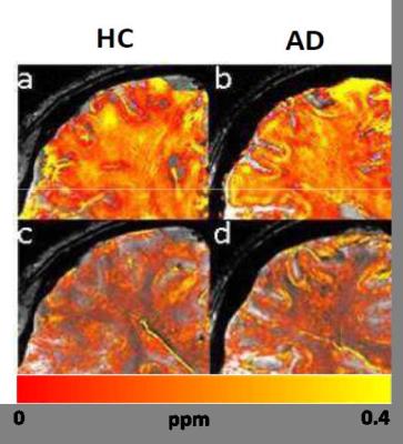 |
In-vivo and ex-vivo R2* and Quantitative Susceptibility Mapping in Alzheimer’s Disease at Ultra-High Magnetic Field compared to Histology
Elisa Tuzzi, Gisela Hagberg, David Balla, Joana Loureiro, Manuela Neumann, Christoph Laske, Rolf Pohmann, Matthias Valverde, Klaus Scheffler
Amyloid-β plaques are classical hallmarks of the post-mortem Alzheimer’s Disease (AD) brain. Ultra-High-Field (UHF) MRI provides a compelling means to investigate pathological processes at an unprecedented level of detail. ß-amyloid plaques can be detected in T2* weighted images at UHF, ex-vivo, due to the local iron content and to the plaque geometry per sè. With this study we aim to explore the source of the observed MR signal changes in AD at UHF using quantitative MRI methods in-vivo and ex-vivo.
|
14:48
|
1171.
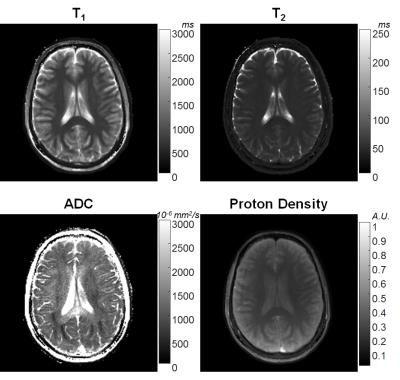 |
Simultaneous T1, T2 and Diffusion Quantification using Multiple Contrast Prepared Magnetic Resonance Fingerprinting
Yun Jiang, Jesse Hamilton, Wei-Ching Lo, Katherine Wright, Dan Ma, Andrew Coristine, Nicole Seiberlich, Vikas Gulani, Mark Griswold
A novel MRF sequence is designed for generating high quality, distortion-free T1, T2 and apparent diffusion coefficient (ADC) maps simultaneously in less than 60 seconds per slice. The method inserts multiple magnetization preparation modules in a FISP-based MRF sequence. The use of magnetization preparation modules shortens the preparation time to achieve variable image contrast. Accurate T1, T2 and ADC quantification is demonstrated in a phantom and in vivo in human brains. This method enables the simultaneous collection of T1, T2 and diffusion maps for tissue characterization without the need to co-register separately acquired maps as in conventional MRI.
|
|