Tuesday, 25 April 2017
| Room 313BC |
16:15 - 18:15 |
Moderators: Kathryn Keenan, Ek Tan |
Slack Channel: #s_diffusion
Session Number: O69
16:15
|
0600.
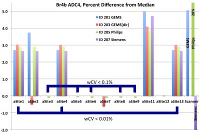 |
Multi-site Concordance of DWI Metrics: Results of the NCI Quantitative Imaging Network ADC Mapping Collaborative Project 
David Newitt, Dariya Malyarenko, Thomas Chenevert, C. Quarles, Laura Bell, Andrey Fedorov, Fiona Fennessy, Michael Jacobs, Meiyappan Solaiyappan , Stefanie Hectors, Bachir Taouli, Kathleen Schmainda, Melissa Prah, Yi-Fen Yen, Jayashree Kalpathy-Cramer, Erin Taber , Christopher Kroenke , Yue Cao, Madhava Aryal, Mark Muzi, Paul Kinahan, Thomas Yankeelov, Lori Arlinghaus, Michael Boss, Amita Shukla-Dave, Nola Hylton
Reproducibility of diffusion metrics is essential given the increasing role quantitative diffusion weighted imaging plays in diagnosis and treatment monitoring. Here we examined the variability in apparent diffusion coefficient (ADC) measures resulting from different post-processing software implementations utilized by researchers across the NCI Quantitative Imaging Network. Agreement between the majority of implementations was good; typical biases for in vivo ADC measures of 2-3%, and lower biases in phantom scans. Higher deviations (above 5%) detected among individual implementations and scanner-generated parametric maps highlighted inadequacies in meta-data and post-processing parameters that need to be addressed in multi-site study settings.
|
16:27
|
0601.
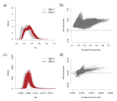 |
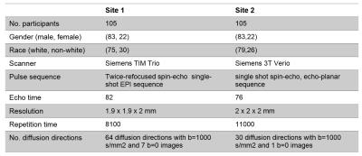
Statistical harmonization of multi-site diffusion tensor imaging data with ComBat
Jean-Philippe Fortin, Drew Parker, Birkan Tunç, Takanori Watanabe, Mark Elliott, Kosha Ruparel, Ruben Gur, Raquel Gur, Robert Schultz, Russell Shinohara, Ragini Verma
Diffusion tensor imaging (DTI) is a well-established magnetic resonance imaging technique to study microstructural changes in the white matter (WM). DTI images suffer from unwanted inter-scanner variability, which is problematic when combining datasets from different sites. In this work, we propose to use ComBat, a location-scale Empirical Bayes model largely used in genomics, to combine and harmonize multi-site DTI datasets. Using a study of 210 subjects with an age range of 8 to 18 years old from two imaging sites, we show that ComBat (1) removes unwanted variation associated with imaging site and (2) improves the power at detecting regions known to exhibit microstructural changes in this age range.
|
16:39
|
0602.
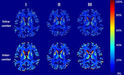 |
Variability and Reproducibility of Multicenter Human Diffusion Measurements with Track-density Imaging Analysis
Qiqi Tong, Ting Gong, Peipeng Liang, Tianyi Qian, Xu Yan, Yi Sun, Chen Li, Qiuping Ding, Hongjian He, Kuncheng Li, Jianhui Zhong
In a multicenter imaging study, it is vital to properly evaluate the variability of image quality among scanners. For diffusion imaging, estimations based on ADC and FA measurements with tensor models have previously been studied. However, such evaluation is not sufficient for studies with more diffusion directions like HARDI or DSI. This study uses track-density imaging to estimate the variability and reproducibility of multi-shell diffusion images from multicenter data acquired in the same subjects.
|
16:51
|
0603.
 |
Normalization of inter-site Structural Connectivity Data for Regression analysis
Takanori Watanabe, Birkan Tunc, Drew Parker, Jean-Philippe Fortin, Mark Elliott, Kosha Ruparel, Ruben Gur, Raquel Gur, Robert Schultz, Russell Shinohara, Ragini Verma
Diffusion tensor imaging (DTI) and tractography have revealed many critical insights about how the human brain is organized as a large-scale complex network. As multisite imaging studies are becoming increasingly popular within the neuroimaging field, it is imperative to develop methods that can correct for inter-site differences, facilitating the combination of data from multiple sites. In this study, we present a normalization scheme that will correct for site-specific differences in diffusion-based structural connectivity data, and demonstrate its efficacy through multivariate regression experiments using the normalized structural connectivity features to predict subject’s age.
|
17:03
|
0604.
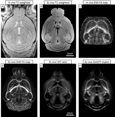 |
MRI vs. X-ray scattering: comparative study of myelin distribution, fiber direction and white matter tracts in mouse brain
Marios Georgiadis, Zirui Gao, Dario Zingariello, Valerio Zerbi, Marianne Liebi, Stefan Sommer, Mark Augath, Oliver Bunk, Manuel Guizar-Sicairos, Aileen Schroeter, Markus Rudin
MRI is the method of choice for brain imaging. However, it uses indirect structural information to infer densities of molecules such as myelin, and water diffusion direction as a proxy for fiber direction. Small-angle X-ray scattering tensor tomography (SAXSTT) provides an alternative approach to assess myelin distribution and fiber direction using directly structural information related to the molecular structure of myelin sheath. We applied SAXSTT to mouse brain to validate and compare different MRI methods (MT, DWI). We found a high degree of similarity with MT macromolecule distribution, and also DWI-derived white matter tracts, but with significant region-specific differences.
|
17:15
 |
0605.
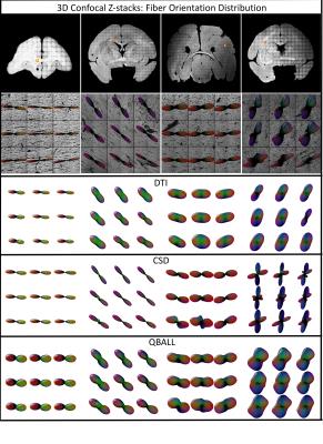 |
Histological Validation of Orientation Dispersion and Fiber Orientation
Kurt Schilling, Vaibhav Janve, Yurui Gao, Iwona Stepniewska, Bennett Landman, Adam Anderson
In this study, using 3D confocal microscopy to extract histological fiber orientation distributions, we validate tract-specific measures of dispersion and orientation derived from high angular resolution diffusion imaging techniques. We find a correlation between histological and diffusion measures of dispersion in q-ball imaging and constrained spherical deconvolution. We also find that an increased dispersion leads to greater error in fiber orientation estimates, as well as the presence of false positive peaks in the diffusion profiles. Future work will validate these measures in more high angular resolution diffusion techniques and microstructural models.
|
17:27
 |
0606.
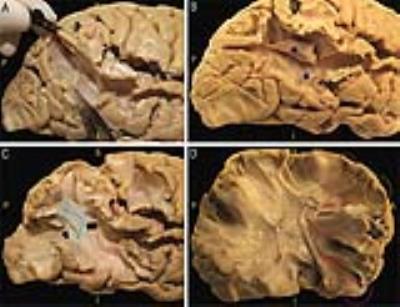 |
Topography of the acoustic radiation as revealed by ex-vivo fiber dissections and in-vivo diffusion-based tractography
Chiara Maffei, Jorge Jovicich, Alessandro De Benedictis, Franco Chioffi, Silvio Sarubbo
The acoustic radiation (AR) is a compact bundle of fibers conveying auditory information from the thalamus to the cortex. Topographical knowledge of this bundle is scarce and its diffusion-based tractographic reconstruction remains hardly achievable, especially for commonly available MRI acquisition protocols. In this scenario, validation of tractography results is particularly important. In this study we used blunt dissection to precisely characterize AR topography and relationships with adjacent bundles. Being aware of the anatomical characteristics of the tract provides us with the underlying ground truth on which methodological decisions, aimed at overcoming the limits of the tractographic reconstruction, can be made.
|
17:39
|
0607.
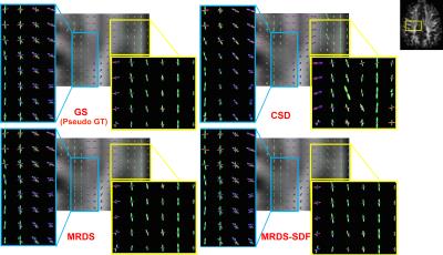 |
Robust Estimation and In-vivo Validation of the Axon Bundle Diffusivity Profiles
Ricardo Coronado-Leija, Alonso Ramirez-Manzanares, Jose Luis Marroquin
A stable, accurate and robust-to-noise general framework for the estimation of the intra-voxel axial and radial diffusivity parameters for diffusion-weighted magnetic resonance imaging is presented. The method estimates the diffusion profiles at multi-fiber voxels, improving the estimation of the intra-voxel geometry at challenging microstructure configurations. It naturally constrains the sparsity on the recovered solutions and exploits the spatial redundancy of the axon packs. A useful evaluation metric is proposed: it combines the information of the success rate of the number of bundles and their angular error. A new evaluation method for the in-vivo estimations on large datasets is also proposed.
|
17:51
|
0608.
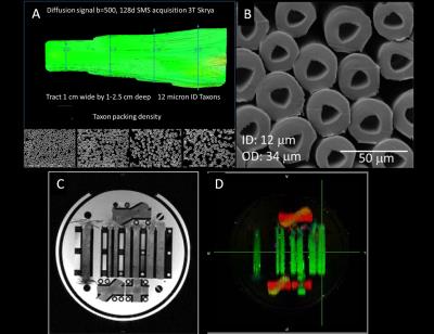 |
Validation of axon diameter and density estimates by TractCaliber MRI in a biomimetic brain phantom
Susie Huang, Qiuyun Fan, Barbara Wichtmann, Aapo Nummenmaa, Walter Schneider, Lawrence Wald
We validate axon diameter and density estimates using a novel biomimetic brain phantom that emulates the microstructural features of white matter, consisting of hollow textile axons (“taxons”) on the micron scale with distinct intra- and extra-axonal compartments and crossing regions. Diffusion data acquired over a range of gradient strengths, directions and diffusion times were fitted with the TractCaliber approach. Taxon volume fractions were accurately estimated, and the diameters were slightly overestimated in areas with less densely packed fibers. Such phantoms may be useful for testing microstructural models of the diffusion signal, and for scanner and protocol calibration.
|
18:03
|
0609.
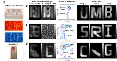 |
Phantom Validation of Diffusion-Relaxation Correlation Spectroscopic Imaging (DR-CSI)
Daeun Kim, Eamon Doyle, Jessica Wisnowski, Justin Haldar
Diffusion-relaxation correlation spectroscopic imaging (DR-CSI) is a novel multidimensional MR imaging approach that infers microscopic tissue compartments using simultaneous diffusion and relaxation information. The approach was previously demonstrated with biological data, although validation was not possible in the absence of a gold standard reference. In this work, we perform simulation and experimental studies, using a gold standard for validation. Specifically, we custom-built a multi-compartment diffusion-relaxation phantom with known characteristics, and performed extensive comparisons between DR-CSI and conventional multi-compartment estimation methods. Our results demonstrate that DR-CSI has good performance, enabled by the combination of multidimensional encoding and constrained spectroscopic image reconstruction.
|
|