Wednesday, 26 April 2017
| Plenary Hall |
11:45 - 12:15 |
Moderators: Ileana Hancu, Valeria Panebianco |
Slack Channel: #s_diffusion
Session Number: O72
11:45
|
0750.
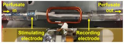 |
Dynamic Diffusion MRI Signal Changes Accompany Electrical Activity in Myelinated Axons 
William Spees, Tsen-Hsuan Lin, Peng Sun, Sam Gary, Sheng-Kwei Song
Because of its exceptionally robust nature, the ex vivo frog sciatic nerve has been the subject of numerous electrophysiology and MRI studies over the years. Here we report on diffusion MRI signal changes resulting from 50 and 100 Hz in-magnet electrical stimulation of perfused bullfrog sciatic nerves. The inexpensive perfusion system we have implemented allows for good long-term in-magnet stability and simultaneous MRI/electrophysiology studies. Decreases in water diffusivity and compound action potential conduction velocities accompany prolonged periods of repetitive electrical stimulation. Both of these changes are consistent with hypothesized microstructural alterations of the PNS myelin.
|
11:50
 |
0751.
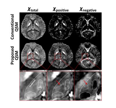 |
Separating positive and negative susceptibility sources in QSM
Jingu Lee, Yoonho Nam, Joon Yul Choi, Hyeonggeol Shin, Taehyun Hwang, Jongho Lee
We proposed a new QSM algorithm that separates positive and negative susceptibility sources within a voxel by utilizing signal relaxation (R2') for dipole inversion. The new method was tested in computer simulated phantoms and in-vivo data, and successfully separated positive and negative susceptibility sources.
|
11:55
|
0752.
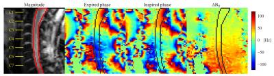 |
Spatiotemporal analysis of breathing-induced fields in the cervical spinal cord at 7T
S. Johanna Vannesjo, Karla Miller, Stuart Clare, Irene Tracey
Time-varying B0 fields related to breathing is one major source of image artifacts in spinal cord imaging at ultra-high field. Here we aim to measure spatial and temporal characteristics of breathing-induced fields in the cervical spinal cord at 7T. We perform a principal component analysis on field measurements based on fast gradient-echo images acquired during free breathing. We observed field variations of about 30Hz at C7 during normal breathing. Furthermore, we observed that a single principal component explained over 90% of the field variance during normal and deep breathing.
|
12:00
|
0753.
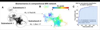 |
Using Machine Learning to study knee Osteoarthritis: the path towards OA Precision Medicine
Valentina Pedoia, Jenny Haefeli , Kazuhito Morioka, Hsiang-Ling Tang, Lorenzo Nardo, Richard Suoza, Adam Ferguson, Sharmila Majumdar
In this study we describe the analysis of a dataset including 178 subjects with and without Osteoarthritis using Topological data analysis (TDA), a machine-learning tool that involves projecting individual patients into the ‘syndromic space’ defined by all outcome variables simultaneously. Demographics, patient reported outcomes Kellgren-Lawrence grading, MRI WORMS morphological grading, cartilage relaxation times, gait kinematics and kinetics during walking were simultaneously considered to define the data topology. TDA shows the presence of subgroups characterized by a strong biochemical signature, showing how this new technique could be used to extract insight from complex data, allowing for more personalized characterization of each individual.
|
12:05
|
0754.
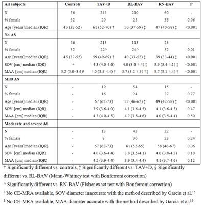 |
Mapping of Abnormal Aortic Hemodynamics in 515 Patients with Aortopathy
Pim van Ooij , Michael Markl, Jeremy Collins, James Carr, S. Chris Malaisrie, Patrick McCarthy, Aart Nederveen, Paul Fedak, Alex Barker
4D flow MRI-derived 3D velocity and wall shear stress (WSS) maps in a large cohort of patients with aortopathy (n=515), stratified for valve morphology and stenosis severity, were compared with age-matched cohort-averaged maps of healthy controls (n=56) to yield maps of abnormally elevated hemodynamics. These maps were projected onto shared geometries and summed to map the incidence of abnormal velocity and WSS. Without stenosis, hemodynamics were significantly increased (Bonferroni corrected Mann-Whitney tests) in patients with bicuspid valves compared to patients with tricuspid valves. Incidence of elevated hemodynamics increased similarly for both cohorts (significant differences disappeared) with increasing stenosis severity.
|
|