Monday, 24 April 2017
| Room 310 |
08:15 - 10:15 |
Moderators: Elizabeth Hecht, Houchun Hu |
Watch the ISMRM 2017 Plenaries and Young Investigator Awards presentations LIVE online for the first time ever!
Note: All times are Hawaiian Standard Time. |
Slack Channel: #s_yia
Session Number: O88
08:15
|
0031.
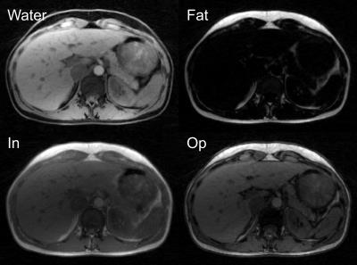 |
Free-breathing volumetric fat/water separation by combining radial sampling, compressed sensing, and parallel imaging 
Thomas Benkert, Li Feng, Daniel Sodickson, Hersh Chandarana, Kai Block
Fat-suppressed T1-weighted gradient-echo imaging is commonly used for abdominal MR examination. However, image quality can be compromised by inhomogeneous fat suppression and imperfect breath-holding. To overcome both limitations, we describe a novel technique for free-breathing fat/water separation (Dixon-RAVE). Motion-robust acquisition is achieved by using radial sampling. A model-based reconstruction, which incorporates compressed sensing, parallel imaging, and fat deblurring, is used to obtain fat and water maps. Two extensions are described that enable motion-resolved fat/water separation (XD-Dixon-RAVE) and dynamic contrast-enhanced fat/water separation (DCE-Dixon-RAVE). The technique is demonstrated for various clinical applications, including free-breathing liver and breast exams in volunteers and patients.
|
08:35
|
0032.
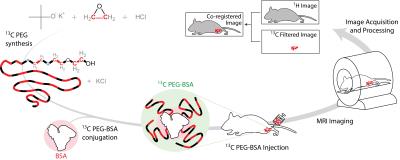 |
Direct Quantitative 13C-Filtered 1H Magnetic Resonance Imaging of PEGylated Biomacromolecules In Vivo
Rohan Alvares, Justin Lau, Peter Macdonald, Charles Cunningham, R. Scott Prosser
We demonstrate a new platform technology in which macromolecular constituents, such as proteins and drug delivery systems, are observed directly and quantitatively in vivo using 1H MRI of 13C-labeled polyethylene glycol (13C-PEG) tags. The 28 kDa 13C-PEG tags are non-immunogenic, and each bears approximately 2500 spectroscopically equivalent 1H nuclei appearing at a single resonance position. By filtering the 1H PEG signal through the directly coupled 13C nuclei, background water and fat signals are largely eliminated. We demonstrate the approach by monitoring in real-time the distribution of 13C-PEG and 13C-pegylated albumin injected into the hind leg of a mouse.
|
08:55
|
0033.
 |
Hybrid MRI-ultrasound acquisitions, and scannerless real-time imaging
Frank Preiswerk, Matthew Toews, Cheng-Chieh Cheng, Jr-yuan Chiou, Chang-Sheng Mei, Lena Schaefer, W. Hoge, Benjamin Schwartz, Lawrence Panych, Bruno Madore
The goal of this project was to combine MRI, ultrasound (US) and computer science methodologies toward generating MRI at high frame rates, inside and even outside the bore. A small US transducer, fixed to the abdomen, collected signals during MRI. Based on these signals and correlations with MRI, a machine-learning algorithm created synthetic MR images at up to 100 frames per second. In one particular implementation volunteers were taken out of the MRI bore with US sensor still in place, and MR images were generated on the basis of ultrasound signal and learned correlations alone, in a 'scannerless' manner.
|
09:15
|
0034.
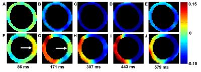 |
Imaging Left-ventricular Mechanical Activation in Heart Failure Patients using Cine DENSE MRI: Validation and Implications for Cardiac Resynchronization Therapy
Daniel Auger, Kenneth Bilchick, Jorge Gonzalez, Sophia Cui, Jeffrey Holmes, Christopher Kramer, Michael Salerno, Frederick Epstein
This study developed methods for imaging left-ventricular (LV) mechanical activation, with application to identifying optimal LV pacing sites for cardiac resynchronization therapy (CRT). Cine displacement encoding with stimulated echoes (DENSE) was used for strain imaging, and mechanical activation time was defined as the time of onset of circumferential shortening (TOS). Active contours were applied to strain data to automatically compute TOS. Results showed a strong correlation between TOS and electrical activation time, heterogeneity of the location of latest activation, and a significant association between TOS at the LV pacing site and CRT response. These methods may enable improved CRT implementation.
|
09:35
|
0035.
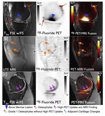 |
PET/MR Imaging of Metabolic Bone Activity in Osteoarthritis
Feliks Kogan, Audrey Fan, Emily McWalter, Uchechukwuka Monu, Edwin Oei, Andrew Quon, Garry Gold
Osteoarthritis (OA) is a leading cause of disability, resulting in reduced quality of life, at tremendous societal cost. New hybrid PET/MR systems allow for simultaneous, sensitive, and quantitative assessments of early bone activity in OA with PET, which can be correlated with high-resolution quantitative MR methods of soft tissues to study the pathogenesis of OA. We demonstrate promising initial results of simultaneous PET/MR hybrid imaging of knee OA. Results suggest that PET/MR may detect metabolic abnormalities in subchondral bone, which appear normal on MRI. These advancements will allow us to detect and track early and reversible changes in OA.
|
09:55
|
0036.
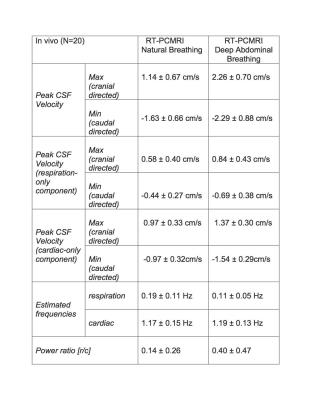 |
Quantifying the Influence of Respiration and Cardiac Pulsations on the Cerebrospinal Fluid Dynamics using Real-Time Phase-Contrast MRI
Selda Yildiz, Suraj Thyagaraj, Ning Jin, Xiadong Zhong, Soroush Heidari Pahlavian, Bryn Martin, Francis Loth, John Oshinski, Karim Sabra
Cerebrospinal fluid (CSF) flow undergoes periodic pulsatile motion driven by cardiac and the respiratory forces. Invasive studies using spinal taps as well as non-invasive studies using phase contrast MRI (PCMRI) sequences have well documented the cardiac-driven CSF flow. PCMRI, however, often uses a conventional cine-phase contrast technique gated to the cardiac cycle, and thus cannot measure the effects of respiration or other non-cardiac transient events such as coughing. Examining these effects requires the ability to perform real-time MRI measurements of continuous CSF flow along the spine and cranial cavity, and determine accurate instantaneous CSF flow velocity values.
|
|