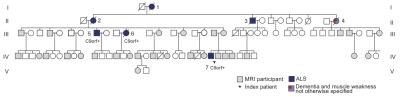
Poster: 7T Neuroimaging
Electronic Power Pitch Poster
Neuro
Monday, 24 April 2017
| Exhibition Hall |
09:15 - 10:15 |
| |
|
Plasma # |
 |
0016.
 |
16 |
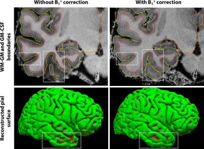
The effects of B1+ correction of MP2RAGE on estimating cortical thickness and T1 at 7T 
Roy Haast, Dimo Ivanov, Elia Formisano, Kâmil Uludag
B1+ inhomogeneities can significantly affect the quantitative T1 values derived from MP2RAGE data and also automatic tissue classification, in particular in the inferior temporal and frontal lobes. Here, we investigated the effects of post-hoc correction at 7T on the T1 and apparent cortical thickness using a B1+ map for the residual transmit inhomogeneities in MP2RAGE data. We found that B1+ correction reduces these inhomogeneities leading to (1) a lower inter-subject variability, (2) enhanced localization of the GM-CSF border and (3) more accurate cortical thickness measurements.
|
|
0017.
 |
17 |
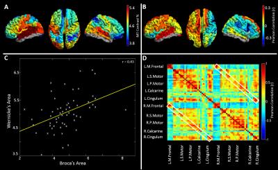
The relationship between cortical myeloarchitecture and functional connectivity in the human brain
Olivier Mougin, Benjamin Hunt, Prejaas Tewarie, Nicolas Geades, Peter Morris, Matthew Brookes, Penny Gowland
The human brain relies upon the dynamic formation and dissolution of functional networks to support ongoing cognition. The goal of this study is to establish a relationship between functional and structural networks. Using ultra-high field MRI, structural network defined by grey matter myelination is measured via quantitative Magnetization Transfer. Magnetoencephalography (MEG) was used to elucidate functional networks representing the major electrophysiological pathways of communication in the brain. Our study sheds new light on the way in which cortical microstructure supports functional networks.
|
 |
0018.
 |
18 |
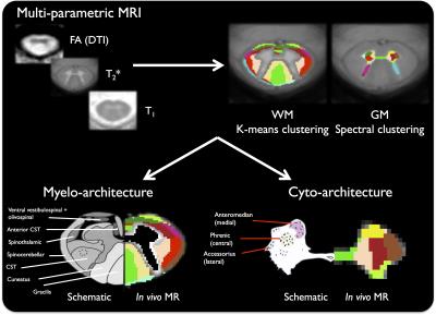
Towards in vivo spinal cord cyto- and myelo-architecture deciphering using multi-modal MRI parcellation at 7T
Manuel Taso, Aurélien Massire, Pierre Besson, Arnaud Le Troter, Maxime Guye, Jean-Philippe Ranjeva, Virginie Callot
Ultra-high-field MRI offers exciting perspectives for the in vivo structural characterization of central nervous system tissues. Based on high-resolution multi-parametric imaging at 7T, this preliminary work focuses on the generation of new spinal cord (SC) templates, hence proposing a high-resolution T2*-w MR average with exquisite anatomical details. Parcellation of the SC substructures into individual WM tracts and motoneurone clusters was also investigated using classification methods and multimodal data (T1/T2*/DTI). Preliminary promising results revealed some insights in the underlying SC cyto- and myelo-architecture. Future developments will extend to the whole cervical cord, holding tremendous promises for studying more specific pathophysiological impairments.
|
|
0019.
 |
19 |
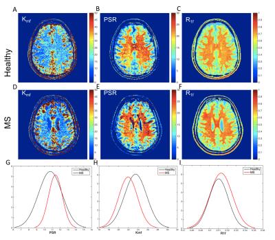
7T Quantitative Magnetization Transfer (qMT) of Cortical Gray Matter in Multiple Sclerosis Correlates with Cognitive Disability
Lydia McKeithan, Bailey Lyttle, Bailey Box, Kristin O'Grady, Richard Dortch, Benjamin Conrad, Seth Smith
Cognitive impairment (CI) is a major manifestation of multiple sclerosis (MS) and is responsible for extensively hindering patient quality of life.1 Cortical gray matter damage is critical to CI, but is poorly characterized by conventional MRI. We employed advanced methods by evaluating SIR-qMT-derived indices for differences between MS patients and healthy volunteers at 7T and derived associations with neuropsychological measures of cognitive impairment. We found significant reduction in kmf in cGM of MS patients, unique association with EDSS score, and strong correlation with cognitive performance indicating that kmf may be a significant biomarker of GM damage in MS.
|
|
0020.
 |
20 |
Changes in structural network connectivity in early-stage multiple sclerosis are associated with cortical demyelination
Atef Badji, Gabriel Mangeat, Russell Ouellette, Constantina Treaba, Tobias Granberg, Elena Herranz, Celine Louapre, Nikola Stikov, Jacob Sloane, Pierre Bellec , Caterina Mainero, Julien Cohen-Adad
Cortical disruption and changes in brain connectomics in multiple sclerosis have been recently investigated; however, the relationship between both processes in early disease remains uncertain. We propose an integrative framework that combines diffusion-based graph theory with high-resolution quantitative T1 and T2* at 7 Tesla to investigate the topological alterations of both structural connectomics and cortical demyelination. We found that both cortical myelin loss and increase in brain connectivity were present in early MS, and that the two processes were spatially anti-correlated. This suggests that the increase in brain connectivity in early MS could represent an adaptative role against initial, mild cortical demyelination, though this would be lost with more severe cortical disease.
|
|
0021.
 |
21 |
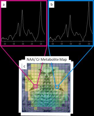
3D magnetic resonance spectroscopic imaging at 7 Tesla of patients with medically refractory focal epilepsy with non-lesional or inconclusive clinical MRIs: First Results
Rebecca Feldman, Madeline Fields, Bradley Delman, Lara Marcuse, Priti Balchandani
SASSI is a B1-insensitive, low-SAR 7T MRSI technique with reduced chemical shift localization errors. We used 3D SASSI to image the hippocampi of patients with medically refractory focal epilepsy who had non-lesional or inconclusive clinical MRIs. Using SASSI at 7T, we detected decreases in the hippocampal NAA/Cr ratios in suspected temporal lobe epilepsy patients, on the same side as the seizure onset zones and/or 7T structural findings.
|
|
0022.
 |
22 |
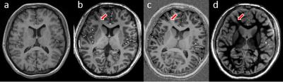
The value of 7T in the clinical evaluation of epileptic patients with focal cortical dysplasia
Kaibao Sun, Xueyuan Wang, Zhongwei Chen, Chang Liu, Jianfei Cui, Zhentao Zuo, Rong Xue, Yan Zhuo, Lin Chen, Shuli Liang, Tao Yu, Bo Wang
Focal cortical dysplasia (FCD) is defined as topical malformations of cortical development and often results in intractable epilepsy. However, many epileptic patients with FCD have not been diagnosed because of the lack of high-quality magnetic resonance imaging. 7T MRI, in comparison with 3T, is assessed in this study to allow better characterization of lesion details and detect previously unrevealed FCD abnormalities. The results of comparison were classified for an accurate and appropriate appraisal.
|
|
0023.
 |
23 |
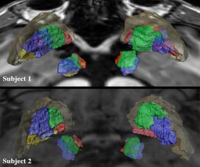
Individualized Tractography-Based Parcellation of the Globus Pallidus Pars Interna using 7T MRI in patients with Parkinson’s Disease Prior to DBS Surgery
Rémi Patriat, Yuval Duchin, Christophe Lenglet, Joshua Aman, Scott Cooper, Jerrold Vitek, Noam Harel
The success of deep brain stimulation (DBS) surgeries for Parkinson’s disease relies on the accurate placement of an electrode within the motor portion of subcortical brain targets. We use 7T MR-tractography to visualize the functional territories of the Globus Pallidus pars Interna. We found that the motor territory is located immediately posteromedially to the associative and limbic territories, akin to the subthalamic nucleus organization. This pattern was reproducible across two DBS patients. These findings shed new light on the functional organization of DBS targets, showing potential for providing valuable information to clinicians for targeting decisions and ultimately enhancing patient’s outcomes.
|
|
0024.
 |
24 |
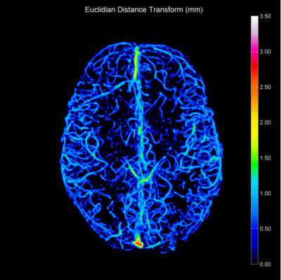
Assessment of cerebral vascular abnormalities in Huntington’s Disease at 7Tesla
Richard Dury, Sarah Mason, Francesca Cicchetti, Janelle Drouin-Ouellet, Roger Barker, Penny Gowland, Susan Francis
Huntington’s Disease (HD) is associated with vascular abnormalities and breakdown in the blood-brain barrier (BBB). Here, we use high spatial resolution time-of-flight magnetic resonance angiography (TOF-MRA) and arterial spin labelling (ASL) to assess vascular abnormalities in HD patients. We develop a pipeline to estimate vessel radii and distribution from TOF-MRA data. A significant decrease in the fractional vessel volume and a higher frequency of narrow vessels (0.15-0.45mm radius) was evident in HD patients compared to healthy volunteers across a number of cortical areas. No significant difference was found in cortical perfusion between the HD patients and healthy volunteers.
|
|
0025.
 |
25 |
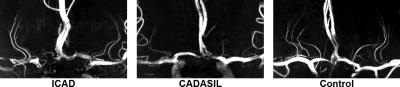
7T TOF-MRA Shows Different Patterns of Perforating Artery in Patients with Intracranial Atherosclerosis Disease (ICAD) and Cerebral Autosomal-Dominant Arteriopathy with Subcortical Infarcts and Leukoencephalopathy (CADASIL)
Qingle Kong, Qi Yang, Zhaoyang Fan, Xianchang Zhang, Yun Yuan, Xiaojing Fang, Jing An, Yan Zhuo, Zihao Zhang
7T high-resolution TOF-MRA has the ability to image perforating arteries of middle cerebral artery (MCA). The distribution patterns of orifices in CADASIL (cerebral autosomal-dominant arteriopathy with subcortical infarcts and leukoencephalopathy) and ICAD (intracranial atherosclerosis disease) patients are still unclear. In this study, for the first time, we investigated the orientation distribution of perforating artery orifices on MCA trunks using 7T TOF-MRA. Specific features are found in the distribution patterns of orifices in patients with CADASIL, ICAD and healthy volunteers. This technique is promising in the pathological studies of intracranial vascular diseases.
|
|
0026.
 |
26 |
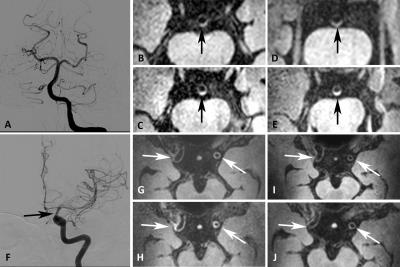
Intracranial vessel wall imaging in suspected cerebral vasculitis: evaluation of diagnostic value and treatment effects using 3T and 7T MRI
Nikki Dieleman, Anja van der Kolk, Catharina Frijns, Anita Harteveld, Jaco Zwanenburg, Hugo Kuijf, Arjen Lindenholz, L. Kappelle, Peter Luijten, Jeroen Hendrikse
Cerebral vasculitis is a rare, but devastating disease that can lead to severe disability or death. Diagnosis is rather challenging, but for treatment purposes, an accurate diagnosis is crucial since different, more aggressive therapy is needed compared with non-inflammatory diseases. In the current study, we investigated the diagnostic value of intracranial vessel wall MRI at 3T and 7T in patients who were suspected of cerebral vasculitis. Our results show that intracranial vessel wall imaging at 3T and 7T MRI should be considered a promising non-invasive diagnostic tool to identify wall enhancement in patients with a suspicion of cerebral vasculitis.
|
|
0027.
 |
27 |
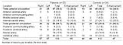
Detection of intracranial vessel wall lesions using 7T MRI: patients with posterior circulation ischemia versus healthy controls
Anita Harteveld, Anja van der Kolk, H. van der Worp, Nikki Dieleman, Peter Luijten, Jaco Zwanenburg, Jeroen Hendrikse
In this study presence and distribution of vessel wall lesions within the intracranial arteries of patients with recent posterior circulation ischemia and matched asymptomatic volunteers were assessed, using intracranial vessel wall MRI at 7T. Overall, vessel wall lesion presence and distribution were comparable between both groups. On arterial segment level, patients showed significantly higher lesion burden in the posterior cerebral artery, suggesting an association between posterior circulation lesion burden and ischemic events. Furthermore, a large amount of lesions showed contrast-enhancement, while the percentage of enhancing lesions was highest in the posterior circulation of the patient group.
|
|
0028.
 |
28 |
Metabolic differences between asymptomatic C9orf72 carriers and non-carriers assessed by brain 7T MRSI.
Henk-Jan Westeneng, Carrie Wismans, Abram D. Nitert, Renée Walhout, Peter R. Luijten, Jannie P. Wijnen, Leonard H. Berg
Amyotrophic lateral sclerosis (ALS) is an incurable and fatal neurodegenerative disease, which is caused by a C9orf72 repeat expansion in 9% of the cases. This mutation may cause changes of brain metabolism in patients but whether it affects brain metabolism in pre-symptomatic mutation carriers was not studied before. We used 7 Tesla magnetic resonance spectroscopic imaging (MRSI) to study brain metabolism in asymptomatic carriers of the C9orf72 repeat expansion and found lower concentrations of glutamate and N-acetylaspartate+N-acetylaspartylglutamate in the left putamen compared to non-carriers. This might indicate asymptomatic neuronal loss, a developmental defect or possibly a protective mechanism against ALS.
|
|
0029.
 |
29 |
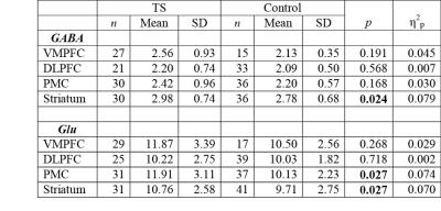
GABA and glutamate in children with Tourette Syndrome: a 7T 1H-MRS study
Nicolaas Puts, Richard Edden, Matthew Ryan, E Mahone, Harvey Singer
Studies have suggested that altered inhibition and excitation contribute to the pathology of Tourette syndrome, especially in cortical-striatal-thalamo-cortical (CSTC) pathways. GABA and glutamate were measured at 7T in large cohorts of healthy children and children with TS in regions of the CSTC network. GABA and glutamate were increased in the striatum. Glutamate was increased in the premotor region and correlated with reduced motor inhibition. These data support involvement of habitual behavioral pathways in TS. Historically the dopaminergic system has been considered to have a dominant role in TS; however, accumulating evidence strongly suggests involvement of GABA and glutamate neurotransmitter systems.
|
|
0030.
 |
30 |
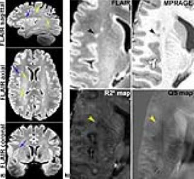
Multi-Parametric MRI at 7 T Enables Differentiation of MS and Age-Related White Matter Lesions
Zahra Hosseini, David Rudko, Jacob Matusinec, Marcelo kremenchutzky , Ravi Menon, Maria Drangova
MRI enables visualization of white matter lesions associated with demyelination in multiple sclerosis (MS). However, subtle white matter hyperintensities are also a sign of normal aging. This study used information collected from multiple 7T MRI contrasts at baseline and four-month follow-up time points to compare signal changes in lesions of five MS patients to those of age-related lesions (ARLs) in five healthy controls.
|
|