Poster: Brain Physiology: Flow, Oxygen, Metabolism
Electronic Power Pitch Poster
Neuro
Tuesday, 25 April 2017
| Exhibition Hall |
09:15 - 10:15 |
| |
|
Plasma # |
 |
0347.
 |
16 |
Long-term Cerebrovascular Dysfunction Following Repeated Mild Traumatic Brain Injury 
Conner Adams, Margaret Koletar, Tina Beckett, Lindsay Cahill, Lydiane Hirschler, Jan Warnking, Emmanuel Barbier, JoAnne McLaurin, John Sled, Bojana Stefanovic
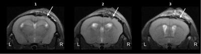
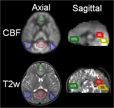
Pseudo-Continuous Arterial Spin Labelling (pCASL) was used to assess cerebrovascular dysfunction of mice having traumatic brain injury (TBI) - which had been induced via serial controlled cortical impacts. Resting perfusion was quantified in absolute units via multiple post-label-delay pCASL experiments, and found to be reduced in the lesion. Furthermore, vascular reactivity to hypercapnic challenge, assessed via pCASL, appears to be enhanced in initial results. These results, in conjunction with immunohistochemical analysis and T2-weighted structural images, imply severe damage due to TBI, with vascular adaptation in the form of angiogenesis as the response from the brain.
|
|
0348.
 |
17 |
Comparative Study of 3D Arterial Spin Labeling and dynamic contrast-enhanced MRI of Nasopharyngeal Carcinoma perfusion imaging
Bohan Xiao, Zhaoxiang Ye, Peiguo Wang, Ying Liu, Yingyu Zhao, Dandan Zheng
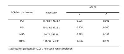
DCE-MRI is already a standard approach to evaluating tumor perfusion in NPC, but it may induce severe side-effects. ASL is a noninvasive MRI technique that has been mainly used to achieve perfusion imaging in central nerve system. In this study, we attempt to evaluate the application of ASL in NPC. Thirty-eight newly diagnosed NPC patients underwent 3D ASL and DCE-MRI perfusion scans on a 3.0-T MRI system. ASL BF value and DCE-MRI parameters were calculated and compared. Statistically significant correlation was found between them. Therefore, 3D ASL may provide a potential alternative to DCE-MRI in NPC diagnosis and therapy evaluation.
|
|
0349.
 |
18 |
Non-contrast vascular compliance mapping using time-resolved VASO CBV imaging
Yang Li, Deng Mao, Jay Pillai, Hanzhang Lu
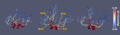
Vascular compliance reflects the stiffness of arterial vessel wall and has been related to a number of diseases, such as cardiovascular disease, cerebral arteriosclerosis, and hypertension. However, the vascular compliance at intracranial arteries has been rarely measured due to limited availability of effective methods. In this work, a 3D VASO-CBV-based MR technique was developed to assess the arterial CBV change with respect to pulsation and thus estimate vascular compliance. With this technique, we were able to map vascular compliance along the intracranial arterial tree.
|
|
0350.
 |
19 |
Propagation Patterns of Cardiac-driven and Respiratory-driven Cerebrospinal Fluid Velocity Waves Characterized by Correlation Mapping in Conjunction with Asynchronous 2-Dimensional Phase Contrast Technique
Satoshi Yatsushiro, Saeko Sunohara, Mitsunori Matsumae, Kagayaki Kuroda
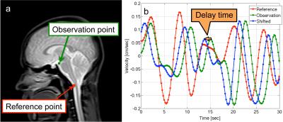
Spatial distributions of the propagation patterns of cerebrospinal fluid (CSF) motion driven by cardiac pulsation and respiration were visualized using velocity waveform correlations based on asynchronous 2-dimensional phase contrast (2D-PC) imaging. These two different driving mechanisms were evaluated using spectral analysis of the velocity waveforms for 11 healthy subjects. Delay time maps and maximum correlation maps showed the spatial distribution differences between the cardiac-driven and respiratory-driven CSF motion propagations. Maximum correlation at the prepontine was 0.83±0.05 for cardiac propagation and 0.74±0.04 for respiratory propagation with a significant difference (p << 0.01). Strong propagation may not necessarily cause large CSF displacement.
|
|
0351.
 |
20 |
Regionally differentiated cerebral blood flow increases during infancy measured with pCASL MRI
Qinlin Yu, Huiying Kang, Minhui Ouyang, Yun Peng, Fang Fang, Hao Huang
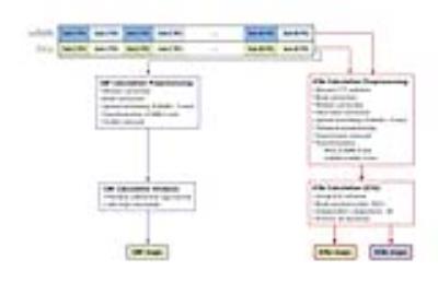
During infant brain development, rapid neuronal growth requires increases of cerebral blood flow. In this study, we quantified cerebral blood flow (CBF) at regional level during infant development by using pseudo-continuous arterial spin labeled (pCASL) perfusion MRI. The CBF maps at different infant stages from 0 to 24 months were revealed. The trend lines of CBF at specific regions were charted. It has been found that the CBF increases linearly at different brain regions, with CBF increasing faster in visual, posterior cingulate, medial prefrontal and inferior parietal cortex than whole brain.
|
|
0352.
 |
21 |
Cerebral blood flow as a marker for cortical parcellation
Roy Haast, Dimo Ivanov, Elia Formisano, Kâmil Uludag
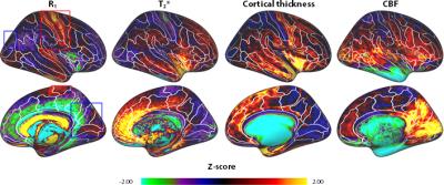
Within-subject differences of T1, T2* and cortical thickness can be used to automatically parcellate the cortex, e.g. to guide functional analyses or increase statistical power. Here, we evaluate the additional benefit of baseline CBF as a marker for brain metabolism to differentiate regions. We demonstrate that CBF data is not redundant with the other quantitative MRI parameters. Therefore, a data-driven parcellation of brain regions that incorporates perfusion information allows delineation of the cortex into smaller units and enhances subsequent anatomical & functional analysis.
|
 |
0353.
 |
22 |
Changes in cerebral blood flow and default mode network connectivity following mTBI observed with pulsed arterial spin labeling
Natalie Wiseman, Armin Iraji, E. Mark Haacke, Zhifeng Kou
Mild traumatic brain injury (mTBI) or concussion disturbs both cerebral blood flow (CBF) and functional connectivity in intrinsic connectivity networks (ICNs). Using pulsed arterial spin labeling (PASL), we derived both CBF and ICNs in mTBI patients and investigated brain CBF responses to network disruptions. We observed that mTBI patients have decreased connectivity within the default mode network (DMN) in two regions as well as increased CBF in a third region which overlaps the DMN. The mismatch of these regions suggests potential repair or compensation for injury.
|
|
0354.
 |
23 |
A Novel Approach to Measuring Cerebral Oxygen Extraction Fraction and Vascular Reserve Using MRI
Charles Cantrell, Yong Jeong, Kevin Midlash, Parmede Vakil, Sameer Ansari, Timothy Carroll
The length scales associated with parenchymal oxygen extraction fraction (OEF), coupled with the near uniformity of normative OEF across the brain dictate the development of an imaging approach that is sensitive to low-spatial frequency imaging behavior. Previous approaches to measure OEF using MRI have utilized high pass-filters—effectively removing much of the signal. We propose a new method to filter out geometric field inhomogeneity, by imaging temporally through the cardiac cycle. In a study in 11 patients with intracranial atherosclerotic disease, we found elevated OEF on the compromised hemisphere as compared to the healthy contralateral side (p<0.0195).
|
|
0355.
 |
24 |
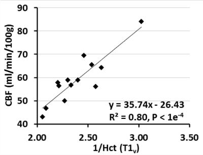
Measurements of Oxygen Delivery and Consumption Using Hematocrit Derived from Blood T1 Quantification
Feng Xu, Wenbo Li, Peiying Liu, Hanzhang Lu, John Strouse, James Pekar, Peter van Zijl, Qin Qin
We verified that venous blood T1 quantified in vivo in humans using fast and non-invasive MRI can be used to derive hematocrit (Hct) values reliably. This Hct information can be used for a more individual estimation of oxygen extraction fraction (OEF) from venous blood T2 measurements. Furthermore, inverse correlation between Hct and baseline cerebral blood flow (CBF) was observed across subjects. Measurement of Hct, OEF and CBF allowed determination of oxygen delivery (OD~CBF·Hct) and consumption (cerebral metabolic rate of oxygen, CMRO2~CBF·Hct·OEF). When compared to CBF, OD and CMRO2 showed less inter-subject variations among normal volunteers.
|
|
0356.
 |
25 |
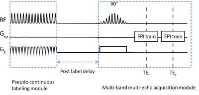
Simultaneous cerebral blood flow and bold oxygen level dependent signal assessments using multi-band multi-echo pseudo-continuous arterial spin labeling (M2-PCASL)
Shiyang Chen, Junjie Wu, Kyle Pate, Xiaodong Zhong, Bruce Crosson, Deqiang Qiu
We proposed a novel multi-band multi-echo pseudo-continuous arterial spin labeling (M2-PCASL) method to simultaneously measure the cerebral blood flow (CBF) and the blood oxygen level dependent (BOLD) signal. Increased spatial resolution was achieved compared to the conventional method, and experiments were also conducted to show simultaneous measurement of CBF and BOLD signal dynamics in response to hypercapnia. M2-PCASL can be a useful tool for measuring cerebrovascular reactivity and studying neurovascular coupling in various conditions.
|
|
0357.
 |
26 |
Measuring Blood Oxygenation and Hematocrit with a Combined T2 and T1 Approach: Initial Experience in Humans
Thomas Christen, Jia Guo, Wendy Ni, Michael Moseley, Greg Zaharchuk
In this study, we propose to obtain simultaneous measurements of blood oxygen saturation (SO2) and hematocrit (Hct) by combining two or more in vivo measurements of blood MR relaxation times. We tested our approach in 5 healthy human subjects using T2+T1 values and calibration curves obtained in vitro. The results found in the superior sagittal sinus vein (SO2=62±5% and Hct=46±1%) are in good agreement with literature values and suggest great potential of the approach once it is further validated.
|
|
0358.
 |
27 |
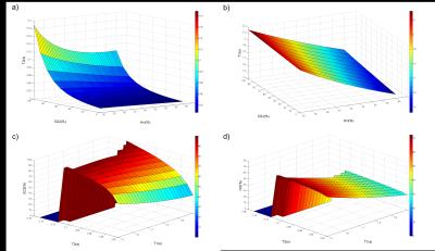
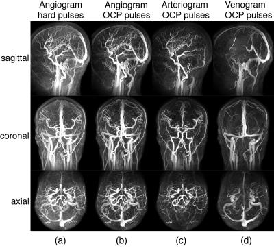
Whole-Brain Arteriography and Venography Using an Improved Velocity-Selective Saturation (VSS) Pulse Trains
Wenbo Li, Feng Xu , Jing Liu, Michael Schär, Taehoon Shin, Peter van Zijl, Ye Qiao, Bruce Wasserman, Qin Qin
Velocity-selective (VS) MRA, a non-subtractive technique and allows large spatial coverage and slow-flow depiction has shown recent promise for cerebral applications at 3T. Here, we improved the velocity-selective saturation (VSS) pulse train by reducing its sensitivity of tissue suppression to B1 inhomogeneity for both the intracranial and cervical regions. Moreover, we propose that arteriograms or venograms can be obtained by placing spatially selective inversion pulses before the acquisition to selectively null signals from venous or arterial blood. The feasibility of these technical advances for VS MRA is demonstrated on healthy volunteers at 3T.
|
|
0359.
 |
28 |
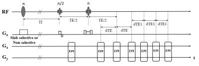
Simultaneous acquisition of oxygen extraction fraction and cerebral blood flow during brain activation
Yayan Yin, Yaoyu Zhang, Yang Fan, Bing Wu, Jia-Hong Gao
The development of neuroimaging methods to obtain oxygen extraction fraction (OEF) and cerebral metabolic rate of oxygen (CMRO2) is crucial for understanding mechanisms underlying physiological function and neuronal activity. In the present study, a novel technique termed FAIR-MGESSE combined flow sensitive alternating inversion recovery (FAIR) and multiecho gradient echo sampling of the spin echo (MGESSE) techniques. It is proposed for acquiring OEF and ASL maps in a single repetition time to determine the relative change in CMRO2 in human subjects. Relative changes in CBF, OEF and CMRO2 were measured during the visual task and the results agree well with expectations.
|
|
0360.
 |
29 |
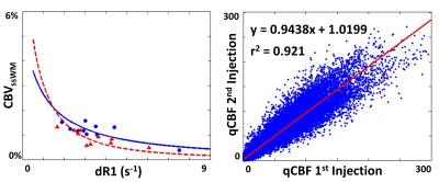
A Method for Quantitative Cerebrovascular Reserve
Yong Jeong, Charles Cantrell, Kevin Midlash, Renee Qian, Parmede Vakil, Sameer Ansari, Gregory Christoforidis, Timothy Carroll
In the occurrence of damage to the hemodynamic system of the brain, a response is elicited by changes in the supply of blood and oxygen to the tissue. Depending on these different changes, the severity of the hemodynamic impairment can be defined. In this study, we propose a method of accurately measuring quantitative cerebrovascular reserve. This is achieved by applying water correction factors to multiple contrast-agent injection cerebral blood flow measurements. In multiple studies involving volunteers, animal model, and patients, we confirm the correction factors and compare against microsphere perfusion.
|
|
0361.
 |
30 |
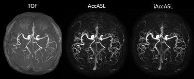
Robust Visualization of MCA Main Trunk by Improved Acceleration-Selective Arterial Spin Labeling (iAccASL) for Intracranial MR Angiography
Yuta Akamine, Makoto Obara, Osamu Togao, Shuhei Shibukawa, Masami Yoneyama, Tomoyuki Okuaki, Marc Cauteren
Improved acceleration-selective arterial spin labelling (iAccASL) was implemented for Intracranial MR angiography and images were acquired in six healthy volunteers. Additional 180?refocusing pulses in control module will efficiently suppress spin dispersion and flow voids in strongly accelerating middle cerebral artery (MCA) main arteries. The vessel visualizations of MCA main trunk and peripheral vessels were compared with our previously reported AccASL and conventional time-of-flight (TOF) approach. We demonstrated iAccASL could produce better MCA main trunk visualization (without venous signal) compared with conventional AccASL technique with better peripheral visualization than TOF.
|
|