Poster: Liver
Electronic Power Pitch Poster
Body: Breast, Chest, Abdomen, Pelvis
Tuesday, 25 April 2017
| Exhibition Hall |
14:45 - 15:45 |
| |
|
Plasma # |
|
0426.
 |
1 |
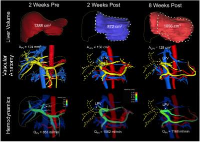
Relationship between hepatic blood flow and segmental liver hypertrophy after portal vein embolization using 4D flow MRI 
Tilman Schubert, Kevin Johnson, Alejandro Roldan Alzate, Scott Reeder
This study used 4D-flow MRI to assess potential correlations between changes in portal-venous blood flow and liver growth after portal vein embolization (PVE). 4 pigs underwent PVE and were examined with 4D flow MRI before and immediately after PVE as well as 1 week and 2 weeks after PVE. The results indicate that a persistent increase in blood flow volume to non-embolized liver segments as opposed to a peak in flow volume that later decrease over time, is likely to influence growth of these segments. Furthermore, 4D-flow MRI is well suited to study changes in portal-venous flow volume after PVE.
|
|
0427.
 |
2 |
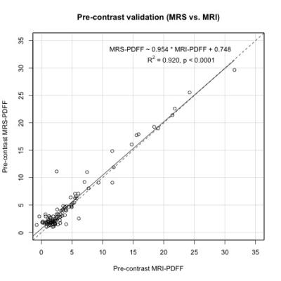
Proton Density Fat Fraction Estimation Accuracy of High-Flip-Angle, Contrast-Enhanced, Magnitude-Based Multi-Gradient-Echo MR Imaging at 3T
Ethan Sy, Cheng Hong, Soudabeh Fazeli Dehkordy, Charlie Park, Alexandra Schlein, Jonathan Hooker, Jennifer Cui, Gavin Hamilton, Claude Sirlin
Proton density fat fraction (PDFF) is a widely-used magnetic resonance imaging (MRI)-based biomarker for noninvasive quantification of hepatic steatosis. PDFF derived from low-flip-angle magnitude-based multi-gradient-echo MRI (MRI-M) without intravenous contrast has been shown to have high accuracy for estimating fat fraction. However, this MRI-M technique has a relatively low signal-to-noise ratio (SNR), which makes it difficult to visualize anatomical features. In this analysis of ninety-two patients, high-flip-angle contrast-enhanced (with either gadoxetate disodium or gadobutrol) MRI-M imaging had no significant difference in fat-grading accuracy from the standard low-flip-angle pre-contrast PDFF, using MR spectroscopy (MRS)-PDFF as the reference standard.
|
|
0428.
 |
3 |
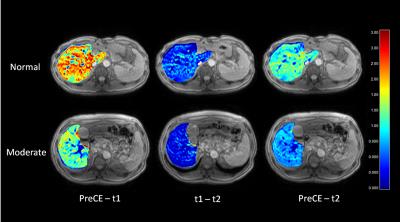
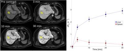
Assessment of liver function using an uptake ratio based on multiple-time points hepatocyte mapping
Tomoyuki Okuaki, Kosuke Morita, Tomohiro Namimoto, Morikatsu Yoshida, Shinya Shiraishi, Yasuyuki Yamashita, Marc Van Cauteren
We calculated the uptake fraction (kHep) based on a pharmacokinetics model of gadolinium ethoxybenzyl-diethylenetriamine pentaacetic acid (Gd-EOB-DTPA) uptake using R1 changes in the hepatocyte. The kHep and ΔT1 values were compared to indexes determined by 99mTc-GSA scintigraphy for three different scan time points. The correlation coefficients of the blood clearance indexes (HH15) and receptor indexes (LHL15) were 0.307 and 0.497 for the ΔT1 and 0.537 and 0.570 for kHep values, respectively. The results indicate that the proposed quantification value of kHep is a more robust index for liver function compared with the ΔT1 value.
|
|
0429.
 |
4 |
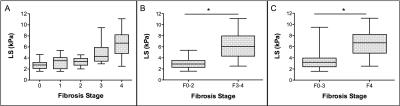
Detection of Advanced Liver Fibrosis and Cirrhosis using MR elastography compared to liver surface nodularity measurement, EOB-DTPA uptake and blood tests
Cecilia Besa, Mathilde Wagner, Grace Lo, Sonja Gordic, Manjil Chatterji, Ashley Stueck, James Babb, Andrew Smith, Bachir Taouli
This study compares the diagnostic performance of multiparametric MRI including qualitative and quantitative assessment of MR-elastography (MRE), liver surface nodularity (LSN) software measurement, hepatic enhancement ratios on Gd-EOB-DTPA (EOB-ER), and serum markers (APRI and FIB4) for the detection of liver fibrosis and cirrhosis. When comparing different MRI methods and serum markers with histologic findings, liver stiffness measured with MRE showed better performance than other methods for detection of advanced liver fibrosis and cirrhosis, especially when combined with blood tests (FIB4).
|
|
0430.
 |
5 |
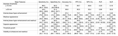
Diagnostic performance of LI-RADS major features, ancillary features, and categories on MRI for diagnosis of hepatocellular carcinoma
Milena Cerny, Catherine Bergeron, Jean-Sébastien Billiard, Jessica Murphy-Lavallée, Damien Olivié, Joshua Bérubé, Boyan Fan, Hélène Castel, Simon Turcotte, Pierre Perrault, An Tang
Diagnosis of hepatocellular carcinoma (HCC) is largely based on imaging. MRI is ideally suited for the non-invasive diagnosis of HCC because it has numerous tissue contrast mechanisms and is the only modality that can assess all major and ancillary imaging features. We evaluated the diagnostic performance of MRI-determined LI-RADS major features, ancillary features, and categories for the diagnosis of HCC. Our results suggest that interpretation that includes ancillary features increases the sensitivity, while preserving a high specificity for definite HCC and a slightly lower specificity for probable HCC. Further, ancillary features in favor of benign entities have high specificity for benignity.
|
|
0431.
 |
6 |
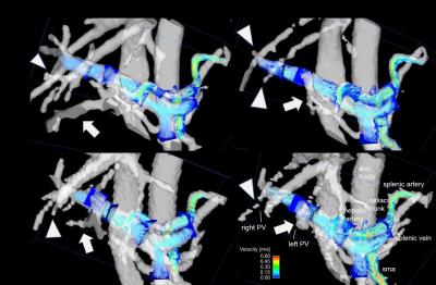
Longitudinal Characterization of Liver Regeneration and Portal Hemodynamics in Living Donor Liver Transplant
Alejandro Roldán-Alzate, David Rutkowski, Luis Fernandez, Scott Reeder
The purpose of this study was to evaluate longitudinal liver regeneration and hemodynamic changes of living donor liver transplant (LDLT) donors in response to surgical liver resection. Five living related liver donors were studied. Subjects were imaged using 4D Flow MRI before and at several times following partial hepatectomy. The ability to longitudinally evaluate liver regeneration and portal hemodynamic changes non-invasively demonstrates that 4D flow MRI is a suitable tool for both surgical planning of LDLT, and for improved understanding of the liver regeneration and hemodynamic changes that occur in the remnant liver of the donor.
|
|
0432.
 |
7 |
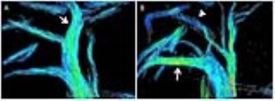
4D flow MRI of Liver Hemodynamics: Influence of Velocity Encoding, Different Field Strength and Contrast Application on Visualization and Quantification of blood flow - permission withheld
Zoran Stankovic, Bernd Jung, Alan Peters, Jelena Surla, Edouard Semaan, Michael Ith, Johannes Heverhagen, Michael Markl, Jeremy Collins
4D flow MRI offers the possibility for complete volumetric and functional assessment of liver blood flow in patients with liver cirrhosis. This study reveals a significant difference when using a lower venc for visualization of the portal vein branches. Similar results are obtained for contrast application and different field strengths. For qualitative assessment of the intrahepatic branches by 4D flow MRI a lower venc is recommended.
|
|
0433.
 |
8 |
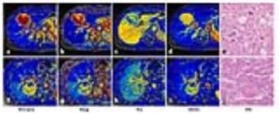
Quantitative Free-Breathing Dynamic Contrast-enhanced MRI in Hepatocellular Carcinoma Using Gd-EOB-DTPA:Correlations With Ki67 Proliferation Status and Histological Grades
Jie Chen, Chenyang Chen, Chunchao Xia, Bin Song
This study aims to validate a free-breathing DCE-MRI in HCC patients using gadoxetic acid, and to determine the relationship between DCE-MRI parameters and histological results. Perfusion parameters (Ktrans, Kep, Ve and iAUC) from 34 patients were compared with CT results and correlated with Ki67 indices and the histological grades of HCC. Significant relationship was found between DCE-MRI and CT results, indicating the validity of this protocol in evaluating the vascular change of HCC. The DCE-MRI derived Ktrans and iAUC were significantly correlated with the histological grades of HCC, suggesting the potential of those parameters in predicting the malignancy of tumors.
|
|
0434.
 |
9 |
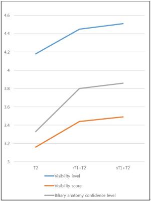
The value of high resolution gadoextic acid-enhanced MR cholangiography for evaluating biliary anatomy of living liver donor: comparison with T2 weighted (T2W) MR cholangiography and conventional gadoxetic acid enhanced MR cholangiography
Hyo-Jin Kang, Jeong Min Lee, Jeong Hee Yoon, Won Chang, Ijin Joo, Joon Koo Han
The addition of gadoxetic acid-enhanced sFOV T1W-MRC to T2W-MRC is able to provide more precise depiction of biliary anatomy, which is crucial for the prevention of post-operative biliary complications of living liver donors.
|
|
0435.
 |
10 |
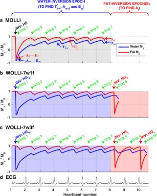
Water-Only Look-Locker Inversion recovery (WOLLI) T1 mapping
Liam Garrison, Christina Levick, Michael Pavlides, Tom Marjot, Ferenc Mozes, Leanne Hodson, Stefan Neubauer, Matthew Robson, Christopher Rodgers
We propose a new “Water-Only Look-Locker Inversion recovery” (WOLLI) sequence, based on MOLLI, that enables water-selective T1-mapping in an 8hb breath-hold at 3T. WOLLI uses a hypergeometric (HG) inversion pulse to selectively invert water with negligible effect on fat. To separate the steady-state fat and water signals, WOLLI adds one or more fat-inversions starting from the plateau of the water T1* recovery. WOLLI uses an extended Deichmann-Haase formula to correct for readout-induced saturation. We validated this approach by simulations, scans in a phantom containing 19 fat/water mixtures, and liver scans in 12 subjects (volunteers and liver disease patients).
|
|
0436.
 |
11 |
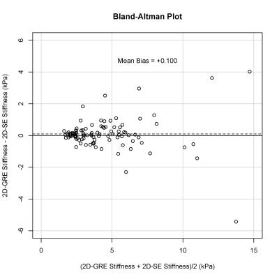
Region-of-interest size of hepatic 2D MR elastography decreases with increasing R2* for gradient-echo but not spin-echo techniques
Cheng Hong, Adrija Mamidipalli, Ethan Sy, Jonathan Hooker, Calvin Tran, Tanya Wolfson, Soudabeh Fazeli Dehkordy, Scott Reeder, Rohit Loomba, Claude Sirlin
MR elastography (MRE) is an emerging technique for the non-invasive assessment of hepatic stiffness and fibrosis, and can be based on gradient-echo (GRE) or spin-echo (SE) acquisition. This study demonstrates that 2D-SE provides significantly larger reliable-wave-quality regions-of-interest (ROIs) than 2D-GRE at 3T. Additionally, 2D-GRE ROI sizes are negatively correlated with hepatic R2*, while 2D-SE ROI sizes are not associated with R2*. This suggests that 2D-SE may be the preferred MRE sequence at 3T and in patients with known iron overload.
|
|
0437.
 |
12 |
Estimating Liver Function in Chronic Liver Disease Patients Using DCE-MRI and Whole-Body Pharmacokinetic Modeling
Markus Karlsson, Mikael Forsgren, Olof Dahlqvist-Leinhard, Nils Dahlström, Bengt Norén, Mattias Ekstedt, Stergios Kechagias, Gunnar Cedersund, Peter Lundberg
Dynamic contrast enhanced MRI with Gd-EOB-DTPA is a promising method for investigating liver function. However, many of the proposed procedures relies on such a high temporal resolution, and low spatial resolution, that the images are rendered unsuitable for radiographic reading. Here we have tested and validated a recently developed procedure using a whole-body pharmacokinetic model in a cohort of patients with chronic liver disease. Moreover we also show that the approach can separate advanced from mild fibrosis.
|
|
0438.
 |
13 |
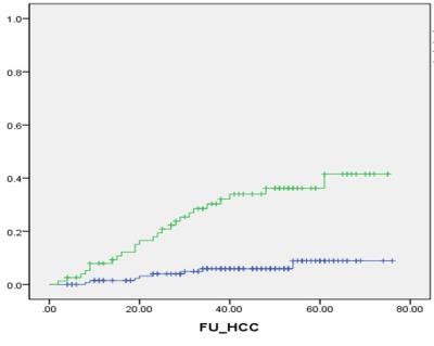
Prognostic Role of Liver Stiffness Measurement Using Magnetic Resonance Elastography in Patients with Compensated Chronic Liver Disease
Dong Ho Lee, Jeong Min Lee
Liver stiffness measurement (LSM) using magnetic resonance elastography (MRE) can estimate the degree of liver fibrosis. We retrospectively evaluate the prognostic role of LSM using MRE in compensated chronic liver disease patients. A total of 217 patients with compensated chronic liver disease who underwent MRE were included. After a mean and median follow-up of 44.5±17.8 months and 46.0 months, LSM value obtained from MRE was turned out to be significant predictive factor for overall survival, development of hepatic decompensation and occurrence of hepatocellular carcinoma.
|
|
0439.
 |
14 |
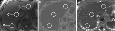
Epidemiology and spatial heterogeneity of hepatic fat and iron deposition: an MRI-based analysis
Daniel Ludwig, Tyler Fraum, Scott Kilian, Kathryn Fowler
MRI has emerged as a reliable, noninvasive means of liver fat and iron quantification. We retrospectively studied 1006 patients that underwent standard-of-care liver MRI at a single tertiary care center. Multivariate analysis was used to identify factors predictive of the severity and spatial heterogeneity of hepatic fat deposition (HFD) and hepatic iron deposition (HID). Greater spatial heterogeneity of HFD and HID generally occurred as the severity of HFD and HID increased, suggesting higher risk for misclassification by biopsy with more advanced disease. Overall, by globally evaluating the liver, MRI constitutes a robust tool for assessing hepatic fat and iron deposition.
|
|
0440.
 |
15 |
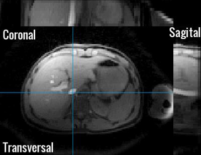
Free breathing T2* mapping of the Liver using a compressed sensing reconstruction - permission withheld
Paul de Heer, Oliver Gurney-Champion, Jurgen Runge, Remy Klaassen, Jasper Schoormans, Bram Coolen, Hanneke van Laarhoven, Gustav Strijkers, Jaap Stoker, Aart Nederveen
Hemochromatosis (iron overload) occurs in a range of liver diseaes.(1,2) Iron content can be measured invasively with liver biopsy but is commonly replaced by non-invasive MR measurements, including T2* mapping. This is commonly done in a breath-hold to reduce the effects of respiratory motion. In this work we show that a stack of stars radial golden angle T2* mapping multi echo sequence during free breathing in combination with CS reconstruction facilitates high resolution imaging in the liver. This approach bears promise beyond liver imaging for visualizing smaller organs and pathologies, e.g. the pancreas and lymph nodes.
|
|