Poster: Breakthrough Methods & Applications in Cancer Imaging
Electronic Power Pitch Poster
General Cancer Imaging
Thursday, 27 April 2017
| Exhibition Hall |
09:15 - 10:15 |
| |
|
Plasma # |
|
0977.
 |
1 |
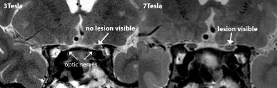
High resolution imaging of the optic chiasm at 7T MRI improves lesion detection and tumour delineation compared to 3T 
Guido van Haren, Lorna Grech-Fonk, Marco Verstegen, Wouter Teeuwisse, Teresa Ferreira, Irene Notting, Wouter van Furth, Alberto Pereira, Gregorius Luyten, Andrew Webb, Jan-Willem Beenakker
The limited resolution of 3Tesla MRI often leads to missed lesions or ambiguities in the tumour environment for patients with a pituitary macro-adenoma. In this study we developed a robust high-resolution 7Tesla MRI-protocol of the optico-chiasmatic system and evaluated its clinical value. The 7T MR-images reveal tiny lesions in the optic nerve or chiasm which are not visible at 3T. These lesions could explain the vision loss for 3 of the 7 evaluated patients and gave the physician new treatment possibilities. Overall, this study shows the great clinical opportunities of 7Tesla MRI for patients with pituitary macro-adenoma or other neuro-ophthalmic conditions.
|
 |
0978.
 |
2 |
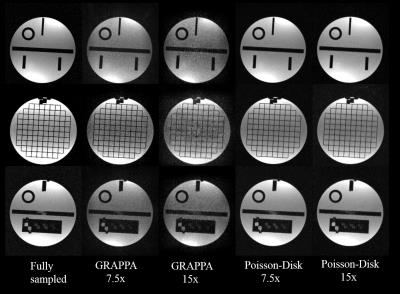
Accelerated 3D bSSFP Imaging for Treatment Planning on an MRI-Guided Radiotherapy System
Yu Gao, Ziwu Zhou, Fei Han, Percy Lee, Daniel Low, Peng Hu, Yingli Yang
The purpose of this work is to introduce a compressed sensing and parallel imaging combined technique to reduce the acquisition time for planning MR. We implemented a variable-density Poisson-Disk under-sampled acquisition along with L1-ESPIRiT reconstruction technique on an MRI-guided radiotherapy system. Phantom study showed that our technique had superior image quality over the conventional GRAPPA approach. Patient and volunteer study demonstrated that comparable images can be acquired with half of the original time. In addition, the proposed technique was able to achieve high resolution imaging where the GRAPPA approach failed due to high noise level.
|
|
0979.
 |
3 |
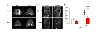
Feasibility of magnetic resonance colonography for an immune check-point inhibitor in orthotopic colorectal rechallenge tumor models
Jinil Kim, Yoon Seok Choi, Dong-Cheol Woo, Chul-Woong Woo, Sang Tae Kim, Jae Im Kwon, Kyung Won Kim
In the immunotherapy research field, establishing appropriate preclinical model is very important to evaluate the complex immune reaction. Orthotopic tumor model is more physiologic than ectopic tumor model, however its use may be limited due to difficulty in evaluating deep-seated tumors, especially in the colorectum having a complex anatomy. MR colonography (MRC) is a new technique in preclinical trial, which uses Fluorinert, a negative contrast agent, to fill the colorectum. Our study demonstrated that MRC is quite feasible to evaluate colorectal tumors and metastatic foci in orthotopic colorectal tumor model, which can be useful in immunotherapy drug development.
|
|
0980.
 |
4 |
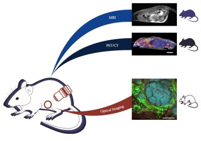
Multi-modal and multi-scale measurement of metabolism in vivo in a breast cancer model
Benjamin Cox, Joseph Szulczewski, David Inman, Erin Adamson, Kai Ludwig, Justin Jeffery, Stephen Graves, Alison Roth, David Mummy, Patricia Keely, Kevin Eliceiri, Sean Fain
Performing MRI, PET/CT, and optical imaging at the cellular scale in vivo provides highly complementary information as a powerful tool to study cancer metabolism. Recent development of implanted optical windows allows for optical imaging in vivo. Here, we demonstrate multi-modal and multi-scale imaging of tumor progression in a mouse model of breast cancer by performing optical imaging through an implanted window, and whole-body MRI and PET/CT in a single day. The challenge of PET-MR registration is also addressed using dual-PET-MR fiducial markers embedded in a custom-imaging tray.
|
|
0981.
 |
5 |
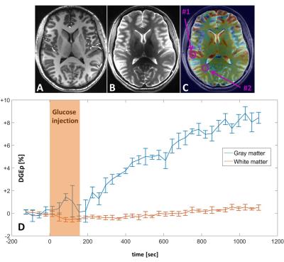
Dynamic Glucose Enhanced MRI - A prospective study in healthy volunteers and glioblastoma patients - permission withheld
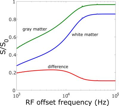
Daniel Paech, Patrick Schuenke, Christina Koehler, Johannes Windschuh, Sibu Mundiyanapurath, Sebastian Bickelhaupt, Philipp Bäumer, David Bonekamp, Martin Bendszus, Wolfgang Wick, Peter Bachert, Mark Ladd, Heinz-Peter Schlemmer, Moritz Zaiss, Alexander Radbruch
Glucose is the main energy source of cancer cells to proliferate and survive. Recently, promising results to assess changes in cellular metabolism using natural unlabeled D-glucose as biodegradable MRI contrast agent, have been reported employing Chemical Exchange Saturation Transfer (CEST) and Chemical Exchange sensitive Spin-Lock (CESL) imaging. In this work, the CESL-based dynamic glucose enhanced (DGE) contrast was investigated in healthy volunteers and a homogenous cohort of newly diagnosed untreated glioblastoma patients at 7 Tesla. DGE MRI allowed for sensitive visualization of physiological glucose uptake in the healthy human brain and pathophysiologically increased glucose enhancement of brain tumors.
|
|
0982.
 |
6 |
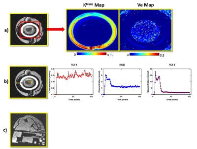
3D printed Breast DCE-MRI phantom to mimic structure and pharmacokinetics
Nithin Vajuvalli, Chethan Kumar M, Amaresha Konar, Shivaprasad Chikop, Darshan Keelara, Ashwini Kumnoor, Ramesh Venkatesan, Sairam Geethanath
DCE MRI plays a critical role in routine clinical breast examination. Current work focuses on the development of Breast DCE MRI phantom using a 3D printer to mimic poor and well perfused regions. The phantom developed was controlled through user entered Ktrans values entered in a GUI which interfaced with a peristaltic pump to control flow rates. The Kep parameters was controlled through the 3D model geometry. Prospective MR images of the phantom were acquired on a 1.5T scanner using the TRICKS sequence; and pharmacokinetic maps based on Tofts model were computed and quantified.
|
|
0983.
 |
7 |
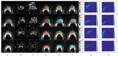
Recurrent Neural Network on DCE-MRI in Prostate Cancer
Xia Li, Vivek Vaidya, Sandeep Gupta, Rakesh Mullick, Oguz Akin, Dattesh Shanbhag
DCE-MRI has become an important protocol in mpMRI analysis of prostate cancer and it has been quantified typically using pharmaco-kinetic modelling and the estimated parameters are then used with other approaches (machine learning or deep learning (DL)) to characterize/discriminate tumor tissue against healthy tissue. However, it is not clear if applying DL to the DCE-MRI time series directly is beneficial for prostate cancer detection. Hence, we propose a DL based method to differentiate prostate tumor from healthy tissues at the voxel level using raw arbitrary signal DCE time-series itself. Overall, DL based tumor characterization provided similar detectability for prostate tumor when compared to Ktrans and ve maps. We also evaluated differences in tumor characterization when contrast agent concentration time-curves were used instead of arbitrary signal curves and found them to provide similar detectability.
|
|
0984.
 |
8 |
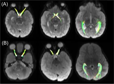
In vivo assessment of tumour invasion of the visual pathway in optic pathway glioma patients using multi-shell diffusion tensor MRI
Patrick Hales, Victoria Smith, Patricia O'Hare, Kshitij Mankad, Felice d'Arco, Jessica Cooper, Ramneek Kaur, Kim Phipps, Darren Hargrave, Christopher Clark
Optic pathway glioma (OPG) is a childhood tumour of the visual pathway. Some OPG patients remain stable, whereas others experience rapid visual decline; however, conventional MRI cannot stratify these patients. We used multi-shell DTI to measure tumour invasion of the optic pathway in 23 OPG patients, in conjunction with visual assessment. A strong correlation was found between fractional anisotropy in the optic nerves and optic radiations, and visual acuity (p=0.00092 and p=0.008 respectively). Our study demonstrates that multi-shell diffusion-MRI offers increased sensitivity over conventional MRI in detecting white matter integrity and function in the visual pathway of OPG patients.
|
|
0985.
 |
9 |
Pipeline for longitudinal assessment of patient-derived mouse xenografts using 3D magnetization transfer-weighted MRI
Kimberly Desmond, David Bakhshinyan, Maleeha Qazi, Parvez Vora, Chirayu Chokshi, Sheila Singh, Nicholas Bock
A pipeline was developed, driven by 3D magnetization transfer-weighted images acquired without contrast agent, to automatically assess mouse models of patient-derived tumours against an atlas of control NOD-SCID mice, for the purposes of longitudinal, high-throughput screening of mice for response to cancer therapy and recurrence.
|
|
0986.
 |
10 |
Tumor Interstitial Fluid Pressure and Hydraulic Conductivity Estimates by DCE-MRI in a Rat Model of Cerebral Tumor
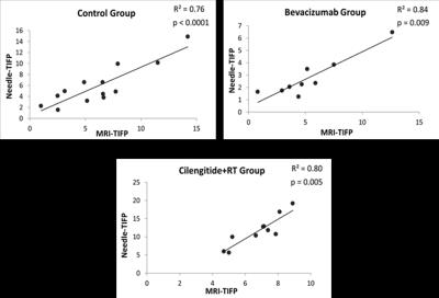
Rasha Elmghirbi, Nagaraja Tavarekere, Stephen Brown, Swayamprava Panda, Kelly Keenan, Glauber Cabral, Hassan Bagher-Ebadian, James Ewing
An elevated tumor interstitial fluid pressure (TIFP) is a critical element for assessing therapeutic response. This study demonstrates the use of DCE-MRI to estimate TIFP, and validates that estimate by an invasive method in a rat glioblastoma model, with and without treatment interventions. Significant positive correlations between MRI-derived TIFP estimates and invasive measures of TIFP were found in all groups (e.g., for untreated group, R2=0.76, p<0.0001). These findings validate an MRI-estimated TIFP as a noninvasive measure of TIFP in embedded cerebral tumors, and suggest that it may be a useful tool in assessing tumor response to therapy.
|
|
0987.
 |
11 |
A preclinical MRI study investigating the impact of the local microenvironment on the progression of diffuse intrinsic pontine glioma in patient-derived xenografts
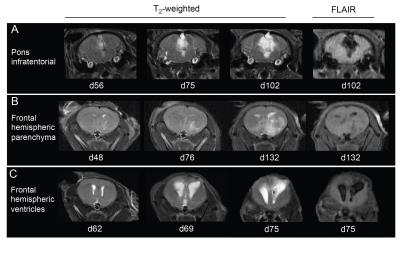
Mariama Fofana, Jessica Boult, Maria Vinci, Valeria Molinari, Kathryn Taylor, Sergey Popov, Alan Mackay, Chris Jones, Simon Robinson
Diffuse intrinsic pontine glioma (DIPG) is a devastating childhood brain tumour with very poor outcome. The local brain microenvironment appears to play an important role in the tumourigenesis of DIPG, and is currently underinvestigated. Infratentorial and hemispheric tumour growth patterns of orthotopic DIPG xenografts were assessed longitudinally by anatomical MRI, and confirmed by immunohistochemical analysis. ADC, T1 and T2 were higher in infratentorial tumours than hemispheric tumours, corresponding to a higher degree of tumour-associated oedema observed histologically.
|
|
0988.
 |
12 |
Multi-parametric MRI Radiomics for Pre-treatment Prediction of the Progression-Free Survival in Advanced Nasopharyngeal Carcinoma
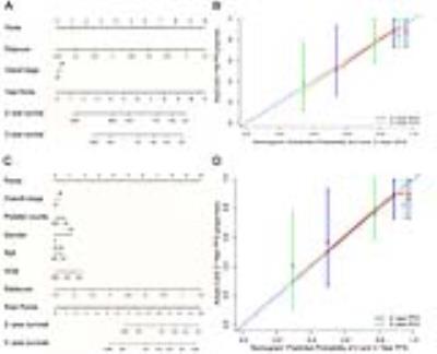
Bin Zhang
To our knowledge, this is the first MRI-based radiomics study to predicting survival of tumor. The results of our study show that multiparametric MRI-based radiomics nomogram significantly improves the 7th edition of AJCC TNM staging system and clinical data in predicting individualized progression-free survival (PFS) in advanced NPC (stage III-IVb). In fact, the radiomics nomogram built in our study could integrate all prognostic biomarkers/signatures that have been published in this area to improve its predictive performance. Besides, for the first time, our radiomics heatmaps showed positive associations between radiomics signature features with overall stage, T-stage and negative associations between radiomics signature features with N-stage. Our radiomics study provides some different insights into the mechanism of hematogenous and lymphatic metastasis of NPC.
|
|
0989.
 |
13 |
MR Elastography and Perfusion MRI for the Early Assessment of Treatment Response in Soft Tissue Sarcomas
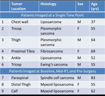
Kay Pepin, Roger Grimm, Soudabeh Kargar, Sarah James, Matthew Howe, Karen Fritchie, Matthew Frick, Doris Wenger, Richard Ehman, Nadia Laack, Michael Herman, Deanna Pafundi
Advanced imaging is a critical component in the development of patient-specific and novel treatment strategies, and the non-invasive evaluation of early response in sarcomas. Our central hypothesis is that changes in sarcoma stiffness quantified with MRE and perfusion with DCE-MRI throughout therapy can predict response. Soft tissue sarcomas are a rare malignancy arising in a wide range of anatomic locations. Anatomy-specific imaging protocols were developed to evaluate soft tissue sarcomas in 9 patients. In 3 patients, we investigated the feasibility to assess response to radiation therapy and observed a decrease in parameters related to tumor stiffness and perfusion metrics.
|
|
0990.
 |
14 |
Quantitative Imaging for Radiotherapy on an MR-Linac Scanner
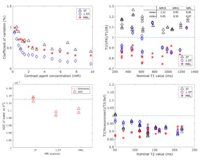
Folkert Koetsveld, Leon ter Beek, Petra van Houdt, Laurens van Buuren, Uulke van der Heide
The MR-Linac integrates and MR-scanner with a radiotherapy treatment machine. Patients undergoing radiotherapy treatment will be imaged daily on the MR-Linac. We investigated the suitability of the MR-Linac as a platform for quantitative imaging. Daily quantitative imaging can be used for imaging biomarker discovery, and give information on tumor treatment response. We did phantom studies of four quantitative MRI techniques: T2 mapping, dynamic contrast enhanced imaging, T1 mapping and diffusion weighted imaging to determine the accuracy of these techniques on the MR-Linac. We tested the repeatability of T2 mapping on a volunteer.
|
|
0991.
 |
15 |
Support vector machine for breast cancer classification using DWI histogram features: preliminary study
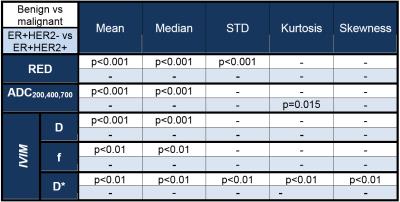
Igor Vidic, Liv Egnell, Jose Teruel, Torill Sjøbakk, Neil Jerome, Agnes Østlie, Hans Fjøsne, Tone Bathen, Pål Goa
In this work we use the machine learning method support vector machine (SVM) to classify malignant and benign tumors, as well as ER+HER2- and ER+HER2+. As feature we use histogram properties of DWI-models (RED, ADC, IVIM) parameters as features. Our study showed that SVM classifiers using combinations of features from different models have predictive power in both analyses, also it performed better than SVM using combination of parameters obtained only from one of the models. The results are encouraging because SVM with DWI parameters can potentialy hinder unnecessary biopsies.
|
|