Poster: Post-Processing & Motion
Electronic Power Pitch Poster
Acquisition, Reconstruction & Analysis
Wednesday, 26 April 2017
| Exhibition Hall |
14:45 - 15:45 |
| |
|
Plasma # |
|
0771.
 |
16 |
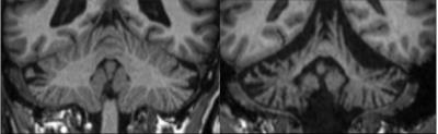
cBEaST: Cerebellar Brain Extraction based on Nonlocal Segmentation Technique – A comparison with state-of-the-art methods 
Daniel Güllmar, Viktor Pfaffenrot, Rossitza Draganova, Xiang Feng, Jürgen Reichenbach, Dagmar Timmann, Andreas Deistung
An automatic segmentation of the cerebellum is required to determine the cerebellar volume and for improving spatial normalization in voxel-based analysis approaches. While existing segmentation approaches typically work quite robust in healthy subjects, errors in segmentation increase with cerebellar atrophy and typically require manual corrections. We introduce a novel cerebellum segmentation approach, referred to as cBEaST, that relies on a dedicated multi-resolution segmentation library with manually edited cerebellar masks of both healthy and diseased subjects in combination with multi-atlas-propagation and segmentation as implemented in BEaST. Finally segmentation of the cerebellum with BEaST is compared with the alternative techniques SUIT and FreeSurfer.
|
 |
0772.
 |
17 |
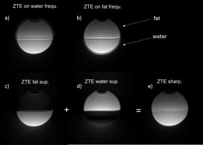
Signal-model-based water-fat separation in Zero Echo Time (ZTE) MRI
Romain Froidevaux, Markus Weiger, Po-Jui LU, Klaas Pruessmann
The separation of water and fat in zero echo time (ZTE) imaging is challenging for several reasons: First, echo-based signal models are violated for fast-relaxing spins and require the use of loud sequences. Second, frequency selective preparation pulses produce large SAR and are inefficient on short-T2 compounds. Finally the off-resonance induced phase vanishes at TE = 0. In this work, we introduce the principles of a technique that allows water-fat separation for a single ZTE acquisition and demonstrate it potential and limits with phantom and in-vitro experiments.
|
|
0773.
 |
18 |
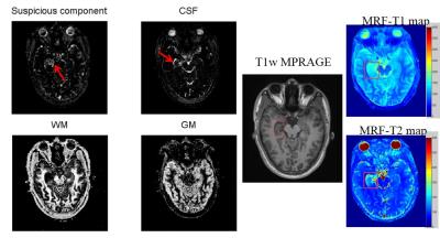
Suspicious Component Segmentation for Identifying Hippocampal Sclerosis Using Regularized Tissue-Fraction MR Fingerprinting
Kang Wang, Congyu Liao, Xiaozhi Cao, Zhixing Wang, Dengchang Wu, Hongjian He, Qiuping Ding, Jianhui Zhong
A relaxometry-based tissue fraction segmentation using MR fingerprinting method was applied for identifying hippocampal sclerosis. The results demonstrated that tissue-fraction MR fingerprinting method could effectively segment multiple tissue components and mark the possible sclerosis regions, which is critical for clinical application including lesions diagnosis and multicomponent analysis.
|
 |
0774.
 |
19 |
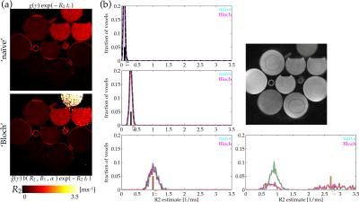
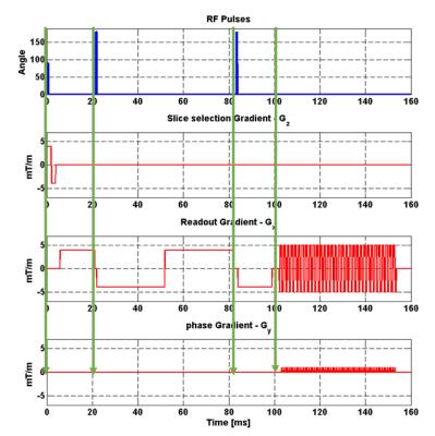
Improved Short-T2* Estimation with Bloch Equation-Modeled Concurrent Excitation and Relaxation
Ethan Johnson, Kim Butts Pauly, John Pauly
Short-T2* magnetization (order of [0.1,1ms]) can relax appreciably during standard-rate excitation pulses, which can bias estimates of relaxation rates formed by fitting to observed signal decay. The effect can, however, be included in an updated model to improve T2* estimation for fast-relaxing signals. Here, a demonstration is presented.
|
|
0775.
 |
20 |
Edge preserving upsampling of image resolution in MRI
Marco Reisert, Elias Kellner
In this work we present a simple and efficient postprocessing method to isotropify the imaging resolution of MRI imagery. In MRI anisotropic voxel sizes are quite common due to several reasons. Typically the trough-plane voxel size is higher than the in-plane resolution. We propose a simple technique to upsample to isotropic image resolution without introducing the typical block like artifacts known from conventional interpolation schemes.
|
|
0776.
 |
21 |
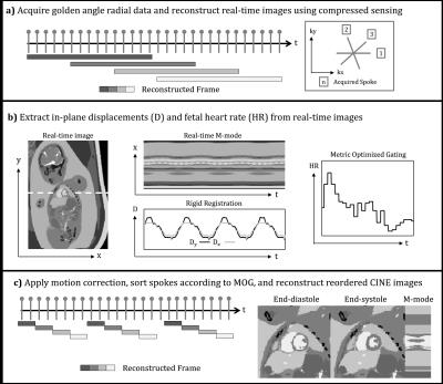
Motion-compensated reconstruction of fetal cardiac MRI using a golden-angle radial acquisition, retrospective gating, and compressed sensing
Christopher Roy, Mike Seed, Christopher Macgowan
Fetal cardiac MRI requires high spatial and temporal resolution but is often limited by stochastic and periodic motion. To compensate for these sources of artifact, a radial golden-angle acquisition was used to acquire and reconstruct real-time fetal cardiac images. In-plane motion and fetal heart rate were then calculated from the real-time images and used to reconstruct reordered CINE images at high spatial and temporal resolution. Using this approach, motion-robust imaging of the fetal heart was successful in seven pregnant volunteers for both short-axis and long-axis multi-slice acquisitions.
|
|
0777.
 |
22 |
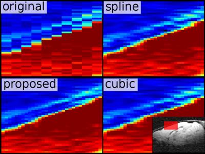
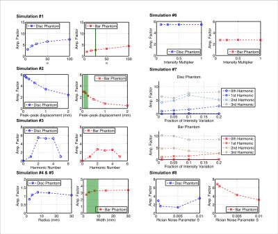
From Visualization to Quantification: Calibrating Motion Magnification by Amplified Magnetic Resonance Imaging
Wendy Ni, Maged Goubran, Greg Zaharchuk, Michael Moseley, Kristen Yeom, Samantha Holdsworth
The brain is constantly in motion. Changes in the cardio-ballistic motion of brain structures can provide invaluable information on natural processes and pathology. We have previously introduced a qualitative visualization technique, Amplified MRI (aMRI), to amplify subtle cardio-ballistic motion in the brain. Now we attempt to quantify the underlying motion through simulation-based characterization of the aMRI technique. By generating calibration curves for a range of motion parameters, we calculated the unamplified tissue displacement in two human subjects. The estimated displacements are higher than literature values. Nevertheless, our simulations are the first steps in benchmarking aMRI’s potential as a quantitative technique.
|
|
0778.
 |
23 |
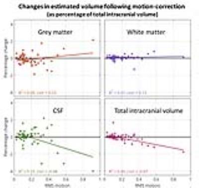
Assessing the effect of head-motion on tissue volume estimates
Daniel Gallichan
We looked at data from 60 subjects undergoing 1mm resolution T1-weighted structural scans at 7T with 3D FatNavs allowing retrospective motion-correction. Motion parameters for 60 subjects were examined for general trends – and typical motion was dominated by translation in the z-direction and a small backwards rotation of the head. Quantitative estimates of tissue volumes were compared before and after motion-correction was applied, suggesting that total intracranial volume tends to be overestimated when the subject moves more – and that this is dominated by an overestimation of the CSF volume.
|
|
0779.
 |
24 |
Prospective motion correction on diffusion weighted imaging: improving data quality with four radio frequency and gradient pulses updates.
Danilo Maziero, Michael Herbst, Thomas Ernst
Prospective Motion Correction (PMC) using fast camera-based tracking systems can dramatically increase DWI data quality and may have an important application in populations where movement is hard to prevent. Here we present images acquired with and without intra-sequence PMC from two subjects realizing fast head rotations up to 30°/s. Our results show that the DWI motion sensitivity can be reduced five-fold by applying 3 intra-sequence PMC updates (prior to each of the two refocusing RF pulse and prior to readout).
|
|
0780.
 |
25 |
Motion corrected high resolution time-of-flight angiography at 7T using Segmented FatNavs - permission withheld
Frédéric Gretsch, Daniel Gallichan
We propose to insert Segmented FatNavs into a time-of-flight sequence, and use it as a multi-purpose module. It allows fat suppression, magnetization-transfer suppression of the tissue signal, and retrospective motion correction. Image quality was always enhanced after correction, even in cases of small motion, thereby making high-resolution brain angiography more reliable.
|
|
0781.
 |
26 |
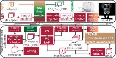
Motion correction on a human PET/MR scanner: Clinical feasibility of a motion correction system in patients – an update report
Thomas Küstner, Christian Würslin, Martin Schwartz, Hadi Fayad, Thibaut Merlin, Christopher Gilliam, Thierry Blu, Petros Martirosian, Fritz Schick, Bin Yang, Holger Schmidt, Nina Schwenzer
The diagnostic accuracy of Positron-Emission-Tomography/Magnetic Resonance (PET/MR) is often reduced in regions affected by respiratory and cardiac motion. These motion-induced artifacts can be corrected by an MR-derived motion model (MM). Here, we improved the previously presented PET/MR motion correction system by two new sampling trajectories for the MR motion imaging and extend it by the usage of an additional Compressed Sensing reconstruction (BART), an optical-flow based registration (LAP) and the incorporation of motion correction into a listmode-based PET reconstruction (CASToR) which are all integrated into the Gadgetron-based reconstruction pipeline for a clinical feasible setup. In-vivo patient data substantiated the improvements.
|
|
0782.
 |
27 |
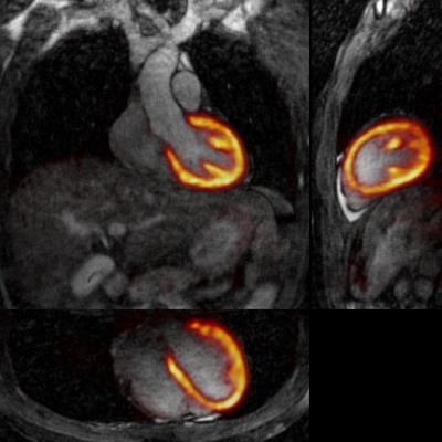
Respiratory motion-corrected simultaneous cardiac PET and coronary MR angiography using a hybrid 3T PET-MR
Camila Munoz, Radhouene Neji, Gastao Cruz, Rene Botnar, Claudia Prieto
Physiological motion remains a major challenge for cardiac PET-MR. Here we propose a framework for non-rigid respiratory motion-corrected simultaneous Coronary MR Angiography (CMRA) and cardiac PET. Motion estimated from low-resolution MR image navigators and from CMRA data itself is used for correcting CMRA and PET datasets to the same respiratory position. The proposed CMRA approach was validated in ten healthy subjects. Results from the PET-CMRA framework on three patients show that motion-corrected PET images have improved sharpness compared to uncorrected reconstructions, whereas motion-corrected CMRA images have improved coronary vessel length and sharpness compared to uncorrected and translational-corrected images.
|
|
0783.
 |
28 |
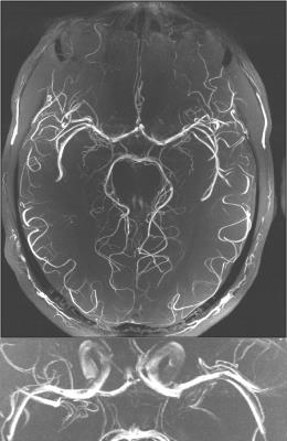
Beyond the biological resolution limit: Prospectively motion corrected Time of Flight angiography at 7T
Hendrik Mattern, Alessandro Sciarra, Frank Godenschweger, Daniel Stucht, Falk Lüsebrink, Oliver Speck
Subject motion limits the potential of high resolution Time of Flight (ToF) angiography at 7T, even small scale, involuntary movements can degrade the image quality. In this study, prospective motion correction was able to overcome the biological resolution limit in a healthy subject population (quality assessment with quantitative and qualitative metrics), and was used to acquire the highest in vivo ToF data set to date with an isotropic voxel size of 0.15mm³.
|
|
0784.
 |
29 |
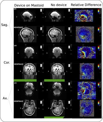
Implementation of a 2.4 GHz wireless sensing platform for transmission of motion data from within a head coil at 3T.
Adam van Niekerk, Andre van der Kouwe, Ernesta Meintjes
Prospective motion correction using external hardware can be compromised by poor marker attachment. In this work we introduce a new attachment site on the mastoid process of the subject. To achieve this, an active wireless marker is implemented that takes advantage of the versatility of an existing method (VectOrient). The effects of the device on the scanner’s operation and visa versa are evaluated. The small 14x16mm2 device shows good MRI compatibility. Any degradation in signal quality is localised and could be further reduced. The link quality is sufficient to stream patient motion parameters, quantifying patient pulse during MEMPRAGE and EPI pulse sequences.
|
|
0785.
 |
30 |
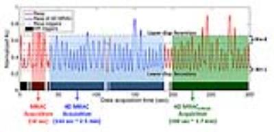
Respiratory Phase-Matched MR-based Attenuation Correction (MRAC) for Four-Dimensional (4D) PET in PET/MRI: A Feasibility Study - permission withheld
Jaewon Yang, Florian Wiesinger, Anne Menini, Jing Liu, Thomas Hope, Youngho Seo, Peder Larson
PET/MRI is capable of simultaneous respiratory motion-resolved four-dimensional (4D) PET/4D MRI data acquisitions. Therefore, it is important to develop a clinically applicable method for respiratory phase-matched MR-based attenuation correction (MRAC) for accurate 4D PET quantification. This study proposed 4D MRAC protocols modifying the current MRAC protocol and evaluated their performance using a patient with liver metastases. For qualitative analysis, 4D MRAC improved phase-mismatch at the lung/liver interface substantially. For quantitative analysis, 4D MRAC improved PET quantification by 10-30% increase of PET-avid tumor uptake values, compared to static MRAC, specifically for the tumors located at the lung/liver interface.
|
|