Poster: Highlights of Multiparametric Acquisition & Reconstruction
Electronic Power Pitch Poster
Acquisition, Reconstruction & Analysis
Monday, 24 April 2017
| Exhibition Hall |
14:45 - 15:45 |
| |
|
Plasma # |
 |
0127.
 |
16 |
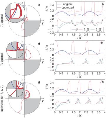
Relaxation in Polar Coordinates: Analysis and Optimization of MR-Fingerprinting 
Jakob Assländer, Daniel Sodickson, Riccardo Lattanzi, Martijn Cloos
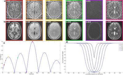
This work analyses relaxation in balanced non-steady-state free precession sequences. Transforming the Bloch equation to polar coordinates gives insights in the spin dynamics and provides the basis for robust numerical optimization of the excitation pattern. The employed optimal control algorithm results in spin trajectories that allow for parameter mapping with considerably reduced noise, as shown in in vivo MR-fingerprinting experiments. The simple shapes of the optimized spin trajectories provide a basis for further analysis of the encoding process of relaxation times for parameter mapping.
|
|
0128.
 |
17 |
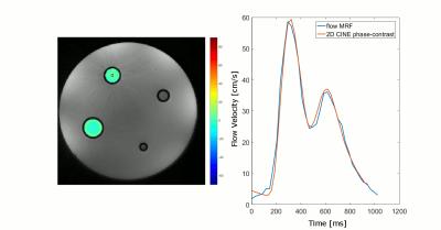
Quantification of Flow by Magnetic Resonance Fingerprinting
Sebastian Flassbeck, Simon Schmidt, Mathies Breithaupt, Peter Bachert, Mark Ladd, Sebastian Schmitter
The goal of this work is to investigate and demonstrate 'flow MRF', a technique that quantifies T1 and T2 within static tissue regions around a vessel (e.g. vessel wall) with high spatial resolution while simultaneously quantifying the 3D blood velocity vector within the vessel with high precision and high spatiotemporal resolution. The results show that simultaneous mapping of pulsatile flow and T1/T2 quantification of static tissue is feasible with MRF. The quantification of flow as an additional parameter will further increase the efficiency of MRF and may provide new diagnostic value in cardiovascular diseases.
|
 |
0129.
 |
18 |
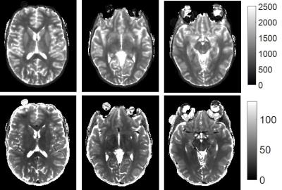
Applications of Low Rank Modeling to Fast 3D Magnetic Resonance Fingerprinting (MRF)
Dan Ma, Eric Pierre, Debra McGivney, Bhairav Mehta, Yong Chen, Yun Jiang, Mark Griswold
The goal of this study is to accelerate the acquisition time of 3D magnetic resonance fingerprinting (MRF) using a low-rank model-based method kt-SVD-MRF. With a total factor of 144 acceleration rate, 3D T1, T2 and proton density (M0) maps can be acquired from a whole brain scan with a resolution of 1.17x1.17x3 mm3 in 2.7 minutes.
|
|
0130.
 |
19 |
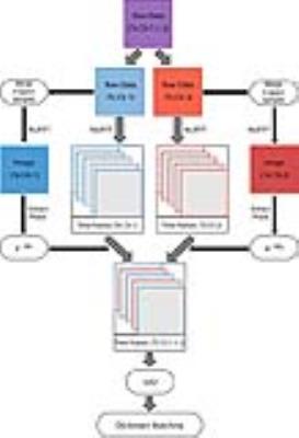
Magnetic Resonance Fingerprint Compression with Multiple Channel Transmission
Riccardo Lattanzi, Bei Zhang, Florian Knoll, Jakob Assländer, Martijn Cloos
Singular value decomposition (SVD) and view-sharing compression can decrease the size of the dictionary without compromising accuracy in magnetic resonance fingerprinting (MRF). While the latter accounts for the B1+ of multiple transmit channels in the dictionary, the SVD compression scheme was designed for single-channel transmission. In this work we extended SVD-based fingerprint compression to the case of two or more independent RF sources and evaluated its performance in simulation. We showed that accurate parametric maps can be achieved using only six SVD components, both in fully-sampled and highly under-sampled MRF experiments. Future work will include optimization of k-space under-sampling.
|
|
0131.
 |
20 |
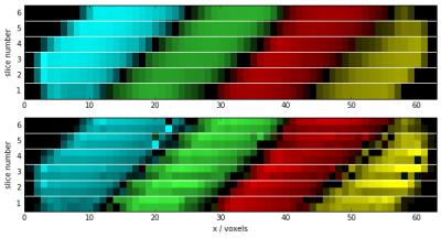
Intra-Voxel Spatial Resolution Using Magnetic Resonance Fingerprinting - permission withheld
Thomas Amthor, Karsten Sommer, Peter Koken, Jakob Meineke, Mariya Doneva
We demonstrate the use of Magnetic Resonance Fingerprinting to retrospectively increase spatial resolution in slice-encoding direction, making use of the non-uniform nature of the excitation slice profile. Assigning individual fingerprints to substances present at different spatial positions within the excited voxel, a multi-compartment analysis recovers the spatial distribution of the components.
|
|
0132.
 |
21 |
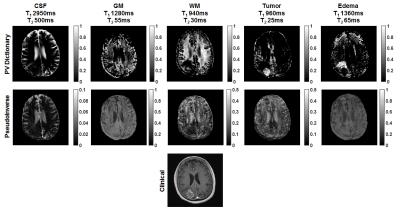
Dictionary approach to partial volume estimation with MR Fingerprinting: Validation and application to brain tumor segmentation
Anagha Deshmane, Debra McGivney, Chaitra Badve, Vikas Gulani, Mark Griswold
MR Fingerprinting signal evolutions can be used to estimate partial volumes of tissues in addition to tissue relaxation times. Two approaches to tissue fraction estimation, a pseudoinverse calculation and dictionary-based or constrained solution, are quantitatively compared in the presence of four potential error sources. The constrained approach is found to more accurately estimate tissue fractions, and also to improve segmentation of pathology.
|
|
0133.
 |
22 |
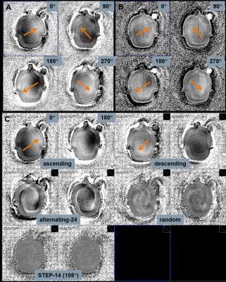
Mitigation of Spiral Undersampling Artifacts in Magnetic Resonance Fingerprinting (MRF) by Adapted Interleave Reordering
Josef Pfeuffer, Argyrios Kechagias, Craig Meyer, Gregor Körzdörfer, Mathias Nittka
In MRF fast spiral scanning is done with variable FA/TR using a highly – up to 48-fold – undersampled trajectory. Spatial undersampling artifacts are mitigated by the MRF dictionary-matching process for the resulting quantitative T1/T2 maps. In this study, we demonstrate how undersampling artifacts can bias MRF results and present strategies to minimize them by spiral-interleave reordering adapted to the specific MRF encoding scheme. An experimental procedure is developed to test for a potential spatial bias in a specific spiral and MRF encoding scheme. The sources of spatial variances are mitigated with appropriate distribution of undersampling artifacts over the temporal MRF encoding.
|
|
0134.
 |
23 |
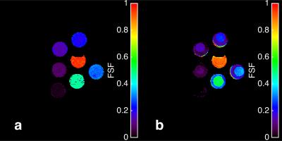
Fat Signal Fraction Determination Using MR Fingerprinting
Jason Ostenson, E. Brian Welch
Magnetic resonance fingerprinting employing multiple echo times is used to quantify fat signal fraction both in phantoms and in vivo on a human 3 Tesla scanner. Reasonable agreement is seen in fat signal fraction maps and intra-class correlation between standard and MRF methods.
|
|
0135.
 |
24 |
Accelerated Magnetic Resonance Fingerprinting using Soft-weighted key-Hole (MRF-SOHO)
Gastao Cruz, Andreia Gaspar, Tom Bruijnen, René Botnar, Claudia Prieto
Magnetic Resonance Fingerprinting estimates multi-parametric maps from a series of highly undersampled time-point images. However, MRF scan times are still long due to the large amount of time-point images (~1000) required to produce accurate multi-parametric maps. Here we propose to exploit redundant information in time-point images with similar contrast to accelerate the MRF scan by further undersampling each time-point image and/or significantly reducing the number of required images in the series. The proposed approach achieved an acceleration factor of 5.7× compared to conventional undersampled MRF while maintaining parametric map quality.
|
|
0136.
 |
25 |
Magnetic Resonance Fingerprinting - Evaluation of Brain Gliomas in Comparison to a Conventional Advanced Tumor Protocol - Preliminary Study
Siegfried Trattnig, Wolfgang Bogner, Bernhard Strasser, Peter Bär, Simone Kitzer, Pavol Szomolanyi, Matthias Nittka, Wolfgang Marik, Martin Zalaudek, Markus Schreiner, Elisabeth Springer
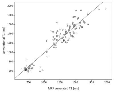
Synopsis: MR Fingerprinting (MRF) was compared to an advanced brain tumor protocol in 10 patients with surgically proven gliomas. The T1 and T2 relaxation times provided by MRF in one scan showed a high correlation with conventionally measured T1 and T2 values. MRF obtained T1 and T2 values allowed a statistically significant differentiation between low and high grade gliomas. MRF with quantitative data and the possibility to generate synthetic MR contrast images may replace conventional MR sequences in the future.
|
|
0137.
 |
26 |
Joint estimation of arterial input function and tracer kinetic parameters from under-sampled DCE-MRI
Yi Guo, Sajan Goud Lingala, R Lebel, Krishna Nayak
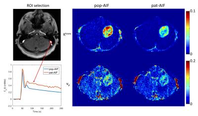
Direct reconstruction of tracer kinetic (TK) parameter maps from under-sampled DCE-MRI has recently been demonstrated. However, this method assumes the arterial input function (AIF) is known or pre-determined. Any mismatches between the assumed AIF and the underlying patient-specific AIF can cause large inaccuracies in the final TK parameters. We propose a novel approach to extract patient-specific AIFs from under-sampled data, while jointly estimating the TK parameter maps. Reconstruction is performed by cycling through the problems of AIF extraction, TK parameter estimation and, data consistency. We demonstrate this approach on brain tumor DCE data sets, where high fidelity AIFs are extracted up to an under-sampling rate of 100x.
|
|
0138.
 |
27 |
Highly accelerated DCE imaging with integrated T1 mapping
R Marc Lebel, Yi Guo, Sajan Lingala, RIchard Frayne, Krishna Nayak
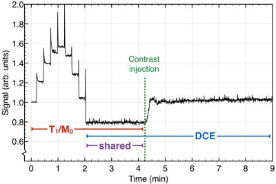
Dynamic contrast enhanced (DCE) MRI requires accurate and precise baseline T1 and M0 maps for pharmacokinetic modeling. Advances in dynamic acquisitions and reconstructions have enabled high resolution DCE imaging with full anatomical coverage. Rapidly obtaining high SNR T1/M0 maps with the same spatial resolution has become a limiting factor for advanced DCE methods. We present a single highly accelerated acquisition that first performs the T1/M0 mapping then dynamic imaging. A model-based reconstruction is used to estimate the T1/M0 maps. This method makes effective use of scan time and generates maps that are superior to separately acquired ones.
|
|
0139.
 |
28 |
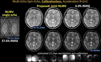
Calibrationless Parallel Imaging in Multi Echo/Contrast Data
Berkin Bilgic, Bo Zhao, Itthi Chatnuntawech, Lawrence Wald, Kawin Setsompop
Parallel imaging relies on fully-sampled calibration data to estimate k-space kernels or sensitivities used to reconstruct subsampled acquisitions. Emerging techniques use low-rank modeling, or joint estimation of sensitivities and image content via nonlinear optimization, to reduce the dependency on calibration data. In a typical study, images at multiple echoes/contrasts are acquired using the same coil sensitivities. Here, we exploit this joint information to dramatically improve conditioning of calibrationless nonlinear inversion and employ joint sparsity to improve reconstruction. To achieve better performance, we also propose complementary k-space undersampling between images to form a composite image with reduced aliasing to initialize the optimization.
|
|
0140.
 |
29 |
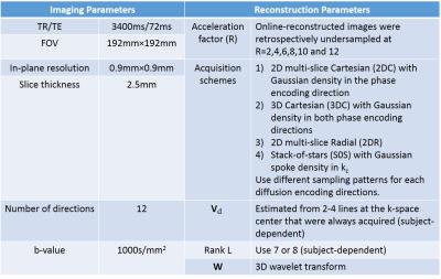
Accelerated Cardiac Diffusion Tensor Imaging Using a Joint Low-Rank and Sparsity Constraint
Sen Ma, Christopher Nguyen, Anthony Christodoulou, Daniel Luthringer, Jon Kobashigawa, Debiao Li
We present an image reconstruction technique to accelerate cardiac diffusion tensor imaging by jointly applying a low-rank and spatial sparsity constraint. We evaluated four acquisition schemes at different undersampling levels on 9 ex vivo diseased human heart, evaluating the reconstruction quality based on the resulting helix angle (HA) maps and helix angle transmurality (HAT) values. A Wilcoxon signed rank test was performed to statistically evaluate changes in HAT to determine the highest achievable acceleration factor for each acquisition scheme. Our framework shows promise in greatly reducing scan time while preserving the fiber architecture features of heart failure.
|
|
0141.
 |
30 |
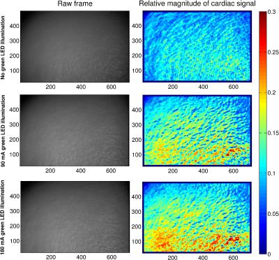
An open-source hardware and software system for video-gated MRI
Nicolai Spicher, Stephan Orzada, Stefan Maderwald, Markus Kukuk, Mark Ladd
The limitations of contact-based hardware (e.g. electrocardiography, pulse oximetry) for cardiac activity measurement pose an obstacle in many ultra-high-field MRI examinations. In this work, we present a freely available hardware and software system for acquisition and processing of video signals from human skin that we developed with the aim to make this contact-free method available to other researchers.
|
|