Poster: Diffusion: Outside the Brain
Electronic Power Pitch Poster
Diffusion
Wednesday, 26 April 2017
| Exhibition Hall |
17:15 - 18:15 |
| |
|
Plasma # |
|
0862.
 |
1 |
Analysis of abdominal movement with Phase Optical Flow: Application to Diffusion imaging. 
Stephan Hahn, Maxime Gérard, Damien Dasnoy-Sumell, Julie Absil, Olivier Debeir, Thierry Metens
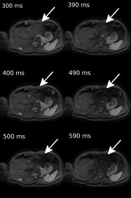
We present a new method, the Phase Optical Flow, which provides an automatic determination of abdominal regional movements along the cardiac cycle and allows a real time determination of the optimal cardiac trigger time to be used in quantitative liver DWI. A phase based motion amplification was applied to real-time BTFE images acquired at 20 images/s. Then optical flow was used to derive the velocity vector field. The optimal cardiac time window was defined as the 100ms-period with minimal absolute vertical velocity. Validation was provided by liver DWI obtained at several cardiac trigger times.
|
|
0863.
 |
2 |
In vivo imaging of mean cell size and density of human breast tumors - permission withheld
Hua Li, Lori Arlinghaus, A. Chakravarthy, Vandana Abramson, John Gore, Junzhong Xu
We report a new MRI method termed IMPULSED (Imaging Microstructural Parameters Using Limited Spectrally Edited Diffusion) to quantitatively characterize mean cell size and density in solid tumors simultaneously and the first application of this method in breast cancer patients.
|
|
0864.
 |
3 |
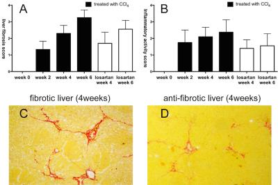
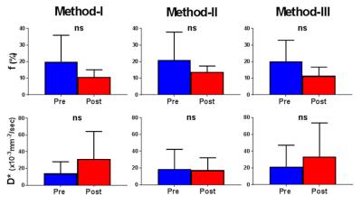
Intravoxel incoherent motion MR imaging for staging liver fibrosis and monitoring anti-fibrotic response to losartan: an experimental study in rat model - permission withheld
Caiyuan Zhang, Yong Zhang, Dengbin Wang
To evaluate the feasibility of intravoxel incoherent motion (IVIM) MR imaging to stage liver fibrosis and its capability for monitoring anti-fibrotic response to treatment, we performed IVIM MR imaging for rats model induced by carbon tetrachloride and for rats treated with losartan. Our studies demonstrated that D* is helpful to stage early and moderate fibrosis, and f is beneficial to discriminate advanced liver fibrosis and cirrhosis. In losartan treated rats, D* and f showed statistical significance compared with CCl4 alone. We concluded that perfusion parameters derived from IVIM have potential to monitor fibrosis progression and evaluate anti-fibrosis response to treatment.
|
|
0865.
 |
4 |
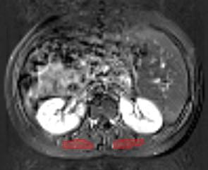
Diffusion-weighted MRI and coherent flow in the kidney
Andreas Weng, Fabian Hilbert, Henning Neubauer, Simon Veldhoen, Thorsten Bley, Herbert Köstler
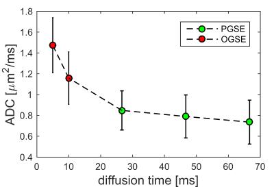
The IVIM model and typical DWI-evaluations assume flow in tissue to be incoherent. However, there is evidence that in some tissues flow might be coherent and thus, the DWI-signal might be influenced by the applied first gradient moment m1. A gradient scheme was implemented that allows applying different m1 for a constant b-value. Moreover, the IVIM model was extended to also include coherent flow in the modeling process of DWI data. It was found that flow in the human kidney is, at least in part, coherent and that the proposed model is able to fit that data very robustly.
|
|
0866.
 |
5 |
An assessment of Intravoxel Incoherent Motion (IVIM) imaging in Detection of Acute Kidney Injury in Rodents
Keisuke Ishimatsu, Shanrong Zhang, Koji Sagiyama, Ming Chang Hu, Orson Moe, Masaya Takahashi
The objective is to investigate the concept of intravoxel incoherent motion (IVIM) for diffusion weight imaging (DWI) in the kidney. We first compared the variability of three methods for bi-exponential fitting applied to the five data sets of DWI obtained in a mouse kidney. Subsequently these three methods were compared with an arterial spin labeling (ASL) in detection of acute kidney injury (AKI) in a rat model. The IVIM imaging did not detect any changes in the AKI model although the ASL clearly demonstrated the reduction of the perfusion.
|
|
0867.
 |
6 |
Diffusion-Weighted MRI Identifies Viable Tissue in Wilms Tumour: Application for Subtype Analysis and Response to Chemotherapy
Harriet Rogers, Patrick Hales, Kathy Pritchard-Jones, Christopher Clark
In Wilms Tumour (WT) Blastemal subtype has the worst prognosis. Diffusion MRI (DWI) can distinguish some histological subtypes. Gadolinium-contrast-injected T1 MRI (T1gad) identifies necrotic tissue. Gadolinium is contra-indicated in renal failure. 27 patients received DWI, 20/27 received T1gad. DWI was fitted with Intravoxel Incoherent Motion providing Dmaps. Viable and necrotic regions identified on T1gad were transferred to corresponding Dmaps. ROC analysis determined a D threshold separating necrotic and viable tissue. ANOVAs showed viable regions separated Blastemal from other subtypes, whole lesions could not. DWI separates necrotic and viable tissue in WT potentially identifying subtypes, assessing chemotherapy, guiding biopsies and surgery.
|
|
0868.
 |
7 |
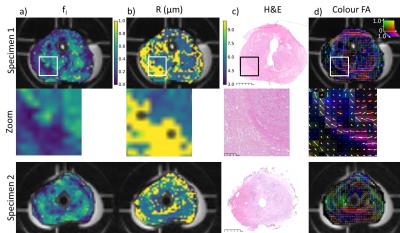
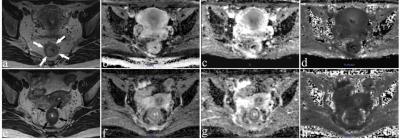
Validation of VERDICT MRI using fresh and fixed prostate specimens with aligned histological slices
Colleen Bailey, Roger Bourne, Bernard Siow, Edward Johnston, Hayley Pye, Susan Heavey, Thomy Mertzanidou, Hayley Whitaker, Alexander Freeman, Dominic Patel, Greg Shaw, Ashwin Sridhar, Shonit Punwani, David Hawkes, Daniel Alexander, Eleftheria Panagiotaki
This study provides the first step in validating the VERDICT diffusion model of tissue microstructure by examining the effects of fixation on tissue microstructure and comparing VERDICT parameter maps to histological features. Fresh and fixed parameter maps showed similar spatial trends: fixation decreased the extracellular volume fraction parameter and decreased the cell radius parameter slightly, consistent with water efflux. Intracellular volume fraction was lower in regions with lower cellularity, such as the peripheral zone, and directions of diffusion anisotropy corresponded with collagen and smooth muscle orientation patterns in the stroma.
|
|
0869.
 |
8 |
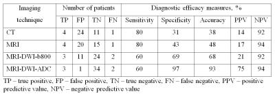
Diffusion-weighted MRI in the evaluation of posttherapeutic residual masses in lymphoma
Siarhei Kharuzhyk, Edward Zhavrid, Nina Sachivko
Residual masses do often present on posttreatment imaging in lymphoma. We conducted prospective study to evaluate diagnostic capabilities of CT, MRI and diffusion-weighted MRI (MRI-DWI) in differentiation residual tumors and non-active posttreatment masses in 40 lymphoma patients. MRI-DWI with visual analysis of apparent diffusion coefficient (ADC) maps was the most effective technique. Quantitative ADC analysis is a promising tool for differentiation of active and non-active residual masses in lymphoma.
|
|
0870.
 |
9 |
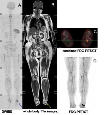
The clinical evaluation of combining DWIBS with whole body T1w imaging for diagnosing bone marrow involvement in lymphoma patients: a comparison with PET/CT - permission withheld
Mengtian Sun, Jingliang Cheng, Yun Meng, Zhizheng Zhuo
This study aimed to evaluate the value of the combination of DWIBS (diffusion weighted imaging with background signal suppression) with whole body T1w imaging for diagnosing bone marrow involvement (BMI) in lymphoma patients, compared to PET/CT imaging. In the first part of this study, patients with newly diagnosed lymphoma were includedand whole body DWIBS, T1w and PET/CT images were acquired for all 20 subjects. For the assessment of individual lesions, DWIBS with whole body T1w has advantages over DWIBS and has similar ability with PET/CT to detect BMI lesions.
|
|
0871.
 |
10 |
Intravoxel incoherent motion diffusion-weighted imaging for discriminating the pathological response to neoadjuvant chemoradiotherapy in locally advanced rectal cancer
Wen Lu, Yu xiaoping, Zhang zhongping
In this study, we investigated the utility of IVIM-DWI in discriminating the pathological response to nCRT in locally advanced rectal cancer (LARC). We found that both the pre-nCRT and post-nCRT IVIM-DWI parametric values for LARC, together with their percentage changes, might benefit the evaluation of pathological response to nCRT, which suggest IVIM-DWI is potentially useful in discriminating pathological response for LARC patients.
|
|
0872.
 |
11 |
The value of diffusion kurtosis imaging in assessing pathological complete response to neoadjuvant chemoradiation therapy in rectal cancer: a comparison with conventional diffusion-weighted imaging - did not present
Fei-Xiang Hu, Tong Tong, Wei Tang, Yi-Qun Sun, Dang Wang, San-Jun Cai, Zhen Zhang, Grimm Robert, Xu Yan, Cai-xia Fu, Wei-Jun Peng
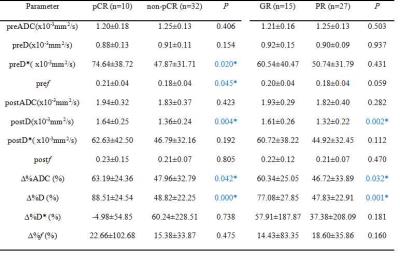
The aim of this study was to evaluate the performance of diffusion kurtosis imaging (DKI) in predicting pathological complete response (pCR) of locally advanced rectal cancer (LARC) to neoadjuvant chemoradiation therapy (CRT) before and at an early stage of the treatment, and in comparison to conventional diffusion-weighted imaging (DWI). Results showed that DKI outperformed conventional DWI in accurately differentiating between pCR and nonPCR patients who received neoadjuvant chemoradiation therapy both before and at an early stage of treatment.
|
|
0873.
 |
12 |
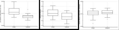
The application of whole lesion IVIM analysis using iZOOM DWI in the diagnosis of thyroid tumor
Yunlong Yue, Lili Zuo, Kaining Shi, Lee Jiang, Jinsong Guo, Yanfang Jin
To explore the ability of IVIM parameters derived from iZoom DWI in differentiating malignant thyroid nodules from benign ones. 40 patients with 45 pathologically proven thyroid nodules were involved. iZOOM DWI with 2D RF was employed to decrease the distortion and carotid coil was used to increase the SNR. 3D ROI was drawn manually to cover the whole lesion. D and f values were significantly lower in malignant nodules than in benign nodules. According to ROC curve analysis IVIM almost reached the upper limit of the accuracy based on US.
|
|
0874.
 |
13 |
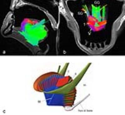
Diffusion Spectrum Imaging Tractography of the Human Tongue
Nahla Elsaid, Maureen Stone, Steven Roys, Rao Gullapalli, Jerry Prince, Jiachen Zhuo
The human tongue is known to have a complex architecture of muscles. To fully understand how the muscle fibers are connected and how their relative position could affect the tongue functionality, diffusion weighted imaging is needed. Diffusion Spectrum Imaging (DSI) is able to characterize the fiber structure on a sub-voxel level including the fiber crossing or branching. As DSI sequences are usually time-consuming, in-vivo studies using DSI can be challenging. In this study, we present for the first time DSI of the post-mortem human tongue. and the associated tractography delineating the tongue muscle fibers. DSI has the ability to identify fiber crossings within the human tongue.
|
|
0875.
 |
14 |
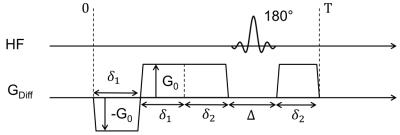
Acquisition at maximum blood velocity overcomes the problem of the ill-posedness of the IVIM model: a demonstration with renal diffusion MRI
Bastien Milani, Jean-Baptiste Ledoux, Menno Pruijm
We explain why diffusion MRI should always be acquired when blood has maximum velocity in the organ of interest. We first give some theoretical arguments to support this hypothesis. We then demonstrate it with numerical experiments and with in-vivo experiments.
|
 |
0876.
 |
15 |
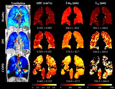
Comparison of in-vivo Lung Morphometry Models from 3D Multiple b-value 3He Diffusion-Weighted MRI
Ho-Fung Chan, Juan Parra-Robles, Guilhem Collier, Jim Wild
The cylinder (CM) and stretched exponential (SEM) models have been proposed as a means of estimating lung alveolar microstructural length scales (Lm for CM and LmD for SEM) from multiple b-value hyperpolarised gas DW-MRI. This work compares Lm and LmD in healthy normals, idiopathic pulmonary fibrosis, and COPD patients. A correlation with a non-linear trend was observed between the Lm and LmD parameters. This suggests that the two models have different operational ranges of length scale estimation accuracy due to inherent differences in their geometrical and mathematical assumptions.
|
|