Poster: Interventional/Safety/Engineering
Electronic Power Pitch Poster
Interventional MRI
Monday, 24 April 2017
| Exhibition Hall |
09:15 - 10:15 |
| |
|
Plasma # |
 |
0001.
 |
1 |
Overcoming Limitations of Virtual Observation Points in pTx using IMPULSE 
Mihir Pendse, Brian Rutt
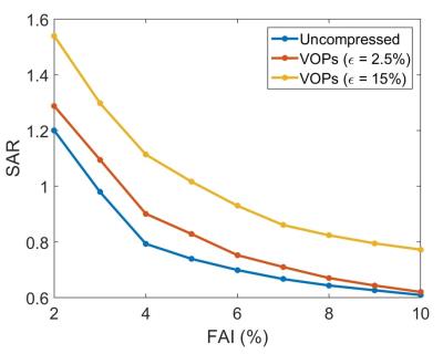
We analyze the performance of the IMPULSE pTx optimization algorithm, which allows SAR-aware pulse design without virtual observation points (VOP) compression. We compare performance of IMPULSE with conventional optimization methods using VOPs and compare different values of the overestimation parameter. We show that IMPULSE results in elimination of the time-intensive compression step without significantly increasing the time for real-time optimization. Additionally by eliminating the overestimation error from the VOP compression, IMPULSE is able to achieve better mitigation of local SAR hotspots after optimization than VOP-based methods.
|
|
0002.
 |
2 |
Optical-based probe for real time assessment of RF electrical field during MRI exam
Isabelle Saniour, Gwenaël Gaborit, Lionel Duvillaret, Anne-Laure Perrier, Olivier Beuf
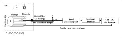
During MRI exam, Specific Absorption Rate (SAR) is essential to be controlled and can be evaluated by measuring either indirectly for instance the rise in temperature or directly the radiofrequency induced electrical E-field. In the current study, we proposed an optical probe based on the Pockels effect for subcentimeter resolution measurements of the E-field without altering the surrounding media. Measurements were performed at 4.7 T and 3.0 T. Results show that the probe has an excellent linear response and allow a real time estimate of the three components of the E-field produced during MRI examination.
|
|
0003.
 |
3 |
Modeling of Peripheral Nervous Stimulation Thresholds in Realistic Body Models
Mathias Davids, Bastien Guérin, Lothar Schad, Lawrence Wald
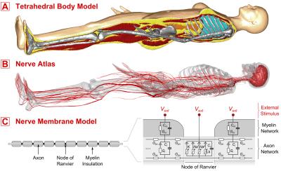
Peripheral Nervous Stimulation (PNS) has become an important limitation in MRI with the latest generation of high-performance gradient systems. We present – to our knowledge for the first time – a model to predict PNS thresholds for arbitrary coil geometries. Our model consists of an accurate body model for EM simulations, a detailed nerve atlas of the human body, and a numerical model to predict nerve responses to induced electrical fields. With this model, we were able to reproduce PNS threshold curves of two leg/arm solenoid coils that were previously evaluated experimentally. We intent to use this PNS model to design high-performance gradient coils with significantly lowered PNS capabilities.
|
|
0004.
 |
4 |
Cardiac Synchronization at Ultra-High Field Using a 3-Lead ECG Trigger Device
Daniel Stäb, Juergen Roessler, Kieran O'Brien, Je Su, Christian Hamilton-Craig, Markus Barth
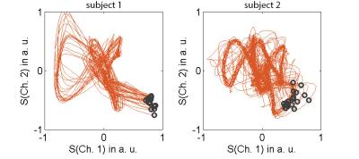
Accurate cardiac synchronization is essential in CMR. At ultra-high field, ECG triggering can be significantly impacted by the magneto-hydrodynamic (MHD) effect. Here, we investigate the performance of a conventional 3-lead ECG trigger device and a state-of-the-art trigger algorithm for cardiac ECG synchronization at 7 T. We show that by appropriate subject preparation and by including a learning phase for the R-wave detection outside of the magnetic field, reliable ECG triggering at ultra-high field is feasible despite severe distortions of the ECG signal. A quantitative analysis in 10 healthy subjects revealed a trigger sensitivity and specificity of 97.6% and 98.4%, respectively.
|
|
0005.
 |
5 |
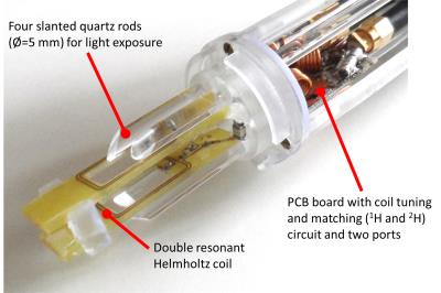
A Combined 7 Tesla MRI/NMR Probe Head for Photochemical Applications.
Jens Groebner, Gernot Heitmann, Marcel Dommaschk, Lukas Huber, Eduard Stadler, Reiner Umathum, Frank Sönnichsen, Rainer Herges
The development of new photoswitchable contrast agents requires analysis with both NMR and MRI. In this work a combined probe head for in situ light exposure is presented which can be used on NMR and MRI systems with both 7 Tesla. A probe head with four slanted quartz rods was fabricated. A dual tuned (1H and 2H) Helmholtz coil was integrated into the probe head. NMR and MRI experiments were performed on photoswitchable solutions (e.g. contrast agents). Results show that photoswitchable solutions can be successfully switched in situ and simultaneously analyzed with NMR or imaged with MRI.
|
 |
0006.
 |
6 |
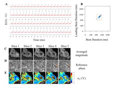
Evaluation of cardiac magnetic resonance thermometry in patients
Valery Ozenne, Solenn Toupin, Pierre Bour, Baudouin Denis de Senneville, Alexis Vaussy, Matthieu Lepetit-Coiffé, Pierre Jaïs, Hubert Cochet, Bruno Quesson
Recent studies have proposed to monitor radiofrequency ablation on the heart using real-time MR-thermometry. Methods rely on ECG triggering which can fail in presence of arrhythmia. This study evaluates the precision of MR-thermometry on patients (N=15) even in presence of cardiac arrhythmia. Phase images were acquired using a single-shot multi-slice echo planar imaging and temperature maps were calculated and displayed on the fly. ECG was recorded simultaneously for further analysis of cardiac rhythm and post-processing of temperature images. Stability of temperature mapping without RF-heating was evaluated in each pixel and correlated to the prevalence of arrhythmia.
|
|
0007.
 |
7 |
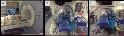
MRI-monitored Anterior Cervical Discectomy and Fusion (ACDF) surgery: Observation of intra-procedural nerve decompression during surgery
Ehud Schmidt, Daniel Kacher, Wei Wang, Mitchel Harris, Thomas Lee, Ravi Seethamraju, Clare Tempany, Jay Zampini
ACDF is a surgical procedure performed when herniated disks produce severe pain, or arm/hand weakness. Severe complications occur in ~9% of cases, mainly due to tissue resection adjoining the spinal canal. MRI imaging, performed at several procedure stages, can visualize the extent of resection, and the degree of nerve decompression. We performed ACDF surgery on the MRI table, using an MRI-compatible tool-set. Imaging at 3T was performed at four procedure phases. Imaging disclosed significant spine decompression immediately after disk resection, with smaller changes after osteophyte and posterior longitudinal ligament removal. MRI-monitored ACDF can revise procedure phases, leading to improved outcomes.
|
|
0008.
 |
8 |
Water diffusivity changes in the brain following exposure to low levels of focused ultrasound energy
Sijia Guo, Jiachen Zhuo, Xin Lu, Su Xu, Rao Gullapalli
MR-guided Focused ultrasound (MRgFUS) for neuro-interventions is gaining popularity. However, the biological effects of low level exposure on the brain tissue are less understood. In this study, we used in-vivo MR diffusion kurtosis imaging (DKI) to measure water diffusion changes in vivo in rats at varying FUS exposure levels in the whole brain. Acoustic simulation and experimental data demonstrate more wide spread tissue diffusion changes and not just local changes with just a single exposure albeit for varied duration. Our results suggest that diffusion changes are mainly due to shearing forces exerted on the brain.
|
|
0009.
 |
9 |
Low Rank plus Sparse Compressed Sensing Reconstruction for PRF Temperature Imaging
Zhipeng Cao, Sumeeth Jonathan, William Grissom
A novel compressed sensing reconstruction method based on L+S separation on complex difference image domain data is demonstrated to perform better than existing methods on major applications in RF heating monitoring and MR guided focused ultrasound intervention.
|
|
0010.
 |
10 |
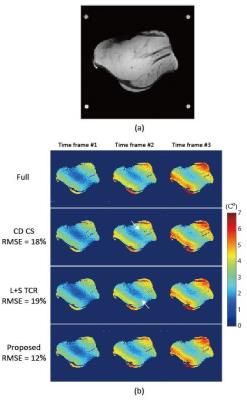
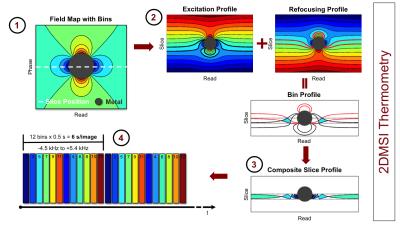
2D Multi-Spectral Thermometry for Monitoring Focused-Ultrasound Sonications Near Metallic Hardware
Hans Weber, Pejman Ghanouni, Aurea Pascal-Tenorio, Kim Butts Pauly, Brian Hargreaves
The lack of a technique for MR thermometry near metallic hardware excludes a growing patient population from MR-guided focused ultrasound treatments. In this work, we explore the temperature-induced signal change in fast two-dimensional multi-spectral imaging for monitoring sonications near metallic hardware. We demonstrate initial feasibility in phantom and ex vivo experiments as well as a patient treatment.
|
|
0011.
 |
11 |
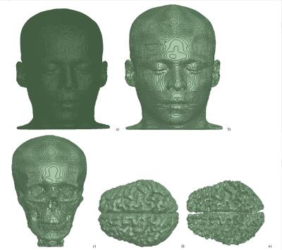
Toward individualized specific absorption rates: Building a surface-based human head model
Mikhail Kozlov, Benjamin Kalloch, Pierre-Louis Bazin, Mario Hlawitschka, Nikolaus Weiskopf, Harald Möller
We built a prototype of a high resolution surface-based human head model that can be simulated in a reasonable time and evaluated the influence of cerebrospinal fluid (CSF) on field propagation estimates of traveling wave excitation at 297.2 and 400 MHz. Combining neighboring triangular faces located in the same plane into a single one is an approach that achieves simulations of high-resolution human models previously not accessible to tetrahedral-mesh-based solvers. If electrical contact between anatomically connected parts of CSF is correctly considered, CSF was found to partially shield brain tissues from the incident RF field.
|
|
0012.
 |
12 |
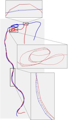
Patient specific modeling of deep brain stimulation patients for MRI safety studies
Bastien Guerin, Peter Serano, Maria Iacono, Todd Herrington, Alik Widge, Darin Dougherty, Giorgio Bonmassar, Leonardo Angelone, Lawrence Wald
We propose a semi-automatic processing pipeline for the generation of realistic radiofrequency models of deep brain stimulation (DBS) patients. The whole process takes ~72 hours for model generation and field computation and models the exact DBS path, without intersections, the internal structure of the implant and the patient’s anatomical structures (e.g., brain, bones, muscles, lungs). We show that simplification of the DBS implant model results in high (up to 75%) differences in the estimation of energy absorption. The proposed framework allows for fast and precise modeling, which may be needed, pending experimental validation, to evaluate MRI RF-induced heating.
|
|
0013.
 |
13 |
Interventional Magnetic Resonance Imaging Guided Carotid Embolectomy using a Novel MRI-Conditional Resonant Catheter: Demonstration of Preclinical Feasibility
Jeffrey Yang, Andre Cote, Caroline Jordan, Aaron Losey, David McCoy, Andrew Chu, Jay Yu, Teri Moore, Carol Stillson, Fabio Settecase, Matthew Alexander, Andrew Nicholson, Mariam Aboian, Daniel Cooke, Maythem Saeed, Dave Barry, Alastair Martin, Mark Wilson, Steven Hetts
MR-guided endovascular interventions can provide real-time imaging biomarkers for procedures such as stroke embolectomy. The purpose of this study is to determine preclinical feasibility and efficacy of imaging wireless resonant circuits embedded in a MR compatible catheter system for in vivo MR-guided carotid embolectomy in porcine stroke models. The resonant catheter system performed effectively under real-time MRI with recanalization rates (TICI 2b/3) similar to reported clinical rates in stroke embolectomy. Qualitative physiologic measures of flow under MRI were comparable to those measured under X-ray, demonstrating feasibility of resonant catheter system for in vivocarotid occlusion and embolectomy under real-time MRI.
|
|
0014.
 |
14 |
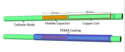
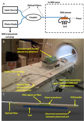
Acousto-optic Based Active MRI Marker for Interventional MRI Devices
Yusuf Yaras, Sarp Satir, Cagla Ozsoy, Rajiv Ramasawmy, Adrienne Campbell-Washburn, Anthony Faranesh, Robert Lederman, Ozgur Kocaturk, Levent Degertekin
Conspicuous and safe MR markers are essential for tracking interventional MRI devices. The RF induced heating on long conductors used in current active MR markers presents a safety risk. In this work, a novel acousto-optic active MR marker with optical fiber connection is proposed to eliminate RF induced heating. The proposed marker consists of a miniature coil coupled to a piezoelectric transducer which in turn modulates the reflected light in the optical fiber. The linearity of the acousto-optic active marker with flip angle is characterized and initial in vitro imaging experiments are performed demonstrating marker visibility under MRI.
|
|
0015.
 |
15 |
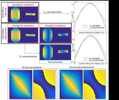
MRI based RF safety characterization of implants using the implant response matrix: a simulation study.
Janot Tokaya, Alexander Raaijmakers, Peter Luijten, Cornelis van den Berg
We introduce a general description of the RF response of an implant, defined as the implant response matrix (IRM). An analytical expression for the IRM is derived through basis functions that depend on a limited number of parameters. This analytical model is validated with a simulation study (Pearson correlation coefficients with simulations R: 0.9979-0.9996). This description allows a significant reduction in unknowns enabling IRM assessment by MRI measurements without hardware modifications to scanner or implant. The feasibility of MRI based IRM/TF measurement is shown in silico. With the simulated complex B1+ fields the IRM is accurately reconstructed (R: 0.974).
|
|