ISMRM 25th Annual Meeting & Exhibition • 22-27 April 2017 • Honolulu, HI, USA

Traditional Poster Session: Acquisition, Reconstruction & Analysis
1272 -1296 Motion Correction
1297 -1316 More Motion
1317 -1331 RF Pulse Design
1332 -1366 Multimodal & Multiparametric
1367 -1386 Elastography
1387 -1406 Artifacts
1407 -1429 Sparse & Low-Rank Reconstruction
1430 -1476 Post-Processing & Analysis
1477 -1511 Pulse Sequences
1512 -1531 Reconstruction
 
Motion Correction
Traditional Poster
Acquisition, Reconstruction & Analysis

 
Monday, 24 April 2017
Exhibition Hall 1272-1296  08:15 - 10:15


1274.  
Quantitative evaluation of prospective motion correction for structural imaging at 7T
Alessandro Sciarra, Hendrik Mattern, Renat Yakupov, Daniel Stucht, Peter Schulze, Frank Godenschweger, Oliver Speck
The problem of motion in MRI scan still remains prevalent for many applications and numerous solutions for motion correction in MRI have been proposed to improve the image quality in presence of motion. Prospective motion correction using an optical tracking system has the advantage that it allows to eliminate motion artifacts without prolonging the overall scan time. In this study, we scanned healthy subjects at 7T with high resolution structural imaging sequences for diverse range of contrasts, which are commonly used in neuroscience applications. The final results show an improved image quality and a great potential of prospective motion correction.


1275.  
Validating the accuracy and effectiveness of Prospective Motion Correction on rsfMRI
Pei Huang, David Hayes, Marta Correia
Prospective motion correction for MRI has been shown to greatly improve image quality for structural scans but its impact on fMRI data is still unclear. In this work, we studied the effectiveness of prospective motion correction by analysing the accuracy of the tracking system and looking at the effects of motion correction on resting-state fMRI with no instructed subject motion. 


1277.  
Free breathing & ungated cardiac cine MRI using joint smoothness regularization on image and patch manifolds (j-STORM)
Ankit Parekh, Sunrita Poddar, Xiaoming Bi, Dingxin Wang, Mathews Jacob
A joint manifold smoothness regularization scheme (j-STORM) is proposed for free-breathing and ungated cardiac CINE imaging. The proposed method assumes that the images and square shaped image patches of the dynamic dataset live on a smooth, but separate, low-dimensional manifolds. We compare the reconstruction of two datasets from their highly undersampled measurements with the proposed j-STORM method to the image manifold smoothness scheme (STORM). The proposed scheme considerably reduces the streaking-artifacts present when only image manifold smoothness regularization is used and not the patch manifold smoothness. 


1278.  
Automatic reference-free detection and quantification of MR image artifacts in human examinations due to motion
Thomas Küstner, Annika Liebgott, Lukas Mauch, Petros Martirosian, Konstantin Nikolaou, Fritz Schick, Bin Yang, Sergios Gatidis
MRI has a broad range of applications due to its flexible acquisition capabilities. This demands profound knowledge and careful parameter adjustment to identify stable sets which guarantee a high image quality for various and, especially in examinations of patients, unpredictable conditions. This complex nature and long examination times make it susceptible to artifacts which can markedly reduce the diagnostic image quality. An early detection and possible correction of these artifacts is desired. In this work we propose a convolutional neural network to automatically detect, localize and quantify motion artifacts. Initial results in the head and abdomen demonstrate the method’s potential.


1279.   
Gating PET data for cardiac imaging with radial MRI data
Daniel Spitzer, Klaus Schaefers, Lynn Frohwein, Bjoern Czekalla, Florian Buether, Cornelius Faber
The increasing availability of MR/PET scanners opens new perspectives for cardiac imaging. However, technical issues for the reconstruction of signal from the moving myocardium remain to be solved.  Using the k-space center in radial cardiac MRI data allows for generating a robust gating signal. Here, MR data acquired with a radial FLASH sequence in a 3 T MR/PET scanner continuously during a 15 minutes PET acquisition were used for retrospective gating. The k-space center magnitude was used to sort MRI and PET data into different cardiac phases while respiration-corrupted data were excluded, resulting in well separated cardiac phases.


1280.   
Subject-specific 3D Modeling of Macaque Brain via Automatic Tissue Registration Based on in vivo MR Images Acquired at 7T
Weidao Chen, Bo Peng, Yi Sun, Gang Chen, Anna Roe, Yakang Dai, Xiaotong Zhang
Accurate subject-specific 3D modeling of macaque brain with anatomical subdivisions is important for neuroscience, neurophysiology and engineering researches. In this study, we have proposed a feasible approach for automatically creating 3D models of macaque brain based on in vivo MR images. A 3D template of macaque brain, consisting of scalp, skull, grey matter, white matter, and cerebrospinal fluid, was firstly constructed from 7T T1w images over an anesthetized macaque; then, by implementing symmetric feature-based pairwise registration method, this template was used to register another in vivo 7T dataset of macaque brain, which enables automatic and subject-specific 3D macaque brain modeling.


1281.  
Estimating the bias associated with image registration in MRI
Riccardo Metere, Pierre-Louis Bazin, Harald Möller
The most promising techniques for understanding the biophysical basis of the MRI signals rely on combining multi-modal quantitative maps as a source of spatially-resolved information. If the MRI maps are not obtained from simultaneous acquisitions, they need to be co-registered in order to ensure a consistent localization of the signal sources for pixel-by-pixel fitting. However, the effects of the co-registration step are notoriously difficult to quantify. Here, we present a method for investigating the bias associated with co-registration for different quantitative MRI acquisitions, and outline the relevance of these biases for multi-parameter analysis.


1282.   
A Retrospective Cardiac Gating Method using Simultaneously Acquired Navigator
Byungjai Kim, Hyunseok Seo, Dongchan Kim, Jaejin Cho, Kinam Kwon, Seohee So, Kyungtak Min, Yoonmee Lee, Youngwoo Park, Hyunwook Park
 A cardiac gating method using a navigator MR signal requires the additional acquisition steps interleaved with the acquisition of image signals. To eliminate the additional acquisitions for navigators, the proposed method obtains an image signal and a navigator signal at the same time, and separates them in post-processing. Cardiac motion information estimated from the separated navigators is used for retrospective cardiac gating. To demonstrate the feasibility of the proposed method, in-vivo experiments were performed for cardiac MRI with short axis view.


1283.   
Prospective Self-Gating For Cardio-Respiratory Synchronised Imaging in the Mouse.
Paul Kinchesh, Boris Vojnovic, Stuart Gilchrist, Robert Newman, Sean Smart
Prospective cardio-respiratory self-gating is demonstrated in the mouse. A gradient echo scan operating at constant TR enabled acquisition of CINE data blocks or maintenance of the NMR steady state depending on the level of a gating control signal that was evaluated within each TR. A portion of the FID during each TR was submitted to a signal processor chain for streaming into a pseudo-continuous analogue cardio-respiratory signal trace and conversion to a series of logic control signals for gating.


1284.   
Fast motion robust abdominal stack of stars imaging using coil compression and soft gating
Tao Zhang, Ty Cashen, Kang Wang, André Fischer, Ersin Bayram
Stack of stars trajectory with golden angle ordering provides better motion robustness than Cartesian imaging for abdominal MRI. However, image reconstruction for non-Cartesian datasets is usually time-consuming, especially for datasets with high-density coil arrays. While additional motion correction methods can improve image quality for stack of stars, they often further increase the reconstruction time. In this work, we aim to reduce the reconstruction time for stack of stars using coil compression and improve motion robustness with a similar reconstruction time using soft gating.


1285.   
Golden-angle Sparse Liver Imaging: Radial or Cartesian Sampling?
Li Feng, Hersh Chandarana, Tiejun Zhao, Mary Bruno, Daniel Sodickson, Ricardo Otazo
This work compares golden-angle stack-of-stars sampling and golden-angle Cartesian sampling for free-breathing liver MRI with eXtra-Dimensional (XD) compressed sensing reconstruction. For Cartesian sampling, the phase-encoding steps in the ky-kz plane are segmented into multiple interleaves that rotate at a golden angle. Each interleave starts from the center (ky=kz=0) of k-space and follows a pseudo-radial pattern on a Cartesian grid. Results from this initial study suggest that golden-angle Cartesian sampling achieves higher effective spatial resolution than radial sampling, but it still suffers from residual ghosting artifacts due to respiratory motion for free-breathing liver imaging. 


1276.  
Quantifying the effectiveness of Prospective Motion Correction using a Visual fMRI task
Pei Huang, Nikolaus Kriegeskorte, Richard Henson, Arjen Alink, Marta Correia
Prospective Motion Correction (PMC) using an optical tracking system has been shown to improve data quality. We conducted a study on 18 subjects using robust visual stimuli to quantify the effectiveness of PMC on task-based fMRI. Our results show that PMC improves voxel-to-voxel registration across time and leads to better contrast-to-noise ratio. This is particularly evident in analyses which are more sensitive to inaccurate voxel registration and motion-induced noise. 


1286.  
Motion-Correction Testing with an Anthropomorphic Brain Phantom
Kyoko Fujimoto, Trent Robertson, Vanessa Douet, Thomas Ernst, David Garmire, V.Andrew Stenger
Motion-correction (MoCo) techniques enable clinicians to obtain high-quality MR images with decreased artifact from patient movement. MoCo methods can be tested on a phantom, but it is difficult to fully observe the benefits of MoCo on a simple phantom. In this study, we propose an anthropomorphic brain phantom with white matter and gray matter structure to work with a pneumatic motion simulator and demonstrate a practical application in a simulated motion study. 


1287.  
How to correct block design task-induced coherent head motion; Evaluation of retrospective motion correction methods in HCP fMRI
Wanyong Shin, Erik Beall, Mark Lowe
Participants in block design finger tapping fMRI have a tendency to have paradigm coherent head motion. This finding appears to be very strong in HCP data, which utilizes spatially and temporally accelerated SMS sequence. We have introduced slice-oriented motion correction method (SLOMOCO1), and found SLOMOCO removed the head motion efficiently in HCP data, especially in the case that head motion pattern is synchronized to task paradigm. In this study, we compared the various motion correction methods in finger tapping fMRI HCP data.


1288.  
Phase-based Motion Detection for Diffusion Magnetic Resonance Imaging
Nahla Elsaid, Steven Roys, Maureen Stone, Rao Gullapalli, Jerry Prince, Jiachen Zhuo
Diffusion-weighted imaging is sensitive to subject motion. Even small subject motion induces dramatic spin phase changes, allowing phase map to have higher sensitivity than the magnitude images to detect subtle motion. In this study, we derived metrics based on the Haralick features in DWI phase maps for automatic motion detection and data rejection in diffusion-weighted imaging data. The motion detection method is validated against motion tracked by an external electromagnetic motion tracking sensor.


1289.   
Motion Artifacts Reduction by Parallel Acquisition with Non-prolonged Deghosting Algorithm (PANDA)
Gaojie Zhu, Xiang Zhou, Hai Luo, Bin Wang, Xia Liu, Ziyue Wu, Leping Zha, Qing-San Xiang
Patient motion produces artifacts in MRI due to k-space data corruption. Ghosted images can be considered as a combination of ghost-free images and ghost masks. If two ghosted images contain the same ghost-free image component and different ghost components, the images and the ghost components can be separated. For images fully sampled with array coils, multiple images can be produced with parallel reconstruction with differently selected raw data subsets. In this work, we propose a new motion artifacts reduction algorithm, which regenerates a new k-space dataset based on data consistency, and then decomposes images into mostly ghost-free images and ghost masks.


1290.   
A Hybrid ECG/Self-Navigation Technique
Artan Kaso, Bernd Wintersperger, Mariana Lamacie, Marshall Sussman
In self-navigation, cardiac and respiratory information is extracted from the MR signal.  ECG and conventional navigator echoes are not required.  Conventional navigators disrupt the steady state and add delays.  Thus, self-navigation provides a clear advantage.  However, ECG is monitored independently of the MR acquisition.  Therefore, eliminating the ECG needlessly throws away information.  In this study, a hybrid ECG/self-navigated sequence is developed.  ECG is used for cardiac triggering.  Self-navigation is used for respiratory compensation, and to provide additional cardiac gating.  In 10 healthy volunteers, the ECG/self-navigation sequence provided equivalent image quality and quantitative cardiac metrics as a reference breath-hold scan. 


1291.   
Correction of Ghosting Due to Respiration-induced B0 Variation in Double Echo Steady State (DESS) Breast Imaging: Initial Validation
Catherine Moran, Brady Quist, Marcus Alley, Akshay Chaudhari, Bruce Daniel, Brian Hargreaves
Double Echo Steady State (DESS) breast imaging for T2 and diffusion-weighted imaging in the breast is limited due to prominent ghosting artifacts. While actual respiratory motion of the breast tissue is minimal due to the prone position of the patient as well as immobilization paddles on the coil, respiratory-induced B0-variation is a source of ghosting. A method based on a per-TR off-resonance estimate and simulated phase data for correction of ghosting artifacts in DESS in the breast is described and validated in eight breast cancer patients. The new method effectively reduces ghosting artifact greatly improving image quality. 


1292.  
Markerless Optical Tracking for Motion Correction in MR and PET/MR Imaging of the Brain
Julian Maclaren, Andre Kyme, Murat Aksoy, Benjamin Zahneisen, Roland Bammer
Optical prospective motion correction using markers attached to the patient’s head has been widely demonstrated to improve image quality in MRI of the brain. To simplify patient workflow, it would be helpful to remove the need for the marker. We have previously demonstrated markerless tracking using a stereo camera system, but this was done outside of the MR environment and used a 6-axis robot to validate tracking. In this work, we demonstrate markerless optical tracking in two volunteers during simultaneous MRI and use the markerless tracking data to retrospectively realign images. 


1293.   
Image registration with structuralized Mutual Information: application to CEST
Bian Li, Huajun She, Shu Zhang, Jochen Keupp, Ivan Dimitrov, Albert Montillo, Ananth Madhuranthakam, Robert Lenkinski, Elena Vinogradov
In image registration, mutual information (MI) has proved to be an effective similarity measure and is widely used for medical image registration. However, the MI algorithm does not consider spatial dependencies of voxels and introduces significant errors when registering images with large intensity changes, like in Z-spectral images of CEST-MRI. This abstract shows that by the incorporation of structural information the SMI algorithm demonstrates robust performance registering Z-spectral images with large and complex intensity variations.


1294.  
Synthetic MRI with Prospective Motion Correction
Murat Aksoy, Julian Maclaren, Dan Rettmann, Roland Bammer, Ajit Shankaranarayanan
Quantification of tissue parameters such as T1, T2 and PD has gained significant interest due to potential applications in characterization of certain pathologies (Alzheimer’s, multiple sclerosis, etc.). One such technique, called “Synthetic MRI”, utilizes a fast spin echo (FSE) readout with inversion recovery in order to simultaneously get T1, T2 and PD maps using a single scan. In this abstract, we demonstrated that application of optical motion correction to simultaneous T1, T2 and PD quantification.


1273.   
Motion Correction for Magnetic Resonance Fingerprinting by Using Sliding-Window Reconstruction and Image Registration
Zhongbiao Xu, Mengye Lyu, Edward Hui, Yingjie Mei, Zhifeng Chen, Wufan Chen, Ed X. Wu, Yanqiu Feng
The recently proposed magnetic resonance fingerprinting (MRF) technique demonstrates to be motion insensitive, but the early motion during the acquisition can still lead to severe errors in parameter quantification. In this study, we present a novel motion correct method for MRF based on sliding-window reconstruction and image registration.


1295.  
Motion-corrected image reconstruction of abdominal DCE-MRI images
Adam Johansson, James Balter, Yue Cao
Respiratory motion of abdominal organs causes motion artifacts, blur and signal loss in DCE-MRI images which can confound liver perfusion quantification. To correct for respiratory motion a motion signal derived using rigid-body image registration was used to group acquired data from a golden-angle stack-of-stars sequence into motion states. These were then aligned using deformable image registration and the resulting deformation vector fields were used to deform complex projection images reconstructed from individual spokes. Deformed projections were finally combined using view-sharing into an image time series. The resulting portal-venous input function exhibited a higher signal enhancement and no breathing-induced intensity ripples.


1296.  
From Hand-Eye Calibration of Optical Tracking Cameras to Motion Correction
James Smith, Olivier Mougin, Maxim Zaitsev, Benjamin Knowles, Richard Bowtell, Paul Glover, Penny Gowland

For motion correction using an optical tracking camera, movements must be converted from the camera reference frame to the MR reference frame. This calibration transform is determined by matching poses recorded in both reference frames. We investigated the impact that errors in one or more of these poses has on the resulting calibration. We then investigated the impact that errors in calibration has upon the apparent motion recorded. Thus we outline the necessary precision in the calibration poses to achieve motion correction of different precisions.


1272.   
Joint Reconstruction of Simultaneous PET/MR Imaging with Motion Correction Using a B-spline Motion Model
Junshen Xu, Yibo Zhao, Kui Ying
Joint Reconstruction (JR) is an important approach to utilize the similarity of PET and MRI in simultaneous PET/MR imaging. For now, almost all the JR models ignore the effect of motion during scan, leading to blurring in images. We propose a motion correction method under the framework of JR, assuming that PET images and MRI images share exactly the same motion field and using a B-spline free deformation model to describe the motion. Both simulation and patient study show that the proposed method can reduce the blurring caused by motion in PET and MR images.
 
More Motion
Traditional Poster
Acquisition, Reconstruction & Analysis

 
Monday, 24 April 2017
Exhibition Hall  08:15 - 10:15


1297.   
Evaluation of prospective and retrospective motion correction exploiting the slice-based acquisition of fMRI
Malte Hoffmann, Stephen Sawiak
Motion artefacts are damaging to fMRI studies, masking real effects or forcing data to be discarded. Standard processing pipelines include linear registration steps between frames, though some groups proposed prospectively exploiting the slice-based nature of acquisition. The improvement this offers is rarely quantified as no “baseline” is available. Here, we simulated MRI acquisitions with a general slice-based navigation method to quantify the accuracy of prospective correction over retrospective registration. Compared to retrospective linear and non-linear techniques, registration of individual slices most accurately matched trial motion trajectories with better image quality than linear methods.


1298.   
Measuring the accuracy of prospective motion correction through retroactive application of estimates
Henric Rydén, Enrico Avventi, Ola Norbeck, Stefan Skare
Several prospective motion correction methods have been presented to address the issue of motion induced artifacts in MRI. We present a method to determine the accuracy of prospective motion correction methods by applying estimates to a PROPELLER reconstruction.


1299.  
Correcting diffusion data for off-resonace effects, movement-induced signal loss and intra-volume movement.
Jesper Andersson, Mark Graham, Ivana Drobnjak, Hui Zhang, Nicola Filippini, Matteo Bastiani
An intra-volume movement model was added to an existing framework for correcting off-resonance distortions, movement-induced signal dropout and subject movement in diffusion data. It was validated on highly realistic simulated data with "normal" and "high" levels of subject movement. The results show that slice-wise movement parameters can be estimated with an accuracy of ~0.2mm and ~0.2degrees for translations and rotations respectively. The simulations also show that the method substantially decreases the difference in fidelity of FA between subjects who move a lot and subjects who move a little. We finally demonstrate how the method corrects telltale signs of intra-volume movement in real data.


1300.  
On the importance of skin color phase variations for video measurement of cardiac activity in MRI
Nicolai Spicher, Stephan Orzada, Stefan Maderwald, Markus Kukuk, Mark Ladd
The limitations of contact-based hardware for cardiac activity measurement in MRI (e.g. electrocardiography, pulse oximetry) might eventually be overcome by using videos of the skin. Currently, a limitation for practical usage is the poor signal-to-noise ratio of the acquired signals, especially in an MR environment. We analyzed pixel intensity variations over time measured on the forehead and observed that 15%-25% of pixels exhibit a different signal morphology and phase than the remaining pixels. This effect is a limitation for many algorithms related and unrelated to MRI: If all pixels within a region-of-interest are used for averaging, different signal types are mixed which biases results.


1301.   
3D Motion Estimation of Head Using Three Orthogonal Navigator Echoes and Coil Sensitivity Profiles
Jiaen Liu, Peter van Gelderen, Jacco de Zwart, Jeff Duyn
Subject motion is a common source of artifact in MRI scans. Various navigator techniques have been developed to monitor motion and correct motion artifacts. In this study, a new approach was developed to extract 3D motion information of the head based on three orthogonal 1D gradient-encoded echoes acquired in about 2 ms. Complementary positional information carried by the individual sensitivity profiles of a receive array was exploited. The method was evaluated in experiments. It suggested the estimated motion parameters were within ±0.5° and ±0.5 mm accuracy in reference to co-registered head images in various positions with rotations up to ±4°.


1302.   
Motion-induced Magnetic Field Changes Inside the Brain
Jiaen Liu, Jacco de Zwart, Peter van Gelderen, Jeff Duyn
In this study, changes of the B0 field inside the brain due to head motion were measured and analyzed. The sources contributing to this change include a static field introduced by the relative position of the head to the body and the shim coils, and a dynamic one from the head’s orientation and its susceptibility. The experimental results suggest that the component from the head and/or body has a strong magnitude and complex spatial pattern, which makes it difficult to be measured with simple navigators. This B0 field change should be properly taken into account for motion correction.


1303.  
High resolution free breathing abdominal imaging
Jan Hendrik Wülbern, Sven Kabus, Holger Eggers, Peter Börnert
The scan resolution of abdominal MR imaging using Cartesian sampling is generally limited by the breath-holding capability of the patient, as the respiratory motion would introduce ghosting artefacts. Here we show that golden-angle stack-of-stars sampling in combination with retrospective motion state binning and elastic registration enables sub millimeter in-plane scan resolutions. The technique allows the patient to breathe freely during the examination, hence improving the patient’s comfort, and simultaneously increasing the achievable scan resolution.


1304.  
A Comparison of Prospective Motion Correction with 19F NMR Field Probes and an Optical Camera
Martin Eschelbach, Ali Aghaeifar, Jonas Bause, Jonas Handwerker, Jens Anders, Axel Thielscher, Klaus Scheffler
This work shows a comparison of prospective motion correction using NMR field probes and an optical tracking system in a phantom with induced motion as well as in an initial in vivo experiment. Tracking results for both systems were recorded concurrently to compare the motion estimates. The prospectively corrected images of a moving phantom and a moving human subject show a comparable correction ability for both systems. However, the lower precision of the field probe based system might prevent an application in highest-resolution imaging.


1305.  
Retrospective motion correction of head rotations in 2D RARE brain images using TArgeted Motion Estimation and Reduction (TAMER)
Melissa Haskell, Stephen Cauley, Lawrence Wald
RARE/TSE/FSE imaging is the most common brain sequence, but can be severely degraded by patient motion. While 2D navigated versions (PROPELLER) and motion-tracking approaches exist, they are not widely used.  We introduced a data-consistency based retrospective method, TAMER, whereby the image and motion parameters are jointly estimated by minimizing data consistency error of a SENSE+motion forward model. We employ reduced modeling techniques which assess only a few targeted voxels at each step to make the large non-linear estimation problem computationally achievable. We demonstrate the approach to mitigating rotations in phantom and human scans in addition to previously reported translation mitigation.


1306.  
Impact of image registration on renal T1 mapping in children with chronic kidney disease
Fabio Nery, Enrico De Vita, Chris A. Clark, Isky Gordon, David L. Thomas
Renal longitudinal relaxation time (T1) is an established indicator of pathophysiological tissue status. We have applied image registration techniques to correct for motion during saturation recovery (SR) acquisitions with multiple recovery times used for T1 mapping in kidneys of paediatric patients. All registration techniques were successful in improving the intra and inter-session repeatability of the T1estimates, as well as the quality of the underlying saturation recovery fits on a challenging patient population, as assessed by intra- and inter-scan repeatability and analysis of the root mean squared error of the SR fits.


1307.  
Ultrasound Monitoring of a Respiratory Phantom for the Development andValidation of Segmented EPI Reconstruction Methods
W. Hoge, Frank Preiswerk, Jonathan Polimeni, Sanjay Yengul, Pelin Ciris, Bruno Madore
In MRI, changes in lung volume can affect the static encoding field during brain imaging, inducing phase modulations in the acquired signal.  In segmented EPI, these phase modulations manifest as ghost artifacts.  To enable rapid development of new segmented EPI image formation methods, this work presents a respiratory phantom that can be monitored using an ultrasound transducer.  Using inexpensive materials, the transducer tracks the thickness of a flexible water bottle as the respiratory phantom inflates/deflates.  Results verify that phase changes induced by the changing air volume can be observed in both MR and ultrasound data, and are well correlated.


1308.   
Comparison of Multishot Readout-Segmented EPI and Interleaved-EPI for Motion Correction
Zijing Dong, Fuyixue Wang, Yishi Wang, Hua Guo
Readout-segmented EPI (RS-EPI) and interleaved-EPI (iEPI) are the two most widely used multishot EPI techniques for high resolution DWI. Many methods have been proposed to correct the motion-induced phase variations and rigid-motion in RS-EPI and iEPI. To compare the performance of motion correction in RS-EPI and iEPI, DWI and DTI simulations were designed. The correction methods we implemented for RS-EPI and iEPI take account of motion-induced phase variation, bulk motion and altered diffusion gradient orientation due to rotation. In our test, iEPI show less artifacts, however the b-matrix correction of RS-EPI is more straightforward with better accuracy.


1309.  
Readout-Segmented EPI with 2D-PACE for the Abdominal Diffusion Weighted Imaging
Wei Liu, Kun Zhou, Fang Dong
The 2D-PACE technique has been proved to be superior to conventional respiratory triggering techniques for imaging the abdominal region because it could detect more accurate respiratory waveform, which is already widely used in single shot EPI for high quality abdominal diffusion weighted imaging. The readout-segmented EPI features much shorter echo spacing compared with single shot EPI and consequently enjoys less distortion. In this study, the 2D-PACE technique is integrated into a readout-segmented EPI sequence and makes high resolution abdominal diffusion images with less distortion and image blurring.


1310.  
Unified Rigid Motion Compensation Using Wireless MR Active Markers for Simultaneous PET/MR Imaging of the Brain
Chang Gao, Chao Ma, Yibo Zhao, Kui Ying, Yoann Petibon, Jerome Ackerman, Chuan Huang, Georges El Fakhri, Jinsong Ouyang
Head motion degrades image quality through loss of resolution in brain PET/MR. This work presents a wireless MR active marker based method to track and correct head motion for both PET and MRI. The proposed rigid motion correction method has been validated using a phantom study on a clinical PET/MR scanner.


1311.   
Motion simulation and correction validation using MR tagging
Ali Aghaeifar, Abbas Nasiraei Moghaddam, Klaus Scheffler
Involuntary subject motion is a well-known problem in MR imaging. Motion simulation is an important step to evaluate correction performance and motion induced artifacts. Here we introduce a new approach based on MR tagging to simulate desired motion pattern on a plain phantom. We employed SPAMM method to generate grid tags with a specified orientation and position. Grid tags were rotated and shifted with a desired pattern per TR. Correspondingly, the imaging slice followed the pattern to compensate the rotation and translation of the tags. Employing this approach, we could simulate motion in 5 DOF.


1312.   
Repeatability and motion-invariance of Zero Echo Time bone maps
Gaspar Delso, Florian Wiesinger, Yiqiang Jian, Shrikant Chikhalkar, Zhe Wang, Chad Bobb, David Goldhaber, Floris Jansen
The goal of the present study was to evaluate the repeatability of a ZTE-based bone imaging method with respect to clinically realistic variations of the ideal acquisition conditions.


1313.   
Rapid continuous multiarterial MRI of the hepatic arterial dominant phase during free-breathing.
Ahmed Othman, Jakob Weiss, Christer Ruff, Manuel Kolb, Marcel Nickel, Peros Martirosian, Konstantin Nikolaou, Mike Notohamiprodjo

Acquisition of continuous multiarterial MRI of hepatic arterial dominant phase during free-breathing using a free-breathing self-gated spoiled gradient-echo sequence with compressed sensing is feasible and yields excellent arterial enhancement with good compensation of respiratory artifacts and can improve robustness of arterial Phase liver MRI


1314.   
19F imaging with physiologic motion using UTE sequence with randomized spokes ordering
Bijaya Thapa, Kyle Jeong, Insun Lee, Eun-Kee Jeong
3D radial, such as ultra-short TE (UTE) MRI, is insensitive to motion-related ghosting artifact, mainly because of heavy over-sampling at the k-space origin. However, using the smooth view ordering, of which direction of the readout gradient smoothly changes, any small portion of FID data with motion-corruption tends to be clustered, while directions of random view ordering are spread out over the entire 3D surface. In this work, we will compare the artifact induced by both motion and missing lines in 3D UTE MRI for smooth and random view orderings.  


1315.  
Application of retrospective motion correction to magnetic resonance fingerprinting
Mauro Costagli, Michela Tosetti, Graziella Donatelli, Guido Buonicontri
The image quality of MRI exams is often poor in patients that cannot lay still or are unable to follow instructions. MR Fingerprinting is  a novel technique with a greatly reduced sensitivity to patient motion and a short scan time. However, its images can still have artifacts when the degree of head movement is substantial. Here, we evaluate a novel motion correction scheme for MRF. Five subjects were instructed to intentionally move their head. In post-processing an iterative motion-correction algorithm was used on the anti-aliased image frames. When using our algorithm, we observed clearer images in all subjects, indicating that our technique can further increase the motion robustness of MRF.


1316.  
Motion detection in spectroscopy using FID navigators
Ryan Kalmoe, Edward Auerbach, Malgorzata Marjanska, Patrick Bolan, Ivan Tkac, Tobias Kober, Gregory Metzger
A free induction decay navigator (FIDnav) was implemented in a spectroscopic sequence to identify motion-corrupted spectra for retrospective rejection.  An optimal channel combination of a weighted sum of channels based on the magnitude of a localized water reference signal allowed for improved identification of motion events. The FID navigator successfully detected motion events in both phantom studies and in vivo for both single voxel spectroscopy (SVS) and 2D chemical shift imaging (CSI). Removal of the motion-corrupted data based on the FIDnav is demonstrated to show improved spectral quality.
 
RF Pulse Design
Traditional Poster
Acquisition, Reconstruction & Analysis

 
Monday, 24 April 2017
Exhibition Hall  08:15 - 10:15


1328.   
New method to characterize and correct with sub-µs precision gradient delays in bipolar multi-spoke RF pulses
Vincent Gras, Alexandre Vignaud, Alexis Amadon, Franck Mauconduit, Denis Le Bihan, Nicolas Boulant
Small gradient delays with respect to radiofrequency (RF) pulses can have disastrous effects on the performance of bipolar spokes RF pulses employed in parallel transmission (pTx) to mitigate RF field inhomogeneity problems. This work reports a new method to characterize this delay with a precision of ~20 ns, shown to appear necessary for high performance pTx. By the same token, the same physics principles underlying the method suggest a way to correct for it by simply phase-shifting every second spoke RF pulse. The technique is validated with measurements on a water phantom and on an adult volunteer at 7T. 


1318.   
Sheared 2DRF Excitation for Improved Off-resonance Robustness in Reduced FOV Imaging
Cagla Bahadir, Suchandrima Banerjee, Ajit Shankaranarayanan, Emine Saritas
Reduced field-of-view (FOV) using two-dimensional spatially selective radio frequency (2DRF) excitation has been widely used for targeted, high-resolution diffusion weighted imaging (DWI). This work proposes a sheared 2DRF excitation scheme to rapidly and efficiently cover the excitation k-space. This approach not only enables extended slice coverage and preserves fat suppression capabilities, but also significantly improves the robustness against off-resonance-induced signal losses of 2DRF pulses.  


1319.   
Double-Spoke Slab-Selective Ramp Pulse Design for UHF TOF MR Angiography
Gaël Saïb, Vincent Gras, Franck Mauconduit, Nicolas Boulant, Alexandre Vignaud, Denis Le Bihan, Laurent Le Brusquet, Alexis Amadon
Recently, the use of TOF with parallel transmission at Ultra High Field demonstrated an improvement in vessel-to-background contrast and spatial resolution. This study further investigates the in-vivo feasibility to correct for blood flow saturation effects in TOF slabs with a new double-spoke ramp pulse design optimization at 7T while mitigating in-plane TOF heterogeneities.


1320.   
2D Outer Volume Suppression with T2-Preparation and Fat Saturation for Coronary Angiography
David Zeng, Corey Baron, Adam Kerr, Dwight Nishimura
A sequence for combined 2D outer volume suppression (OVS) with T2-preparation and fat saturation is proposed. This sequence provides a uniform passband robust to inhomogeneities, significantly lower SAR than existing methods, and flexibility to be used as a standalone OVS sequence. Numerical simulation and phantom results verify the sequence in theory and in vivo results corroborate the performance in practice.


1329.   
Validation of the universal pulse concept at 7 Tesla on the Nova 8Tx/32Rx head coil with parallel transmit kT-point RF pulses
Vincent Gras, Franck Mauconduit, Alexandre Vignaud, Alexis Amadon, Markus Boland, Tony Stöcker, Nicolas Boulant
At ultra-high field, the use of parallel transmit (pTx) kT-point RF pulses can greatly improve the excitation uniformity of non-selective radiofrequency pulses but this approach generally requires additional pre-scans to map subject-specific transmit field sensitivities and compute optimal waveforms thereupon. Alternatively, quasi-optimal RF pulses can be obtained by replacing subject-specific field maps by a so-called field database resulting from the accumulation of field maps acquired in a small cohort. This concept is validated here at 7 Tesla on the Nova 8Tx/32Rx head coil with the implementation of a MP-RAGE protocol integrating universal kT-point pTx pulses.


1321.  
An Efficient Minimum-Time VERSE Algorithm
Graeme McKinnon
A computationally-efficient constrained minimum-time VERSE algorithm is presented which is sufficiently fast that it could be used for real time VERSE’ing of RF pulses during pulse sequence prescription.


1326.   
Local SAR minimization of Turbo Spin-Echo sequences by Dynamic RF Shimming
Alessandro Sbrizzi, Arian Beqiri, Hans Hoogduin, Joseph Hajnal, Shaihan Malik
Turbo Spin-Echo sequences (TSE) are frequently characterized by high local specific absorption rate (SAR), a limiting factor for their application. Here we show that the direct signal control (DSC) framework can drastically reduce the local SAR response of a TSE sequence by expanding the search-space for the amplitude and phase RF weights. A solution is found which enforces optimal contrast behavior and local SAR limits across different shim settings. A cardiac exam at 3 Tesla MRI is used as simulated test case.  


1322.   
Selective excitation using Multix and Active contouR Technique
Chennagiri Padma, Jayashree Ganguly, Hemanth Thayyullathil, Naveen Bajaj, Yogesh Mariappan , Sairam Geethanath
Selective excitation using Multix and Active contouR Technique (SMART) proposed here, is an optimization framework for the joint design of k-space trajectories and radio frequency pulses. The combination of active contour and the multix has been done for the first time which allows the use of arbitrary k-space trajectories.  SMART has been prospectively implemented on two channel 3T Philips Ingenia system for different geometric shapes and organs to demonstrate arbitrary volume selective excitation and has shown improved excitation profile. Current and future work involves implementation on in-vivo studies.


1327.   
Squeezed Variable Density Spiral Trajectory for SAR Reduction in Parallel Transmission 2D RF Design
Qing Li, Congyu Liao, Huihui Ye, Ying Chen, Hongjian He, Qiuping Ding, Jianhui Zhong
A squeezed variable density spiral (SVDS) trajectory was proposed to reduce SAR and peak RF power in 2D RF pulse design using parallel transmission (pTX). SVDS was generated by applying a pointwise squeezing factor to conventional variable density spiral (CVDS) trajectory. Compared to CVDS, SVDS can reduce peak RF and SAR by close to 40%, with minimal increase in the normalized root mean square error (NRMSE) of the excitation profile and pulse duration.


1330.   
Probabilistic analysis of the SAR intersubject variability safety factor in parallel transmission MRI
Morgane Le Garrec, Vincent Gras, Marie-France Hang, Guillaume Ferrand, Michel Luong, Nicolas Boulant
Electromagnetic simulations remain to date the preferred method to assess the Specific Absorption Rate (SAR). Within that framework, taking into account the SAR intersubject variability by using a multiplicative safety factor on generic model results remains attractive due to its computational simplicity. Here we report a probabilistic analysis based on the unscented transform sampling scheme followed by the reconstruction of a polynomial approximation of the SAR with respect to head geometrical and position variables. Probabilities of exceeding a given SAR value in the population are returned and safety factors can be deduced based on risk over benefit ratio assessments.    


1331.   
B1+ maps intersubject variability study for universal pulses applications in parallel transmission MRI
Morgane Le Garrec, Vincent Gras, Michel Luong, Nicolas Boulant
Despite its power to mitigate B1+-inhomogeneity, subject-specific tailored parallel transmission (pTx) suffers from a cumbersome workflow involving measurement of field maps as well as online pulse design. Recently however, it was shown that RF inhomogeneity-mitigating (universal) pulses could be found offline to work robustly over a given B1+ maps-database, thus potentially sparing the user the time-consuming calibration. To gain further performance with improved database matching for universal pulses, in this work we investigate with electromagnetic simulations the intersubject B1+ map variability by systematically varying position (in Z and Y), head length and head breadth of a reference head model. 


1324.   
RF pulse design by optimal control with physical constraints
Armin Rund, Christoph Aigner, Karl Kunisch, Rudolf Stollberger
The design of customized RF pulse and slice selective gradient shapes gives rise to an optimal control problem for the Bloch equation with different inequality constraints. A state-of-the-art method of optimal control is designed especially for this problem class. The optimization model and method is applied to recent test examples. The results are validated on a 3T scanner with phantom and in vivo measurements.


1317.  
Analytic description of the magnetisation phase during excitation
Bahman Tahayori, Zhaolin Chen, Gary Egan, N. Jon Shah
An approximate analytic expression is calculated for the phase of the transverse magnetisation during the excitation period. A spherical Bloch equation is used to find this expression. Simulation results are in agreement with the analytic solution.  This analytic solution can be used where the phase information is required and, therefore, may be used to improve the performance of imaging through optimal pulse sequence design. 


1323.  
The Effect of RF Exposure Duration in RF Pulse Design Using Temperature Constraints
Cem Deniz, Giuseppe Carluccio, Christopher Collins
There is an increasing interest in using temperature to ensure safety in MRI. We designed parallel transmission RF pulses using either SAR or temperature constraints and compared to each other and unconstrained RF pulse design in terms of excitation fidelity and safety for four different RF exposure durations (from 6 mins to 24 mins). We found that the benefit of using temperature correlation matrices on RF pulse design diminishes as RF exposure duration increases. However, safety of the subject is always guaranteed (the maximum temperature was equal to 39°C). This trend was observed in both head and hip regions, where the perfusion rates are very different.


1325.   
Combined flip angle and echo scaling modulation for optimal fast spin echo
Li Zhao, David Alsop
Fast spin echo acquisition plays an essential role in a worthy of MRI applications. But the flip angles of refocus pulses are conventionally designed intuitively. In this work, we proposed a global scheme that can provide a comprehensive framework for flip angled design and proposed the requirement with a well-proposed optimization problem. The performance of the proposed method was demonstrated on the correction of T2 blurring with numerical simulation.
 
Multimodal & Multiparametric
Traditional Poster
Acquisition, Reconstruction & Analysis

 
Monday, 24 April 2017
Exhibition Hall  08:15 - 10:15


1332.   
Water and Fat Based Partial Volume Correction for PET/MRI
Hyungseok Jang, Alan McMillan
In this study we propose to develop methods that improve the resolution of PET images by utilizing water and fat-based partial volume correction. These methods are expected to be particularly useful in simultaneous breast PET/MR imaging as white adipose tissue is known to be minimally FDG avid.


1333.  
Simultaneous Estimation of ADC, T2-relaxation, Perfusion and 11C-acetate PET Uptake in Prostate Cancer
Mikael Skorpil, Patrik Brynolfsson, Axel Hartwig, Mathias Engström
Multiparametric MRI is the standard to evaluate suspected prostate cancer. T2-weighted and DWI are essential, while DCE is less crucial. We here demonstrate that simultaneous quantification of ADC, T2-relaxation and perfusion fraction f, which was calculated from non-IVIM low b-value data, is feasible in combination with 11C-acetate PET/MR imaging. ADC and T2-values differed significantly between healthy tissue and cancer, while f was more inconsistent. An important benefit of simultaneous acquisition is the lack of image mismatch between T2-maps and DWI. This enables more objective tumor grading, decreased inter-rater variability and using mathematical/statistical approaches or computer-aided detection to estimate cancer probability.


1334.  
Zero-echo-time PET/MRI attenuation correction shows good correlation between 15O-water PET and simultaneously acquired ASL in standard regional flow territories
Markus Fahlström, Karolina Lindskog, Lieuwe Appel, Mathias Engström, Johan Wikström, Gunnar Antoni, Eva Kumlien, Elna-Marie Larsson, Mark Lubberink
Zero-Echo-Time (ZTE) MRI for attenuation correction (AC) in hybrid PET/MRI-systems is a promising method. This study aims to examine reproducibility between 15O-water PET and simultaneously acquired Arterial Spin Labelling (ASL) using ZTE-MRI AC and the reproducibility between to subsequent ASL measurements. Measurements were performed on six subjects on an integrated 3.0 T PET/MRI-system. Regional cerebral blood flow (CBF) values from standard flow territories were compared. ASL showed good correlation with 15O-water PET, presenting another advantage of ZTE-MRI AC. A significant decrease between ASL measurements was detected, which may be important to consider when designing PET/MRI-studies.  


1335.  
Hybrid ZTE/Dixon MR-based Attenuation Correction for Knee PET/MR Sodium Fluoride Studies
Andrew Leynes, Valentina Pedoia, Florian Wiesinger, Anand Venkatachari, Sharmila Majumdar, Peder Larson
This study introduces a new hybrid ZTE/Dixon MR-based attenuation correction (MRAC) method including bone density estimation for PET/MRI and quantifies the effect of bone attenuation on sodium fluoride uptake in the knee.


1336.   
Impact of new attenuation correction methods on whole-body PET/MR
Mark Oehmigen, Marcel Gratz, Verena Ruhlmann, Lale Umutlu, Matthias Fenchel, Jan Ole Blumhagen, Harald Quick
Recent developments in MR-based whole-body PET/MR attenuation correction allow for adding bone information and for eliminating truncation artefacts along the patients’ arms using the HUGE technique. 43 patients underwent a PET/MR whole-body examination. The PET SUVmax of 57 active lesions were measured for PET data reconstructed with four different µmaps: standard, standard+bone, standard+HUGE, and standard+bone+HUGE. Compared to the standard-µmap, the mean SUVmax of all 57 lesions increases by 14%±12% when adding bone, by 17%±12% when adding HUGE, and by 24%±19% when adding bone+HUGE. These results are an important step towards improved MR-based attenuation correction in whole-body PET/MR hybrid imaging.


1337.  
Comparison of UTE-based Attenuation Correction Methods for Simultaneous PET/MR Imaging of the Children's Brain
Chang Gao, Junshen Xu, Bowen Fan, Jiajin Liu, Kui Ying
In simultaneous PET/MR imaging, PET attenuation correction is based on MRI, unlike PET/CT systems, which directly use CT measurements. Various approaches have been developed based on templates, atlas information, direct segmentation of T1-weighted MR images. In the present study, we introduced two approaches of UTE-based attenuation correction for simultaneous PET/MR imaging focusing on children’s brain, including segmentation-based method and Support Vector Machine (SVM) regression method. The results have been compared with Gaussian Mixture Regression (GMR) model method.


1338.   
A Non-invasive Method for Quantifying Cerebral Blood Flow by Hybrid PET/MR
Tracy Ssali, Udunna Anazodo, Jonathan Thiessen, Frank Prato, Keith St Lawrence
While PET with [15O]H2O is the gold standard for imaging CBF, quantification requires measuring the arterial input function (AIF), which is an invasive and noisy procedure. ASL is an attractive alternative, however, its accuracy is limited by low SNR. Considering these limitations, we propose a hybrid PET/MRI approach using global CBF measurements from phase contrast MRI to convert [15O]H2O PET data into CBF maps. To test this method, using a large animal model, CBF was measured by this hybrid approach and by PET only, where the AIF was measured. Good agreement was found over a CBF range (20-100 ml/100g/min).


1339.  
Effects of B1 Correction on the Accuracy of T1, T2 and ADC Values Measured with a Diffusion-Weighted Dual-Echo Steady-State (DW-DESS) Sequence
Rachel Chan, Aaron Boyes, Masoom Haider
Diffusion-weighted dual-echo steady-state (DW-DESS) imaging allows multiple MR parameters to be quantified without image distortion. In this work, we investigate the effects of B1 correction on the accuracy of T1, T2 and ADC parameters estimated from DW-DESS. We extend the quantification to species with ADC and T2 values that are similar human cancers by using a novel phantom mixture. The accuracy of parameter estimates measured with DW-DESS is improved after B1 correction, with correlation coefficients of 0.912, 0.997 and 0.778 without B1 correction to 0.993, 0.998 and 0.947 after B1 correction (for T1, T2 and ADC, respectively).


1340.   
Improved clinical workflow for simultaneous whole-body PET/MRI using high-resolution CAIPIRINHA-accelerated MR-based attenuation correction
Martin Freitag, Matthias Fenchel, Philipp Bäumer, Thorsten Heußer, Christopher Rank, Marc Kachelrieß, Klaus Kopka, Antonia Dimitrakopoulou-Strauss, Frederik Giesel, Uwe Haberkorn, Klaus Maier-Hein, Ralf Floca, Mark Ladd, Heinz-Peter Schlemmer, Florian Maier
The present study assesses the value and reproducibility of a novel CAIPIRINHA-accelerated T1-weighted Dixon-based prototype for whole-body PET/MRI in comparison to the clinical standard. This prototype allows the aquisition of an MR-based attenuation correction and a high-resolution T1w DIXON stack that may be used for diagnostic correlation of PET findings in one single step. Voxel-wise intra-individual differences, intermethod-agreement using regression and bland-altman plot analysis, inter-reader agreement for image quality and a repeated measurement experiment in a healthy volunteer without tracer injection were peformed. The novel prototype demonstrated a high reproducibility of standardized uptake value quantification compared to the standard and excellent image quality for all body regions. Smaller breathing artifacts in the lungs may transfer on the PET µmap and thus influence the attenuation correction. Therefore, physicians and the technicians need to assess the µmap to veriy artifact-free acquisition. The novel protoype is useful for clinical PET/MRI studies towards time-efficient protocols as a separate T1w-sequence may be omitted.


1341.  
Preliminary Clinical Experience with FDG-PET/MRI in Plasma Cell Dyscrasias
Tyler Fraum, Daniel Ludwig, Ephraim Parent, Farrokh Dehdashti, Michelle Miller-Thomas, Monica Shokeen, Keith Stockerl-Goldstein, Ravi Vij, Kathryn Fowler
Multiple myeloma (MM) is an attractive target for FDG-PET/MRI, given the inherent limitations of both MRI and FDG-PET/CT when obtained separately. We performed FDG-PET/MR in a total of 36 patients with clinical diagnoses of a plasma cell dyscrasia (PCD), including a subset that underwent PET/CT and PET/MRI on the same day as part of a research protocol. FDG-PET/MRI was feasible for both the initial staging and subsequent treatment response assessment of PCDs and provided additional useful information compared with PET/CT. Sagittal T1-weighted images of the spine should be incorporated into standard protocols to improve spinal lesion detection.


1342.  
Application of MR-based truncation correction (HUGE) in whole-body PET/MR hybrid imaging
Maike E. Lindemann, Mark Oehmigen, Jan Ole Blumhagen, Harald H. Quick
In quantitative PET-imaging, it is essential to correct for the attenuation of photons in tissue. In combined PET/MR-imaging the attenuation correction (AC) is based on MR-data and subsequent tissue class segmentation. The MR-FOV is limited due to B0-inhomogeneities and gradient nonlinearities. Therefore, the AC-map is truncated and reconstructed PET-data are biased. HUGE (B0-Homogenization using gradient enhancement), which determines an optimal readout gradient to compensate gradient nonlinearities, was applied in whole-body PET/MR examinations of 24 oncologic patients. The extension of the MR-FOV for MR-based AC showed an improvement of PET-quantification in integrated PET/MR-imaging by reducing the truncated areas of the AC-map.


1343.   
3D Printed Phantom for PETMR Attenuation Correction
Derrick Gillan, Thomas Hope, Peder Larson
This experiment sought to explore PET/MRI attenuation correction with a 3D printed skull phantom used to mimic bone attenuation and MR characteristics.


1344.   
Optimal MRI sequences for 68Ga-PSMA-11 PET/MRI in evaluation of biochemically recurrent prostate cancer
Spencer Lake, Kirsten Greene, Antonio Westphalen, Spencer Behr, Ronald Zagoria, Eric Small, Peter Carroll, Thomas Hope
On 68Ga-PSMA-11 PET/MRI for biochemically recurrent prostate cancer, small PSMA-positive lesions are detected on some, but not all MRI sequences.  To determine the most effective sequences to obtain for a 68Ga-PSMA-11 PET/MRI protocol, the sensitivities of small FOV T2, T1 post-contrast, and diffusion-weighted sequences for identification of small abdominopelvic nodes were evaluated.  In addition, multiphasic contrast-enhanced Differential Subsampling with Cartesian Ordering (DISCO) images were evaluated for detection of prostate bed recurrence.  Examination of 48 consecutive patients indicates that small FOV T2 images are most sensitive for small abdominopelvic nodes and DISCO images are most sensitive for prostate bed recurrence.


1345.   
T1 and T2 mapping using highly accelerated radial data acquisition and alternating direction method of multipliers
Zhiyang Fu, Zhitao Li, Mahesh Keerthivasan, Diego Martin, Maria Altbach, Ali Bilgin
Quantitative MRI requires accurate parameter estimation but long acquisition time limits the use of conventional techniques in the clinic. Recently, several T1 and T2 mapping methods based on highly accelerated radial trajectories have been proposed. The reconstruction problems in these works are formulated as unconstrained optimizations and solved using the non-linear conjugate gradient method. We propose an alternative formulation based on the alternating direction method of multipliers which reduces reconstruction time without compromising reconstruction quality.


1346.   
Eye Tracking System for Prostate Cancer Diagnosis Using Multi-Parametric MRI
Haydar Celik, Baris Turkbey, Peter Choyke, Ruida Cheng, Evan McCreedy, Matthew McAuliffe, Naji Khosravan, Ulas Bagci, Bradford Wood
Medical images have been studied using eye tracker systems from visual search and perception perspectives since 1960’s. However number of studies for the multi slice imaging is very limited due to the technical challenges. We developed a software to overcome the difficulties, and enable visual search/perception studies using multi-parametric MRI of prostate cancer. Multiparametric MR images (T2w, DWI, ADC map, and DCE) were synchronized with the eye tracker system and visual-attention maps were successfully created for each image types using gaze information. This is the first multiparametric MR study using an eye tracker system.  


1347.  
A Constrained Least Squares Approach to MR Image Fusion
Nicholas Dwork, John Pauly, Jorge Balbas
Fusing a lower resolution color image with a higher resolution monochrome image is a common practice in medical imaging. By incorporating spatial context and/or improving the Signal-to-Noise ratio, the fused image provides clinicians with a single frame of the most complete diagnostic information.  In this paper, image fusion is formulated as a convex optimization problem which avoids image decomposition and permits operations at the pixel level. This results in a highly efficient and embarrassingly parallelizable algorithm based on widely available robust and simple numerical methods that realizes the fused image as the global minimizer of the convex optimization problem.


1348.  
A Comparison of Optimised Single-Shot MR Fingerprinting Pulse Sequence Designs.
Jack Allen, James Kennedy, Peter Jezzard
The parameter mapping accuracy of MR Fingerprinting (MRF) relies on specific signal patterns for each set of parameters. Choice of sequence design and acquisition parameters are two aspects that affect this. We optimised the TR and Flip Angle (FA) of a selection of simulated single-shot MRF designs, based on Inversion-Recovery Gradient Echo (IR-GRE), GRE, Spin Echo (SE) and IR-SE sequences. We compared the parameter assignment accuracy of each optimised design. Our results suggest that GRE-based sequences should be used for single-shot MRF.


1349.   
First results from an MRI compatible small animal PET insert operating in a clinical 3T PET/MRI
Matthew Fox, Vanessa Palmer, Graham Schellenberg, Jarod Matwiy, Scott King, Andrew Goertzen, Jonathan Thiessen
Reaching sub-millimeter limits in spatial resolution and improved temporal resolution, simultaneous PET/MRI using a pre-clinical MR compatible PET insert enables new discoveries in science and medicine. We recently evaluated a versatile MR compatible small animal PET insert in collaboration with Cubresa. Presented here are the first simultaneous images obtained using both imaging modes within our clinical 3T MRI.


1350.  
Quality Factors for Efficient and Precise MRF Imaging
Danielle Kara, Mingdong Fan, Jesse Hamilton, Nicole Seiberlich, Mark Griswold, Robert Brown
With the invention of MRF imaging, there is considerable freedom in input parameter selection, but it is difficult to determine how each choice affects the resulting parameter maps. Quality factors are introduced as a means of comparing MRF sequences with various input parameters (FA, TR, TE, N) on their abilities to precisely quantify T1 and T2. Simulations, fully sampled, and undersampled experiments verified that sequences with higher quality factors result in lower standard deviations in R1 and R2. With quality factor analysis, researchers and clinicians can readily determine the appropriate MRF input parameters to image more efficiently and precisely.


1351.   
Cross-Modality MR Image Reconstruction: CT-Constrained Anisotropic Diffusion to Preserve Edge Information in MRI of an Ancient Mummified Hand
Johannes Fischer, Ali Özen, Dmitry Kurzhunov, Marco Reisert, Agazi Tesfai, Frank Rühli, Ute Ludwing, Michael Bock
Constrained reconstruction is making use of additional image information to improve the precision in the reconstruction of undersampled MR data. Here we use co-registered CT-data as an anisotropic diffusion constraint to improve sharpness in short T2* images reconstructed from undersampled 3D UTE data of a mummified human hand. The results are compared to other established reconstruction methods.


1352.  
Contrast matching of ultra-high resolution minimum deformation averaged MRI models to facilitate computation of a multi-modal model of the human brain
Julie Munk, Nina Jacobsen, Maciej Plocharski, Lasse Østergaard, Markus Barth, Andrew Janke, Steffen Bollmann
A contrast matching algorithm was developed to enable non-linear coregistration of multi-modal minimum deformation averaged MRI models using cross correlation. The registration results show that the contrast conversion enables non-linear multi-modal coregistration.


1353.   
Fast Analytical Solution for Extreme Unaliasing of MR Fingerprinting Image Series
Eric Pierre, Mark Griswold, Alan Connelly
A non-iterative analytical solution to the MRF image reconstruction problem is presented. The method performs a direct interpolation of the acquired k-space points based on a singular-vector basis of fingerprints from a first-pass MRF estimate. The method is shown to drastically reduce spatio-temporal aliasing, allowing accurate T1 and T2 measurements for a single slice with a 7.6s acquisition, without need for auto-calibration data, and with computational overhead an order of magnitude faster than previously reported iterative image-based methods.    


1354.  
Removing the estimation bias due to the noise floor in multi-parameter maps
Karsten Tabelow, Chiara D'Alonzo, Lars Ruthotto, Martina Callaghan, Nikolaus Weiskopf, Joerg Polzehl, Siawoosh Mohammadi
We demonstrate the bias effect due to the low signal-to-noise ratio of ultra-high resolution (0.5μm isotropic) Multi-Parameter Mapping acquisitions of quantitative R1, R2* and PD maps and develop a method for improved parameter estimation.


1355.   
Analyzing the Bayesian Approach to Partial Volume in Magnetic Resonance Fingerprinting
Debra McGivney, Anagha Deshmane, Yun Jiang, Mark Griswold
We present work to optimize the parameters used in the Bayesian approach to partial volume in MRF. Care needs to be taken when choosing parameter values to balance effects from noise and over regularization of the solution. MRF brain data from a normal volunteer is analyzed to determine the optimal parameters in separating white matter from gray matter and gray matter from CSF. Parameter choices are confirmed by examining the results from our algorithm in regions of pure white matter.


1356.  
Accelerated Magnetic Resonance Fingerprinting Reconstruction using Majorization-Minimization
Yang Li, SHUAI Wang, Edward Hui, Di Cui, Hing-Chiu Chang, Yik-Chung Wu
Magnetic resonance fingerprinting (MRF) is a novel and efficient method for the estimation of MR parameters, such as off-resonance (DB­0), proton density (PD), T1 and T2. Because of the highly undersampled readout that is conventionally used, large number of dynamics (e.g. <1000) are often acquired for maintaining the fidelity of MR parameter estimations (a.k.a. dictionary matching). In this study, we propose a new algorithm, MRF reconstruction using majorization-minimization (mmMRF), such that fidelity of dictionary matching can remain similar even when significantly less number of dynamics are available. 


1357.   
Toward an Optimized Dictionary for Pattern Matching in Magnetic Resonance Fingerprinting
Kun Yang, Yun Jiang, Mark Griswold, Vikas Gulani, Debra McGivney
An important issue in magnetic resonance fingerprinting (MRF) is the precision of pattern matching.  The sensitivity of inner product between the signal and dictionary can be corrupted by closely spaced dictionary entries.  In order to make MRF more sensitive and precise, four modifications of the MRF dictionary are proposed. The performance of each method is tested and compared over 30 repetitions in a phantom scan.  Some of the methods demonstrate a significant reduction in the error over the original MRF dictionary. 


1358.  
Proton Density Mapping and Receiver Bias Correction for Absolute Quantification with MR Fingerprinting
Anagha Deshmane, Debra McGivney, Yun Jiang, Dan Ma, Mark Griswold
MR Fingerprinting (MRF) can simultaneously map multiple parameters and can be used to compute estimates of tissue fractions.  However, MRF-derived M0 maps contain information about both proton density (PD) and the receiver sensitivity profile (RP).  Here we estimate relative PD and RP from MRF M0 and T1 maps.  Relative PD and tissue fractions are combined for absolute quantity mapping of CSF, gray matter, and white matter as a fraction of the voxel equilibrium magnetization.


1359.   
In vivo MR blood oximetry based on $$$T_2$$$-prepared bSSFP
Michael Langham, Ana Rodríguez-Soto, Hyunyeol Lee, Nadav Schwartz, Felix Wehrli
To develop in vivo MR oximetry based on T2-prepared bSSFP constant refocusing-pulse interval ($$$\tau_{180}$$$) T2-preparation, three-parameter signal fitting for T2 extraction and magnetization reset for shortened pulse sequence cycle were implemented and evaluated against a multi-spin echo sequence using static and flowing MnCl2 solutions with known T2 values. The whole-blood T2 was also quantified to investigate the effect of varying . The phantom results support three-parameter fitting, and <5% error incurred in T2 quantification with shortened T2-preparation cycle and presence of constant flow. Approximately 10% longer whole-blood T2 was observed with constant  relative to varying .


1360.   
Comparison of Renal R2* Analysis Methods
Inge Kalis, Axel Krafft, Michael Bock
BOLD MRI can be applied as an indirect measure of the oxygenation level changes in the kidneys while performing an experiment with a functional renal challenge. These changes are detected by the relaxation rate R2* in the renal cortex and medulla. For R2* analysis different methods are proposed, such as the conventional manual ROI method and a compartmental method. Here, these methods and two further compartmental methods are compared to each other by analyzing full time-resolved renal BOLD MR experiments in healthy volunteers.


1361.   
Development of contrast agents for simultaneous PET/MRI of murine tumor models
Samuel Gilmore, Abbie Shepard, Christine Howison, Joshua Goldenberg, Julio Cárdenas-Rodríguez, Mark Pagel
PET/MRI contrast agents represent a new field of molecular imaging that provide an outstanding opportunity to employ simultaneous PET/MRI instrumentation for quantitative imaging. Over 90 responsive MRI contrast agents have been reported that change their contrast based on a biomarker and agent concentration, limiting their utility in vivo because a change in signal could arise from the presence of the biomarker and/or a change in the concentration of the agent.  We propose to use PET to quantify the agent concentration, and a comparison of PET and MRI contrast can quantitatively evaluate the biomarker in a concentration-independent manner.  Herein we describe the synthesis and characterization of 18F-radiolabeled responsive MRI contrast agents that are designed to measure pH and redox state using simultaneous PET/MRI for small animal studies.


1362.  
STEAM-CPMG: A method for localized Pulsed-Field-Gradient-Stimulated-Echo CPMG acquisitions
Ericky Caldas de Almeida Araujo, Olivier Scheidegger
The CPMG method has been long applied for multi-component T2 studies which have been shown to reveal the micro-anatomical compartmentation of water in biological tissue. A new method for localized CPMG acquisitions that makes use of the Stimulated Echo Acquisition Mode technique is presented. Besides offering localized T2-relaxation data within less than 10 s, the method is suited for performing Diffusion-Relaxation-Correlation-Spectroscopy studies. Such studies allow evaluating the translational diffusion of the different T2-compartments observed in vivo and shall offer new insights on diffusion and relaxation processes in biological tissues. The method has been validated in vitro.


1363.   
Early changes of irradiated parotid glands evaluated by T1rho-weighted imaging: a pilot study
Zhengyang Zhou, Jian He, Weibo Chen
Twenty-six NPC patients underwent serial T1rho-weighted imaging to evaluate the dynamic changes of parotid glands in patients undergoing intensity-modulated radiation therapy. Parotid volumes, T1rho values, mean radiation doses, and xerostomia degrees were recorded. Change rates of T1rho values were correlated with atrophy rates, mean radiation doses and xerostomia degrees. During RT, parotid volume decreased and parotid T1rho values increased significantly. The change rate of T1rho value correlated with the atrophy rate significantly at post-RT. Intra- and interobserver reproducibility of T1rho measurements were excellent. Dynamic changes of radiation-induced parotid damage in NPC patients underwent IMRT could be evaluated by T1rho-weighted imaging.


1364.   
A short protocol for determining apparent kurtosis validated in a hybrid MR-PET clinical environment
Ricardo Loucao*, Ana-Maria Oros-Peusquens*, Karl-Josef Langen, Hugo Ferreira, Nadim Jon Shah
Mean kurtosis (MK) obtained from the kurtosis tensor is often associated with acquisition protocols that may be long for clinical demands. Apparent kurtosis (Kapp), obtained from the direct fit of the signal to an exponential decay, is faster to acquire and may provide with similar information. Directional averaging is required to preserve spherical invariance; however, in clinical applications the trace of diffusion tensor measured with 3 directions is often used as tissue marker with good results. In this study we investigate Kapp derived from trace data in forty brain tumour patients and compare it to mean kurtosis. Kapp was found to be underestimated but the two metrics show a significantly high degree of correlation.


1365.  
68Ga-PSMA dose reduction for imaging the pelvic region with simultaneous PET/MR
Edwin ter Voert, Hannes Nagel, Gaspar Delso, Irene Burger
When performing simultaneous single station PET and MR scans in the pelvic region, the PET acquisition time could be increased to match the usually more extensive MR protocol acquisition time. The gain in detected coincidences could be applied to decrease the PET tracer dose and thus the patient’s radiation burden, while maintaining the same image quality. In this study we investigate the minimal 68Ga-PSMA dose for a 15-minute single station PET(/MR) scan to match image quality of the standard 2 minutes scan at full dose.


1366.   
Comparison of Myelin Water Fractions from Multi-Echo Spin-Echo and Multi-Gradient Echo Techniques
Eva Alonso Ortiz, Ives Levesque, G. Bruce Pike
Myelin Water Fraction (MWF) imaging is typically achieved using a Multi-Echo Spin-Echo (MESE) sequence that has a long acquisition time. The Multi-Gradient Recalled Echo (MGRE) sequence on the other hand, is fast, has multi-slice and 3D imaging capabilities, high temporal sampling of the signal decay curve, and low SAR. In this study, we imaged 11 healthy volunteers using MESE and MGRE sequences to perform a method-comparison study for the MWF. Our results suggest that the MGRE approach to MWF imaging is highly promising.
 
Elastography
Traditional Poster
Acquisition, Reconstruction & Analysis

 
Monday, 24 April 2017
Exhibition Hall  08:15 - 10:15


1367.  
High Resolution 3D GRE Brain MR Elastography is Feasible with a High Performance Compact 3T Scanner
Arvin Arani, Roger Grimm, Joshua Trzasko, Paul Weavers, Brandon Nelson, Richard Ehman, Clifford Jack, Matthew Bernstein, John Huston III
Several groups have investigated the role of magnetic resonance elastography (MRE) for the diagnosis of neurological diseases, which has primarily been done using 2D imaging acquisitions. The objective of this study is to determine if 3D FGRE MRE can localize small regions of elevated stiffness for brain MRE applications on a high performance compact 3T head only scanner. This study demonstrated that a compact 3T scanner has sufficient gradient performance to successfully acquire high resolution 3D FGRE MRE exams, and localize inclusions as small as 1.75cm in diameter, which is not possible on a conventional 3T scanner.


1368.  
Robust MR elastography stiffness quantification using a localized divergence free finite element reconstruction
Daniel Fovargue, Ralph Sinkus, David Nordsletten
The increasing use of MR elastography demands that fast, robust, and out-of-the-box reconstruction methods are available. A new local direct method is presented here to fit these needs. The method utilizes the finite element method, assumes that stiffness is locally homogeneous, and includes additional advancements that improve quality and robustness. Efficacy of the proposed method is demonstrated across phantoms and volunteer data in the brain and breast. The method is contrasted with other standard techniques, showing similar or improved accuracy and robustness, improved computational cost and an independence from regularization parameters.


1369.   
A novel MR Elastography transducer concept based on a rotational eccentric mass: the gravitational transducer
Jurgen Runge, Stefan Hoelzl, Jelizaveta Sudakova, Ayse Dokumaci, Jules Nelissen, Jack Lee, Jaap Stoker, Aart Nederveen, David Nordsletten, Ralph Sinkus
Several transducer designs are in use for MR Elastography, based mainly on either pneumatic (or compressed) air or on electromagnetic engine-coils fixed to a lamella. These technologies have enabled significant development within the MR Elastography community and shaped its clinical application. In this abstract, we build on these concepts to introduce a novel transducer that limits image artefacts, limits resonant frequencies and vibrational impurities, and importantly preserves transducer amplitude with frequency; effectively improving the quality of the wave data that can be encoded with MR.


1370.   
Fast Magnetic Resonance Elastography using a DENSE approach with Multi Phase Offset Readout
Johannes Strasser, Lukas Pirpamer, Franz Fazekas, Stefan Ropele
In MRE, motion encoded phase images are acquired to calculate mechanical tissue parameters based on shear wave propagation. We here propose a fast multi readout MRE imaging concept based on the displacement encoding via stimulated echo acquisition (DENSE). In this proof of concept study, phantom experiments yielded excellent clear wave images. The results indicate that the proposed technique could be used to acquire images using short echo times and accelerate the total acquisition time of MRE examinations.


1371.   
Reproducibility Study of Direct and Non-Linear Inversion High-Resolution Magnetic Resonance Elastography (MRE) of the Hippocampus
Lucy Hiscox, Mike Perrins, Curtis Johnson, Matt McGarry, Eric Barnhill, John Huston III, Ingolf Sack, Jürgen Braun, Edwin van Beek, John Starr, Neil Roberts
Certain neurological disorders may not be detected with current clinical imaging modalities. Magnetic Resonance Elastography (MRE) combines acoustics with MRI to provide maps of tissue mechanical properties and may be sensitive to subtle tissue pathologies. Two published approaches for performing high-resolution MRE (so-called Direct Inversion and Non-Linear inversion) were applied to enable comparison of test-retest reproducibility of the hippocampus. Intraclass correlation coefficient found DI and NLI to display fair (0.42) and excellent (0.95) reproducibility, respectively, for measuring the magnitude of the complex shear modulus |G*|. Future work will assess the relative magnitude of technical and biological variance including both sex and ageing effects. 


1372.   
Anisotropic shear modulus estimation in ex vivo white matter of the brain using magnetic resonance elastography and finite element modeling
John Schmidt, Dennis Tweten, Ruth Okamoto, Andrew Badachhape, Joel Garbow, Philip Bayly
White matter in the brain is thought to be mechanically anisotropic and vulnerable to mechanical strain. By studying mechanical shear wave propagation in ex vivo brain tissue using magnetic resonance elastography (MRE), and comparing to results from computer models, shear moduli and shear anisotropy were estimated. This method allows the relaxation of assumptions of isotropy and homogeneity in traditional MRE inversions. The ratio of shear moduli governing shear parallel and perpendicular to fiber direction was approximately 1.25, indicating mild anisotropy in shear. This quantitative characterization of shear anisotropy in white matter  has important implications for traumatic brain injury modeling.


1373.  
Utilization of MR Elastography for selective boost to dominant intraprostatic lesions
Lumeng Cui, Paul Babyn, Francis Bui, Niranjan Venugopal
MRE has the ability to distinguish between elastic properties of the tissue. Furthermore, it is maturing as a technique to differentiate normal and cancerous tissue. In this work we present a strategy to establish high quality MRE data, and present a method to incorporate this information into a radiation treatment planning framework. This new information is used to evaluate the efficacy of dose escalation to dominant intraprostatic lesions using a volumetric modulated arc therapy technique.    


1374.  
Waveguide Effects in Cardiac Magnetic Resonance Elastography: A Finite Element Study
Armando Manduca, Timothy Rossman, David Lake, Kevin Glaser, Arvin Arani, Shivaram Arunachalam, Phillip Rossman, Joshua Trzasko, Dan Dragomir-Daescu, Richard Ehman, Philip Araoz
MR elastography is increasingly being applied to thin or small structures in which wave propagation is dominated by waveguide effects, which can bias stiffness results with common processing approaches. Finite element simulations of a realistic cardiac geometry were used to investigate the importance of waveguide effects, and to study the ability of the curl operator to remove these effects. The results establish that waveguide effects in a cardiac geometry can strongly bias stiffness results, but can be correctly handled by application of the curl operator to the measured displacement field, followed by true 3D inversion.


1376.  
Introduction of IVPD-MRE: Quantitative Assessment of Shear Stiffness without Spatial Derivative Operators
Shreyan Majumdar, Pakhi Chaturvedi, Dieter Klatt
Intravoxel phase dispersion (IVPD) in MRE describes signal loss of the MR magnitude due to spin dephasing imposed by voxel deformation. The extensity of IVPD is, among other parameters, dependent on the tissue stiffness. Therefore, tissue stiffness can be quantified by analyzing IVPD-imposed signal loss within a single voxel. The new approach does not rely on the use of spatial derivative operators as in conventional MR Elastography. In the present work, we examine in simulations the extensity of IVPD for varying experimental conditions and present stiffness maps of an inhomogeneous phantom, which were calculated by numerically fitting the IVPD equation.


1377.  
Comparison of mechanical properties of porcine brain tissue in vivo and ex vivo using MR elastography
Charlotte Guertler, Ruth Okamoto, John Schmidt, Andrew Badachhape, Curtis Johnson, Philip Bayly
Computational models of traumatic brain injury (TBI) require accurate estimates of brain tissue mechanical properties, however properties of brain tissue ex vivo may differ from corresponding propertiesin vivo. To characterize possible differences, we performed MR elastography (MRE) on four Yucatan mini-pigs in vivo. Brain tissue samples from the same animals were later tested using ex vivo MRE. Shear moduli of both in vivo and ex vivo brain tissue were estimated using local direct inversion. Our results suggest that in vivo tissue is stiffer than ex vivo tissue at the same frequency, which has important implications for TBI model development.


1378.   
Hadamard Encoding for Magnetic Resonance Elastography
Christian Guenthner, Jurgen Runge, Ralph Sinkus, Sebastian Kozerke
We propose the use of Hadamard encoding for Magnetic Resonance Elastography (MRE) of the full displacement vector field (4D-MRE). To this end, motion is encoded along the four diagonals of the regular cube spanned by the main gradient axes. This allows for a factor four higher phase accumulation compared to classical, unbalanced four-point encoding within the same acquisition time. In this abstract, we demonstrate the increase in phase-to-noise ratio for a gel phantom and show the direct benefit of using Hadamard encoding in-vivo to capture high-quality wave displacement maps in the brain.


1379.  
Development and validation of spin-echo planar imaging (SE-EPI) based MR Elastography on 3T: A phantom and volunteer study
Hui Wang, Suraj Serai, Tom Cull, Jonathan Dillman, Charles Dumoulin, Andrew Trout
While MRE based on 2D gradient recalled echo (GRE) MRI is FDA approved at 1.5T, the utility of 2D GRE MRE in the liver at 3T is limited by susceptibility effects and relatively long echo times that results in less SNR.  MRE performance, particularly at 3T, can be improved by developing a faster technique that is less sensitive to liver T2* effects. In this work, we describe the development of SE-EPI MRE and its validation with respect to 2D GRE MRE in phantoms and healthy volunteers.


1380.  
Two novel low-cost 3D-printed mechanical actuators for MR elastography using exact end-to-end motion and centripetal force
Wiebke Neumann, Lothar Schad, Frank Zöllner
For reliable quantification of the shear modulus of soft tissues, MR elastography (MRE) needs consistent methods of low-frequency wave induction to the region of interest in the human body. This work proposes two novel designs of 3D-printed mechanical actuators powered by compressed air. Driver A offers constant and specific actuation amplitude independent of the chosen frequency of wave induction. Driver B employs centripetal force for wave induction. Contrary to conventionally used air cushions, the amplitude increases at higher frequencies, thus, making it suitable for high frequency MRE and multi-source wave induction.


1381.   
Integration of MR Elastography and Fat/Water Separation Imaging
Tomokazu Numano, Daiki Ito, Takaaki Onishi, Kazuyuki Mizuhara, Koichi Takamoto, Hisao Nishijyo, Masaki Misawa, Naotaka Nitta
In the present study, we developed a method to combine a simple MRE technique with a fat/water separation method (Dixon method) based on a conventional gradient-echo type multi-echo MR sequence (GRE-MultiEcho-MRE). Because the proposed method used the GRE-multiecho MRE, it is possible to select shortest in-/opposed-phase TE in the 1st TE. Thus, the proposed method allows to increase signal-to-noise ratio of fat/water images.


1382.  
Stacked Frequency Wave Inversion (SFWI): Heterogeneous And Edge-Preserving Direct Inversion For Magnetic Resonance Elastography (MRE)
Eric Barnhill, Monika Bahl, Florian Dittmann, Sebastian Hirsch, Jürgen Braun, Ingolf Sack
A new reconstruction method for MR elastography exploits a recent finding of mathematical uniqueness to develop a heterogeneity-accommodating and edge-preserving direct inversion method that uses first order derivatives. We investigate this new method on FEM models and a cohort of in vivo brain acquisitions by comparing it with a previously published method known as ESP. In models the method removes artefacts from use of second derivatives and homogeneity assumptions. In brain images appear better conditioned, but shows values differing from ESP in more anisotropic brain regionss.


1383.   
Validation of MRE measurements of shear modulus in both annulus fibrosus and nucleus pulposus within in two animal species
Delphine Perie, Lauriane Jugé, Alexandra Mlodzinski, Lynne Bilston
Magnetic resonance elastography (MRE) is an effective method to measure the shear modulus of the nucleus pulposus, and its changes with degenerescence. However, MRE was not used for the annulus fibrosus. This study validated the use of MRE for the measurements of the shear modulus variations within the intervertebral discs between regions (annulus fibrosus versus nucleus pulposus) and animal species (bovine versus kangaroo). Shear dynamic mechanical tests and MRE showed equivalent tissue differences. This study also highlighted that the loading history of the intervertebral disc has to be considered when choosing an animal model.


1384.   
Magnetic Resonance Elastography based on Finite Deformation Imaging and Topology Optimization
Luyao Cai, Claus Pedersen, Corey Neu
We developed an inverse modeling approach for magnetic resonance elastography of tissues undergoing finite (large) deformations at physiologically-relevant loading rates. Inverse modeling was designed to directly incorporate displacement-encoded MRI with topology optimization to reveal stiffness distributions. The approach was validated using forward simulations with known material properties and boundary conditions, and sensitivity analyses. Inverse modeling may enable noninvasive characterization of material stiffness for complex tissues like articular cartilage in disease and repair.


1375.  
In vivo cerebral MR elastography in a mouse model of Alzheimer’s disease: preliminary results
Shreyan Majumdar, Dieter Klatt
In vivo magnetic resonance elastography (MRE) experiments on Alzheimer’s disease (AD) mouse model were conducted. The AD and Control mice were in the age group of 3-4 months (n = 4 for both). Median stiffness values for the overall mouse brain (central axial slices) and for the hippocampus region were calculated. No differences were observed between the two groups for the overall brain. For the hippocampus region, a trend of cerebral stiffness decrease in AD was measured. The small sample size did not allow for statistically significant conclusions. Further experiments are underway.


1385.  
Optimal spatial resolution for accuracy and precision in simulated and experimental micro-MRE at 11.7 T
Felicia Julea, Jin Long Yue, Tanguy Boucneau, Marion Tardieu, Benoit Larrat, Claire Pellot-Barakat, Xavier Maître
MRE outcomes depend on various factors, which include SNR, spatial resolution, mechanical frequency, induced shear wave amplitude, and reconstruction method. It was formerly shown in a simulation study that 7 to 10 voxels were needed to properly resolve the shear wavelength, λref, and both accurately and precisely quantify the mechanical properties of the targeted tissue by inversion of the equation of motion. The purpose here is to experimentally reproduce the conditions defined by the simulation to determine the actual influence of the acquisition voxel size, a, on MRE acquisitions and validate the predicted λref/a conditions for optimal MRE reconstruction.


1386.   
Alteration of mechanical tissue parameters during progressive formalin fixation measured by broadband Magnetic Resonance Elastography using a compact and portable tabletop scanner
Jürgen Braun, Heiko Tzschätzsch, Clara Körting, Marika Jenderka, Angela Ariza de Schellenberger, Toni Drießle, Michael Ledwig, Ingolf Sack
A compact tabletop MR elastography (MRE) device was employed for rheological tests of soft tissue samples to measure the change of viscoelastic powerlaw constants in liver and brain tissue during progressive fixation. Shear-modulus dispersion functions were acquired from 300 to 5700Hz in animal tissues at different states of formaldehyde fixation and fitted by the rheological springpot-powerlaw model. Formalin fixation reduced viscosity and increased elasticity of liver tissue faster and to a higher degree than in brain tissue similar to the alteration of mechanical properties observed by in vivo elastography of hepatic fibrogenesis.
 
Artifacts
Traditional Poster
Acquisition, Reconstruction & Analysis

 
Monday, 24 April 2017
Exhibition Hall  08:15 - 10:15


1388.   
Characterization of k-space trajectory error using time delay correction for EPI
Yi-Cheng Hsu, Ying-Hua Chu, Maxim Zaitsev, Fa-Hsuan Lin
The ghosting artifacts in echo-planar imaging can be greatly reduced by aligning the k-space coordinates between odd and even lines based on their relative time delay. However, we found this approach is limited in artifact reduction. Specifically, we found that the image still has prominent artifacts and high spatial frequency k-space coordinates for even and odd lines still differ from each other significantly after time delay correction. This result suggested the origin of the ghosting artifact is beyond the time delays between neighboring readouts. The image artifact, however, can be greatly reduced using trajectory correction. 


1391.  
Characterising and correcting for MR signal drift in dynamic SPGR oxygen-enhanced MRI acquisitions
Adam Featherstone, James O'Connor, Geoff Parker, Julian Matthews
Dynamic oxygen-enhanced (OE)-MRI, in combination with dynamic contrast-enhanced (DCE)-MRI, shows use in identifying hypoxic regions in tumours, but relies on an accurate knowledge of baseline (pre contrast-agent administration) tissue characteristics. We present a method of characterising baseline signal drift in an oxygen-enhanced MRI study of preclinical tumour xenografts, where the drift would otherwise impede quantitative analyses. We then demonstrate the utility and necessity of our methods through a comparison of calculated ?R1 values (reflecting tissue oxygen delivery) with and without our baseline drift correction.


1392.  
Comprehensive analysis of MR geometric distortion of multiple pulses sequence for radiotherapy applications
Max W.K. Law, Jing Yuan, Oilei Wong, Ben Yu
This study evaluated three-dimensional geometric distortion of six selected potential MR sequences for radiotherapy applications, acquired from a 1.5T 700mm-wide bore MR-simulator. Every sequence was investigated in three acquisition-orientations and multiple receiver-bandwidths, under various diameter-sphere-volume (DSVs). A large geometric accuracy phantom was constructed to quantify the distortion within the largest field-of-view allowed. Detailed distortion statistics, evaluation based on distortion requirements of different radiotherapy applications and comparison among sequences were reported. Results showed that sequence-types and acquisition-orientations were more influential to distortion than receiver-bandwidths. The distortion statistics are also a valuable guideline for sequence selection and optimization for radiotherapy. 


1394.  
Robust On- and Off-Resonance constant amplitude spin-lock at the presence of B1 RF and B0 field inhomogeneity
Weitian Chen
T1rho is often measured by constant amplitude spin-lock, which can be played out either on-resonance or off-resonance. A major challenge to T1rho imaging with constant amplitude spin-lock is its susceptibility to B1 RF and B0 field inhomogeneity. A method to improve the robustness of on- and off-resonance constant amplitude spin-lock is presented in this work. The experimental results indicated that the proposed method can achieve superior image quality and improved quantification accuracy compared to the conventional approach in the presence of system imperfections. 


1390.   
Performance assessment of EPI-distortion correction of brain images; which plane and phase encoding direction should be chosen?
Hengameh Mirzaalian, Benoit Scherrer, Onur Afacan, Ali Gholipour, Simon K. Warfield
Echo-Planar-Imaging (EPI) is often used in DW-MRI to acquire a full volume in a short period of time. These images, however, show substantial local geometric and intensity distortions due to the susceptibility artefact. A popular method to correct for these distortions is to acquire a pair of images with opposite phase-encoding-directions (PED) but same slice-encoding-direction (SED) from which the distortion field can be estimated. While the choice of SED and PED likely impacts the correction effectiveness, it has never been rigorously quantitatively evaluated. In this work, we acquired scans with all combinations of SEDs and PEDs and evaluated the correction quality of three different distortion correction implementations.


1389.  
Susceptibility-induced local ?B0 variations are essential for predicting EPI distortions in the breast
Michael van Rijssel, Frank Zijlstra, Peter Seevinck, Peter Luijten, Dennis Klomp, Josien Pluim
Applications involving EPI readouts, such as diffusion weighted imaging and functional imaging, are hampered by geometrical distortions caused by static field inhomogeneities (ΔB0). Pixel shift maps can be inferred from ΔB0 maps. Though it is common practice to smooth these maps before calculating pixel shifts, doing so reduces susceptibility-induced local ΔB0 variations. This study investigates the importance of local ΔB0 changes in correctly predicting EPI distortions. Preliminary data obtained from the human breast in-vivo shows that susceptibility-induced changes in ΔB0 are essential in accurately predicting EPI distortions.


1401.  
Validation of an ASL processing pipeline accounting for low SNR and the presence of an EPI artifact, using simulated and real data.
Maria-Eleni Dounavi, Aneurin Kennerley, Esben Petersen, Iain Wilkinson
This study aimed to optimize a processing pipeline for QUASAR ASL. We have focused on 3 aspects: the assignment of AIFs in voxels; dealing with voxels having excessive values potentially due to an EPI artifact; and minimization of partial volume effects. Simulations showed that GM CBF values closer to the ground truth are obtained by using AIFs in a distance double than the nearest-neighbor AIF to every voxel. In terms of an EPI artifact present in the analysis, we have shown that identification and exclusion of influenced voxels with a developed algorithm, results in values closer to the expected ones.


1395.  
Correct Shaking Artifact in Diffusion Spectrum Imaging Using Estimated Maximum Likelihood
Chang-Le Chen, Yu-Jen Chen, Yung-Chin Hsu, Wen-Yih Tseng
Unexpected phase errors occurred in the k-space of the diffusion spectrum EPI sequence would lead to abrupt shifts in the phase-encoding direction across different diffusion gradient directions which cause shaking artifact of the image. The shaking artifact can lead to errors in image registration and diffusion index calculation. Here, we developed an estimated maximum likelihood method to detect and correct image shifts. After correcting the shaking artifact, the performance of registration and the diffusion index calculation were significantly improved comparing to the images without correction.


1396.  
Improving Apparent Diffusion Coefficient Accuracy on a Compact 3T MRI Scanner using Gradient Non-linearity Correction
Ashley Tao, Yunhong Shu, Ek Tan, Joshua Trzasko, Shengzhen Tao, Paul Weavers, John III Huston, Matt Bernstein
Errors are introduced into apparent diffusion coefficient quantification of diffusion weighted imaging (DWI) due to imperfect gradient linearity. A post-processing gradient non-linearity (GNL) correction algorithm can alleviate this problem on a conventional whole-body MR scanner equipped with a symmetrical gradient system. A compact 3T (C3T) scanner with a high-performance gradient was recently developed and exhibits more complex GNL than conventional whole-body gradients due to its asymmetric design. Here, we test the robustness of this GNL correction on the C3T using phantom and in-vivo experiments, and demonstrated improved accuracy of quantitative maps for DWI on the C3T using this algorithm.   


1397.   
Compensation for Distribution of Receiving Sensitivity in Body Coil
Shinji Kurokawa, Yasuhiro Kamada, Masahiro Takizawa, Yoshitaka Bito
We propose a new method to compensate for distribution of receiving sensitivity in a body coil. It corrects a shading artifact that remains after compensation based on sensitivity ratio of surface and body coils. The method utilizes body coil sensitivity taken in advance with an uniform phantom. The body coil sensitivity is transformed by referring the ratio of intensity between channels. The method is intrinsically insensitive to tissue contrast because it is cancelled by taking ratio. The method is useful when the reference image has strong tissue contrast. The effect of proposed method is examined by phantom and volunteer study.


1398.   
Shimming for BOLD sensitivity in the brain
Yuhang Shi, Signe Johanna Vannesjo, Karla Miller, Stuart Clare
Whole brain resting state fMRI benefits greatly from shimming due to increases in BOLD sensitivity. This work presents a rapid shim calculation method using prior knowledge of nonlinear optimization results on a large database of field maps, which can significantly reduce the time spent on shim determination for BOLD sensitivity optimization, whilst delivering shim quality that is closer to the results of nonlinear optimization. 


1387.   
Robust k-space trajectory mapping with data readout concatenation and automated phase unwrapping reference point identification
E. Brian Welch, Ryan Robison, Kevin Harkins
A fundamental challenge when mapping k-space trajectories that require unwrapping of phase proportional to position in k-space is the accrual of phase during gradient prewinders and phase encodes, which are not sampled. To overcome this challenge, we present a simple method using data readout concatenation and automated phase unwrapping reference point identification to robustly map a broad range of trajectories. This approach works for any k-space trajectory for which data readouts can be connected to provide a path crossing near the center of k-space from which 1D phase unwrapping can be performed.


1393.  
Longitudinal Monitoring of MR Image Distortion of a dedicated MR-Simulator for Radiotherapy over a 6-month Period
Max W.K. Law, Jing Yuan, Oilei Wong, Ben Yu
This study investigated the variation of three-dimensional geometric distortion of geometric distortion caused by B0 inhomogeneity and gradient nonlinearity throughout a 6-month period of a 1.5T 700mm-wide bore MR-simulator. A large customized geometric accuracy phantom was constructed and a program was developed for distortion quantification. Experiment results showed that the distortion varied throughout the testing period. Regular validation of geometric accuracy might be needed if prior distortion information is used for distortion correction. Nonetheless, the variation of distortion had minimal effects on the sequences when considering radiotherapy distortion requirements. 


1402.  
Patch-based super-resolution for arterial spin labeling MRI
Cédric Meurée, Pierre Maurel, Elise Bannier, Christian Barillot
Partial volume effects (PVE) are an important limitation of arterial spin labeling (ASL) acquisitions, impacting the validity of quantitative cerebral blood flow (CBF) estimations. This abstract presents a super-resolution algorithm, which includes information of high resolution (HR) structural images to reconstruct HR CBF maps from low resolution ASL series, without increasing the acquisition time. Compared with nearest neighbor, trilinear and 3rd order spline interpolations, the proposed algorithm is found to generate a CBF image closer to the one obtained with a reference HR ASL acquisition. CBF calculations can therefore be improved by using this algorithm, which reduces the PVE.


1400.   
Partial Volume Correction and Transit Time correction effect in absolute perfusion quantification with 3D Pseudo-Continuous Arterial Spin Labelling.
Juan Hernandez-Tamames, Eva Manzanedo, Virginia Mato, Pablo Garcia-Polo, Marion Smits
This work studies partial volume correction and transit time correction in CBF assessment with 3DPCASL sequence. It is shown how important are these corrections depending on brain regions and the amount of gray matter inside the voxel.


1399.   
Single-Scan GRE Myelin Water Imaging with Macroscopic Field Inhomogeneity Compensation
Doohee Lee, Jingu Lee, Jongho Lee, Yoonho Nam
In this study, we propose a single-scan GRE-MWI method that corrects for the effects of macroscopic field inhomogeneity using a modified z-shimming. In addition, a new three-component magnitude model corresponding to the modified sequence is proposed. Compared to the conventional method, the results showed an improved MWF estimation, particularly in frontal lobe regions. 


1403.  
Automated segmentation of Intramuscular Connective Tissue (IMCT) from skeletal muscle in presence of artifacts: Application to Changes in IMCT in an Unilateral Limb Suspension Induced Acute Atrophy Model in the Plantarflexors
Vincent Ugarte, Usha Sinha, Vadim Malis, Shantanu Sinha
Chronic muscle atrophy can be induced by limb suspension and is characterized by a loss of muscle mass and force.  However, there could also be changes to the connective tissue volume that may contribute to the loss of muscle function. We studied the changes in connective tissue volume in a model of chronic atrophy induced by Unilateral limb suspension (ULLS) using ultralow TE sequences.  We integrated an artifact correction algorithm to a 3D fuzzy segmentation algorithm to automatically segment the connective tissue from dual echo UTEs images corrupted by artifacts.  Percent connective tissue increased post-ULLS but not the absolute values.


1404.   
Contrast and Resolution Mixing for Magnetic Resonance Fingerprinting
Gregor Körzdörfer, Mark Griswold, Dan Ma, Yun Jiang, Josef Pfeuffer, Thorsten Feiweier, Thomas Kluge, Mathias Nittka

Quantitative parameter maps obtained from Magnetic Resonance Fingerprinting (MRF) are sensitive to B1+ inhomogeneities. Extending a dictionary by an additional B1+ dimension is a promising approach to account for this. In order to improve the differentiation of data in the B1+ dimension, we implemented a novel B1+ sensitive encoding. This approach employs the extension of the conventional FISP encoding by RF spoiled parts. Since high undersampling factors prevent a direct implementation of this technique, the undersampling pattern is varied during the acquisition. We use a spiral acquisition scheme which samples most of the FISP encoded parts of the fingerprint with high resolution. RF spoiled parts as well as a small fraction of the FISP encoded parts are being sampled with low resolution.


1405.   
Background Field Removal Technique using Non-regularized Variable Kernels Sophisticated Harmonic Artifact Reduction for Phase Data for Quantitative Susceptibility Mapping
Hirohito Kan, Nobuyuki Arai, Masahiro Takizawa, Kazuyoshi Omori, Harumasa Kasai, Yasujiro Hirose, Yuta Shibamoto
QSM is relatively new biochemical and quantitative reconstruction method which directly estimates the iron distribution. It needs a precision of separation of local field from background field for measurement of accurate susceptibility value. We introduced a novel method using non-regularized variable kernels sophisticated harmonic artifact reduction for phase data (NR-VSHARP). The proposed method utilized multiple kernel sizes and minimization of norm only inside mask without any regularization parameter. NR-VSHARP enabled to estimate high accurate local field, compared with VSHARP. NR-VSHARP method provides high accurate local field map with saving cortical information. 


1406.   
Noise and Artifact Reduction in 3D Abdominal and Thoracic Imaging using CAIPIRINHA
Timothy Colgan, Karl Vigen, Curtis Wiens, Scott Reeder
This study investigated Controlled Aliasing In Parallel Imaging Results IN Higher Acceleration (CAIPIRINHA) in combination with Rotated Slab Excitation (ROSE) to reduce parallel imaging artifacts and residual aliasing artifacts using a coronal reconstruction and sagittal excitation.  Images acquired using the CAIPIRINHA sampling pattern had fewer parallel imaging artifacts than using a conventional sampling pattern.  G-factor analysis also demonstrated improved SNR performance using the CAIPIRINHA sampling pattern.  The CAIPIRIHNA sampling pattern when combined with ROSE reduced residual aliasing artifacts and parallel imaging artifacts. This enables higher acceleration factors for shorter scan times, without sacrificing image quality.
 
Sparse & Low-Rank Reconstruction
Traditional Poster
Acquisition, Reconstruction & Analysis

 
Monday, 24 April 2017
Exhibition Hall  08:15 - 10:15


1407.  
Continuous domain compressed sensing (CD-CS): application to accelerated dynamic MRI
Arvind Balachandrasekaran, Greg Ongie, Mathews Jacob
We introduce a novel continuous domain compressed sensing (CD-CS) framework for the recovery of MRI data. We formulate the recovery of the high-resolution continuous domain Fourier coefficients of the image from few of its samples as a structured low-rank matrix completion problem. We also introduce novel algorithms to solve this matrix completion problem in run-times that are comparable with discrete CS formulations. The application of this algorithm to (2D+time) dynamic MRI problems is observed to yield significantly improved reconstructions compared to state of the art CS methods.


1408.  
Novel annihilation filter framework for accelerated parameter mapping
Arvind Balachandrasekaran, Mathews Jacob
Quantitative parameter maps offer valuable information about various tissue attributes, which are early markers for many neurological disorders. However the long acquisition time of the associated image time series puts a restriction on the achievable spatial resolution. In this work, we introduce a novel framework, which exploits the exponential nature of the time profiles at every pixel and spatial smoothness of the exponential parameters to recover the images from highly under-sampled measurements. Our preliminary results clearly demonstrate the potential of the proposed algorithm.


1409.  
Task-based Optimization of Regularization in Highly Accelerated Speech RT-MRI
Jieshen Chen, Sajan Goud Lingala, Yongwan Lim, Asterios Toutios, Shrikanth Narayanan, Krishna Nayak
Speech RT-MRI has recently experienced significant improvements in spatio-temporal resolution, through the use of sparse sampling and constrained reconstruction. The regularization parameters used for balancing data consistency and object model consistency were often chosen by visual assessment of image quality. Here, we perform task-based optimization of regularization in highly accelerated speech RT-MRI, focusing on the production of consonants and vowels, and analyzing the articulatory features, using both qualitative and quantitative methods. Results drawn from different methods help determine proper regularization parameters for the reconstruction of specific speaking tasks.


1410.   
Study on regularization paremeter tuning in compressed sensing using no-reference image quality assessment
Kihun Bang, Jinseong Jang, Dosik Hwang
In Magnetic Resonance Imaging system, acquiring fewer measurements is required to reduce scan time, but it leads the aliasing artifact. Compressed Sensing is exploited to reconstruct image from undersampled data without artifacts by solving the optimization problem. However, It has some difficulites in selecting regularization parameters and this abstract propose the way to select regularization parameters by evaluating image quality. The quality of reconstructed image from proposed method is much better than the image from manual parameters. This study also has potential to be helpful in fast MR imaging.


1411.  
Compressed Sensing MRI Using Bunched Phase Encoding
Jingxin Zhang, Kazi Islam, Kai Zhu
This abstract presents a novel method for compressed sensing (CS) MRI. This method combines the variable density random undersampling and iterative image reconstruction in CS-MRI with the regularly reduced bunched phase encoding (BPE) and linear equation based image reconstruction of BPE-MRI to further reduce data acquisition time and improve image quality of CS-MRI. Simulation results demonstrate the effectiveness and advantage of the presented method. 


1412.  
A Novel Hybrid Total Variation Minimization Method to MRI Reconstruction from Highly Undersampled Data
Hongyu Li, Yong Wang, Dong Liang, Leslie Ying
This abstract presents a novel hybrid total variation minimization algorithm to reconstruct MR images from reduced measurements. The method combines the benefits of both L1 and homotopic L0 minimization algorithms for sparse signal reconstruction in the sense that substantially fewer measurements are needed for exact reconstruction. The algorithm minimizes the conventional total variation when the gradient is small, and minimizes the L0 of gradient when the gradient is large. An auto-adaptive threshold determines the transition between L1 and L0 of the gradients. The experimental results show the proposed algorithm outperforms either L1 or homotopic L0 minimization when the same reduction factor is used.


1413.   
High resolution 3D MRI reconstruction using 3DMDTV regularization
Yue Hu, Xin Lu, Kuangshi Zhao, Mathews Jacob
Three-dimensional (3D) MRI plays an important role in many clinical applications due to its ability to provide the full geometry of the targeted region of the body. However, speed limitation remains the key challenge to 3D MRI. Here, we present a compressed sensing reconstruction scheme using the 3DMDTV regularization. The experiments demonstrate that for high acceleration factors, the proposed method has better performance than other schemes by providing more accurately recovered images with more subtle details preserved.


1414.  
Accelerated 3D Arterial Spin Labeling using Cartesian Acquisition with Spiral Reordering and Compressed Sensing
Huajun She, Joshua Greer, Xinzeng Wang, Elena Vinogradov, Ananth Madhuranthakam
Arterial spin labeling (ASL) is a non-contrast perfusion imaging method for MRI. However, 2D ASL suffers from low signal to noise ratio. 3D ASL is favorable to overcome the limitation of 2D ASL, but 3D acquisition is time-consuming, so acceleration of 3D ASL is highly desired. The new compressed sensing (CS) theory allows perfect reconstruction far below Nyquist rate. We implemented a novel 3D TSE acquisition using Cartesian Acquisition with SPiral Reordering (CASPR), which can be undersampled and combined with CS. Preliminary results show improved image quality using 3D Sparse-BLIP reconstruction that is comparable to fully sampled acquisition.


1415.   
Accelerated MR Diffusion Tensor Imaging Using Partial Fourier Compressed Sensing
Chia-Chu Chou, Frank Ye, Cecil Yen, Behtash Babadi, Rao Gullapalli, David Leopold, JiaChen Zhuo
3D-DTI are often used in ex vivo imaging to achieve superior spatial resolution and to map fine white matter structure. However, image acquisition time is long especially when many diffusion directions are used to better define orientation profiles and resolve crossing fibers. In this study we apply a new imaging acceleration technique – Partial Fourier Compressed Sensing (PFCS) on DTI acceleration. We demonstrated PFCS provide satisfactory reconstruction with only half of the raw data while retaining fine anatomical details on DTI parameter maps.


1416.   
Beyond Low-Rank and Sparsity: A Manifold driven Framework for Highly Accelerated Dynamic Magnetic Resonance Imaging
Ukash Nakarmi, Konstantinos Slavakis, Jingyuan Lyu, Chaoyi Zhang, Leslie Ying
The state-of-the-art methods in accelerating dynamic Magnetic Resonance (dMR) Imaging rely on sparse and/or low-rank priors. We propose a novel manifold driven framework that exploits the manifold smoothness priors to highly accelerate data acquisition in dMR. We postulate that images in dMR lie on or close to a smooth manifold and learn the manifold geometry from the navigator signals. Capitalizing on the learned manifold, we develop two regularization loss functions and subsequently build a framework to reconstruct dMR images from highly undersampled k-space data. The proposed method is shown to be superior than competitive methods in different data sets. 


1417.   
Application of Iterative Reconstruction for MR Digital Subtraction Angiography: Toward Better Visualization of Small Vessels and Reduction of Gadolinium-Based Contrast Media.
Yasutaka Fushimi, Tomohisa Okada, Akira Yamamoto, Tsutomu Okada, Aurelien Stalder, Christoph Forman, Michaela Schmidt, Kaori Togashi
Improved signal and temporal footprint for dynamic MRA due to the retrospective iterative reconstruction may better visualize small peripheral vessels with reduced GBCA dose. CE-MRA-TWIST images were reconstructed twice from the same raw data, with the standard product reconstruction and with iterative reconstruction (IT) by using L1 wavelet regularization in space and time. CE-MRA-IT-TWIST nicely visualized hemodynamics in the brain even with 20% GBCA dose administration compared with CE-MRA-TWIST, especially in the arterial phase. Enhancement slope of CE-MRA-IT-TWIST was statistically higher than that of CE-MRA-TWIST.


1418.   
A low rank Hankel matrix reconstruction approach to recover hybrid time and frequency data in non-uniformly sampled magnetic resonance spectroscopy
Hengfa Lu, Xinlin Zhang, Tianyu Qiu, Jian Yang, Di Guo, Zhong Chen, Xiaobo Qu
Magnetic resonance spectroscopy has many important applications in bio-engineering while the acquisition of high dimensional spectroscopy is usually time consuming. Non-uniformly sampling can speed up the data acquisition but the missing data points have to be restored with proper signal models. In this work, a specific two dimensional magnetic resonance signal, of which the first dimension lies in time domain while the second dimension lies in frequency domain, is reconstructed with a proposed low rank enhanced Hankel matrix method. Results on realistic magnetic resonance spectroscopy shows that proposed method outperform the state-of-art compressed sensing method on recovering low intensities spectral peaks.


1419.   
Accelerating 3D Head-and-Neck MR Imaging Using Compressed Sensing with Structure-Guided Total Variation for MR-Guided Multi-Fractional Radiotherapy
Yihang Zhou, Jing Yuan, Oi Lei Wong, Winky Wing Ki Fung, George Chiu, Kin Yin Cheung, Siu Ki Yu
MR image-guided radiotherapy (IGRT) holds potentials on outcome improvement in the head-and-neck (HN) radiotherapy. Patients receiving MR-guided multi-fractional HN IGRT are immobilized in each treatment fraction and set up to the exact position as of the treatment planning scan. Inter-fractional MR images are supposed to show highly correlated anatomy structure and edge information which can be incorporated into compressed sensing (CS) based MR reconstruction to shorten the scan time while preserve image quality in multiple fractions. In this study, we investigated the feasibility of accelerating high spatial resolution 3D MRI using CS with structure-guided total variation for multi-fractional HN radiotherapy.


1420.   
Accelerate multi-dimensional magnetic resonance spectroscopy with low rank tensor and Hankel structures
Jiaxi Ying, Hengfa Lu, Qingtao Wei, Jiang-Feng Cai, Di Guo, Jihui Wu, Zhong Chen, Xiaobo Qu
Non-uniformly sampling is an effective way to accelerate high-dimensional magnetic resonance spectroscopy (MRS). The spectrum is usually reconstructed with proper prior knowledge. In this work, we exploit the intrinsic N-D exponential signals  of multi-dimensional MRS to reconstruct the spectrum. A low rank tensor representation of multi-dimensional MRS and the exponential structure of the associated factors are simultaneously explored. Results on 3-D MRS data shows that the proposed method can faithfully reconstruct the spectrum from a small amount of measurements, allowing a significant reduction of acquiring time in real applications.


1421.  
A novel compressed sensing inspired approach for flow reconstruction
Felipe Cortés, Carlos Sing-Long, Sergio Uribe
High scan times are one of the most important drawbacks in 4D flow scans and multiple solutions have been proposed to solve this issue. We propose a novel method for undersampled flow reconstruction inspired on the ideas of compressed sensing. By considering the magnitude and complex phase as separate variables, we were able to impose independent properties on each, such as having a constant magnitude over all flow enconding acquisitions and enforcing low phase values on low magnitude areas, thus directly reducing the resulting images' noise. Our method was able to successfully reconstruct flow data with negligible error from undersampled data.


1422.   
Quantitative Susceptibility Map Reconstruction from MR Phase Data Using Morphology-Adaptive Total Variation
Li Guo, Yihao Guo, Yingjie Mei, Jijing Guan, Wufan Chen, Yanqiu Feng
MEDI reduces streaking artifacts in QSMs by minimizing total variation in smooth regions in the susceptibility map. However, MEDI still contains artifacts near image edges because this method does not impose any constraint on voxels near edges. We aim to improve the reconstruction of quantitative susceptibility map from MR phase data by introducing morphology-adaptive TV regularization which imposes the TV constraint on the whole susceptibility map but with different weights in smooth and non-smooth regions. The performance of the proposed method is demonstrated in both simulation and in vivo data sets.


1423.  
Multi-contrast image guided graph representation and its application in compressed sensing MRI reconstruction
Zongying Lai, Xiaobo Qu, Jiaxi Ying, Hengfa Lu, Zhifang Zhan, Di Guo, Zhong Chen
Under-sampling the k-space data and reconstructing images with sparsity constraint is one efficient way to accelerate magnetic resonance imaging However, achieving high acceleration factor is challenging since image structures may be lost or blurred when the sampled information is not sufficient. In this work, we propose a new approach to reconstruct magnetic resonance images by learning the prior knowledge from multi-contrast images with graph-based sparsifying transform. To incorporate extra information from multi-contrast image, registration is included in a bi-level optimization frame as well as the sparse reconstruction. Experiment results demonstrate that the proposed method outperforms the state-of-art with high accelerating factor.


1424.   
Image quality impact of randomized sampling trajectories: implications for compressed sensing
Melissa Jones, Richard Frayne, Robert Lebel
Compressed sensing (CS) has the potential to drastically reduce MR acquisition times, however image quality of prospectively implemented CS is not as good as predicted by retrospectively under-sampled data. This may be due to the sensitivity of appropriate (randomized) CS k-space sampling to eddy currents. We show the existence of these detrimental interactions in full but randomly-sampled k-space, and quantify these interactions in under-sampled CS image reconstruction. We demonstrate how sorting the acquisition order to minimize the total k-space trajectory length mitigates this issue and improves image quality.


1425.   
Optimization of Reconstruction Parameters of Compressed Sensing STIR SEMAC for Metal Artifact Reduction MRI of Hip, Knee and Ankle Arthroplasty Implants: How many Iterations and how much Regularization is needed?
Gaurav Thawait, Dharmdev Joshi, Esther Raithel, Mathias Nittka, Wesley Gilson, Jan Fritz
Compressed sensing-(CS)-based Slice Encoding for Metal Artifact Correction (SEMAC) turbo spin echo (TSE) pulse sequences achieve high–quality metal artifact reduction MRI around arthroplasty implants. Compressed sensing-based undersampling of k-space permits the time-neutral use of SEMAC, but requires iterative reconstruction algorithms, which are time consuming. We determined minimum number of iterations and regularization required for diagnostic image quality of STIR CS-SEMAC data sets of hip, knee and ankle arthroplasty implants. Our results show that 15-17 iterations and 0.0035 regularization results in optimal image quality of STIR CS-SEMAC images, which currently requires 4-5 minutes of reconstruction time.


1426.  
Flexible Prospective Compressed Sensing Acceleration of Prostate DCE-MRI with Quantized CIRCUS
James Rioux, Nathan Murtha, Allister Mason, Chris Bowen, Sharon Clarke, Steven Beyea
Improving the temporal resolution of dynamic contrast-enhanced (DCE) MRI sequences often requires a reduction in image spatial resolution or quality.  We propose an acquisition and reconstruction strategy, Quantized CIRCUS, which allows reconstruction of prospectively accelerated DCE-MRI data with desired spatial and temporal resolution, similar to golden-angle radial acquisition schemes but using Cartesian sampling.  We demonstrate that this approach allows improved temporal resolution compared to standard clinical methods, without significant degradation of image quality or resolution, which may provide more accurate information for diagnosis of diseases like prostate cancer.


1427.   
Flexible convex optimization with non-smooth regularizations for accelerated MRI reconstructions
Renjie He, Ruobing He, Guobing Li, Nan Liu, Renkuan Zhai, Ding Yu, Qi Liu, Jian Xu, Weiguo Zhang
Convex optimization with non-smooth regularizers has recently gained increased interest as it has shown excellent performance and the ability to facilitate most of reconstruction problems in MR convincible. While there are many approaches towards its fulfillment, a flexible yet easy and comprehensive to realize method is always beneficial.  One of the algorithms is proposed in this abstract. and we demonstrate that this algorithm can be easily adapted to many reconstruction problems in MRI with accelerated performance.


1428.  
Quantitative Evaluation of Temporal Sparse Regularizers for Compressed Sensing Breast DCE-MRI
Dong Wang, Lori Arlinghaus, Thomas Yankeelov, David Smith
We quantitatively evaluate temporal sparse regularizers for breast DCE-MRI data under standard compressed sensing schemes. We consider five temporal regularizers on 4.5x retrospectively undersampled Cartesian in vivo breast DCE-MRI data, namely Fourier transform (FT), Haar wavelet transform (WT), total variation (TV), second order total generalized variation (TGV$$$_{\alpha}^{2}$$$) and nuclear norm (NN). Both signal-to-error ratio and concordance correlation coefficients of the derived pharmacokinetic parameters $$$K^{\text{trans}}$$$ (volume transfer constant) and $$$v_\mathrm{e}$$$ (extravascular extracellular volume fraction) are estimated. Results show that NN produces the lowest image error while TV/TGV$$$_{\alpha}^{2}$$$ produce the most accurate pharmacokinetic parameters.


1429.  
Assessing a radial multi-spin-echo sequence for robustness to motion artefacts in quantitative NMR imaging
Bertrand Coppa, Benjamin Marty, Pierre-Yves Baudin, Noura Azzabou, Pierre Carlier
Fast parametric NMR imaging such as T2 and fat fraction mapping can be performed with a multi-spin-echo (MSE) sequence and an EPG-based model. Here, we compared a radial MSE encoding scheme to a standard Cartesian acquisition to monitor these two parameters in area subject to respiratory motion artifacts. Results show that the radial sequence was less affected from motion than the cartesian one and then improved the confidence of parameters estimation at these locations.
 
Post-Processing & Analysis
Traditional Poster
Acquisition, Reconstruction & Analysis

 
Monday, 24 April 2017
Exhibition Hall  08:15 - 10:15


1430.   
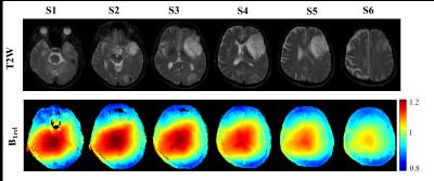
Evaluating effect of B1 field Inhomogeneity on DCE-MRI Data Analysis of Brain Tumor Patients at 3T
ANIRBAN SENGUPTA, RAKSHIT DADARWAL , RAKESH GUPTA, ANUP SINGH
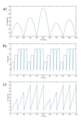
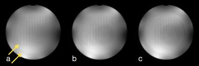
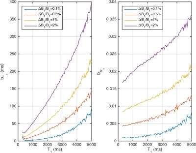
In the current study, transmit B1 field inhomogeneity(B1FI) distribution and propagation of flip-angle(FA) related errors to Dynamic-contrast-enhanced(DCE) MRI data analysis in brain tumors of human subjects at 3T MRI were studied. Experimental and simulation studies were performed to evaluate the propagation of these errors to DCE-MRI data analysis and its correction were performed during signal intensity(S(t)) to concentration-time-curve(C(t)) conversion. This study show that B1FI introduced substantial errors in DCE-MRI data analysis(tracer-kinetic and hemodynamic parameters) and these errors were mitigated by correcting FA using B1 map. B1FI related error also showed dependence on concentration of contrast agent and length of concentration-time-curve.


1431.  
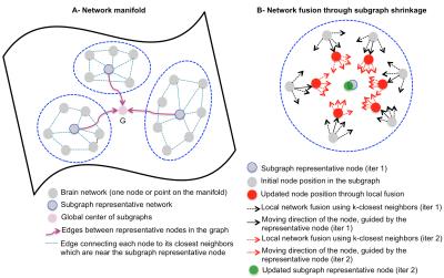
Brain Network Atlas Estimation using Centered Graph Shrinkage with Application to Developing and Aging Brains
Islem Rekik, Gang Li, Minjeong Kim, Weili Lin, Dinggang Shen
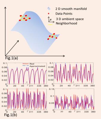
Learning how to average brain networks (i.e., build a brain network atlas) constitutes a key step in creating a reliable ‘mean’ representation of a set of normal brains, which can be used to spot deviations from the normal network atlas (i.e., abnormal cases). However, this topic remains largely unexplored in neuroimaging field. In this work, we propose a network atlas estimation framework through a non-linear diffusion along the local neighbors of each node (network) in a graph. Our evaluation on both developing and aging datasets showed a better ‘centeredness’ of our atlas in comparison with the state-of-the-art network fusion method.


1432.   
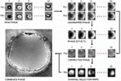
Robust and fast phase unwrapping strategy to improve SWI quality
Yongquan Ye, Jinguang Zong, Weiguo Zhang
A robust and fast phase unwrapping strategy was developed for multi-echo SWI, to improve image quality where classic SWI fails, such as cavity vicinity regions, as well as to provide pristine field map for subsequent QSM applications. A smoothing and a seed prioritizing method were proposed, which was demonstrated to provide very robust unwrapping on phase difference map between neighboring echoes. And with a high quality unwrapped phase difference map, the phase of all echoes can be robustly unwrapped in seconds. 


1433.   
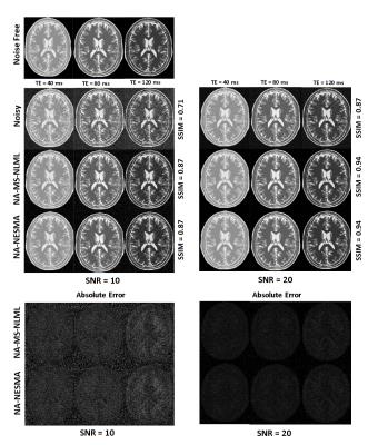
A Fast Adaptive Multispectral Nonlocal Denoising Filter
Michael Maring, Mustapha Bouhrara, Richard Spencer
We introduce a new high-performance nonlocal filter, NESMA, for noise reduction in multispectral (MS) MR imaging.  Through extensive analysis, we show that the NESMA filter demonstrates a high degree of overall image denoising while preserving edges and small structures. We compared the performance of the NESMA filter to the multispectral nonlocal maximum likelihood (MS-NLML) filter.  Although the MS-NLML filter is highly efficient, it requires extensive computational time. NESMA markedly decreases computation time while maintaining comparable levels of noise reduction and feature preservation.  Finally, we show that adaptive selection of similar voxels further improves filtering quality.    


1434.   
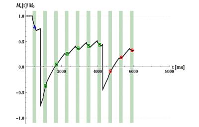
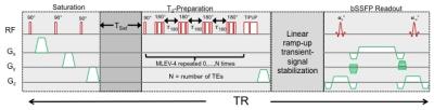
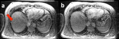
Inversion quality independent robust $$$T_1$$$-quantification of MOLLI sequence data
Thomas Kampf, Theresa Reiter, Wolfgang Bauer
Quantitative mapping of the longitudinal relaxation time has gained increasing interest as it allows monitoring of important structural and functional information of the myocardium. The MOLLI sequence commonly used in clinical research requires a high quality of the inversion pulses for unbiased quantification which is non-trivial especially at high fields. In this work we present a simple modification of the MOLLI sequence which in combination with the recently introduced IGF post processing solves the problem of insufficient inversion quality as demonstrated in phantom experiments.


1435.  
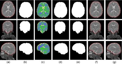
Improved Infant MRI Brain Extraction utilizing Clustering and Morphological Approaches
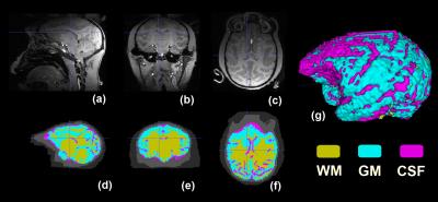
Yao Wu, Sonia Dahdouh, Marine Bouyssi-Kobar, Manoj Kumar, Josepheen Cruz, Wonsang You, Catherine Limperopoulos
Accurate brain extraction is a key procedure in neuroimage analyses. This paper aims to solve the intracranial cavity overestimation issue inherent to existing brain extraction methods when applied to infant brains. We applied k-means clustering method and morphological approaches to improve the accuracy of previously published brain extraction techniques. Evaluation of our proposed method on 28 preterm MR images showed more robust and effective infant brain extraction compared to previous methods.


1436.   
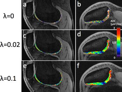
SNR-Weighted Regularization of ADC Estimates using Double-Echo in Steady-State
Bragi Sveinsson, Garry Gold, Brian Hargreaves, Daehyun Yoon
Double-echo in steady-state (DESS) is a 3D sequence which offers both morphological images and quantitative parameter maps (SNR-efficient 3D maps of T2 and apparent diffusion coefficient (ADC)) in various applications, such as breast imaging or knee cartilage imaging. The sequence has less sensitivity to ADC than to T2, sometimes leading to noisy ADC maps. Here, we investigate the effects of using regularized fitting of the signals, with a penalty in ADC variability, to produce less noisy ADC maps. The method is designed to apply less regularization to regions with high SNR. The approach makes use of a recent analytical expression for a ratio between DESS signals.


1437.   
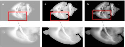
Optimal contrast enhancement of blockface images for MRI guided reconstruction of mouse brain volumes
Christoffer Gothgen, Catharina Holland, Christos Zoupis Schoinas, Karine Mardon, Maciej Plocharski, Lasse Østergaard, Andrew Janke
Blockface imaging can improve atlases of the rodent brain by supplying high resolution images. This study compares three different contrast stretching methods for enhancing the information in blockface images together with a registration to a 16.4 T atlas of the mouse brain. Contrast enhancement technique used was histogram equalization, adjusting the image intensity values by stretching them between the bottom 1% and the top 1% and CLAHE with a clip limit of 0.01 and a uniform histogram. Registrations was rigid, affine and SyN. By using CLAHE as contrast stretching method a high similarity to the MRI was found. 


1438.  
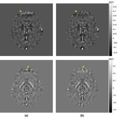
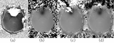
B0 Field Inhomogeneity Corrected Quantitative Susceptibility Mapping
Young-joong Yang, Jong-Hyun Yoon, Hyeon-Man Baek, Chang-Beom Ahn
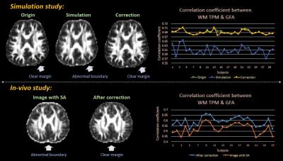
QSM is a method that generates internal susceptibility distribution of subject using material’s intrinsic magnetic susceptibility property. Bo inhomogeneity affects magnitude and phase images. In this study, QSM with B0 field inhomogeneity correction is proposed. Using numerical simulation and in-vivo experiment, proposed method is verified. In simulation, improved susceptibility map is obtained with less root mean square error. In in-vivo experiment, signal loss and non-uniformity at frontal lobe are reduced. As field inhomogeneity increases according to the increase of main field strength, this method would be a more important element for QSM.


1439.  
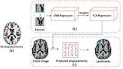
Real-time large-scale anatomical landmark detection with limited medical images
Jun Zhang, Mingxia Liu, Dinggang Shen
Landmark detection based on deep neural networks has achieved state-of-the-art performance in natural image analysis. However, it is challenging to detect anatomical landmarks from medical images, due to limited data. Here, we propose a real-time large-scale landmark detection method with limited training data. We train our model with image patches and test it with the entire image, inspired by fully convolutional networks. Also, we develop a weighted loss function in our model to increase the correlations between image patches and their nearby landmarks. The experimental results of detecting 1741 landmarks from brain MR images demonstrate the effectiveness of our method.


1440.   
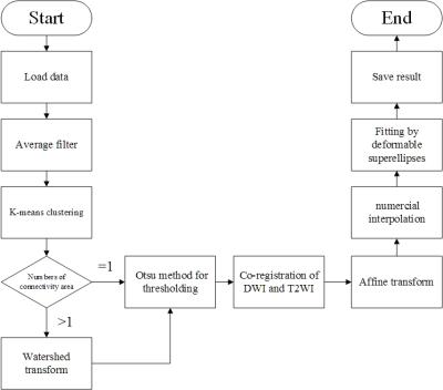
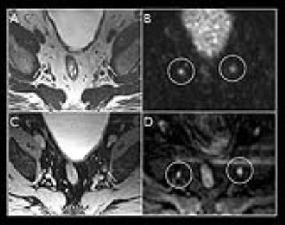
A fully automatic prostate segmentation method for both DWI and T2WI
Yi Zhu, Rong Wei, Ge Gao, Yajing Zhang, Xiaoying Wang, Jue Zhang, Jing Fang
Automatic prostate segmentation in MR images is a meaningful work, not only can be used in the first step of the Prostate Imaging Reporting and Data System, but also helps to predict pathologic stage of disease by determining the prostate volume. Here we show a novel new method can get the prostate contour for both T2WI and DWI fast and accurately without any manual intervention. The segmentation accuracies for 60 images are 83.7% (T2WI) and 87.1% (DWI). Even in some cases, such as prostate hyperplasia, our method shows good robustness.


1441.   
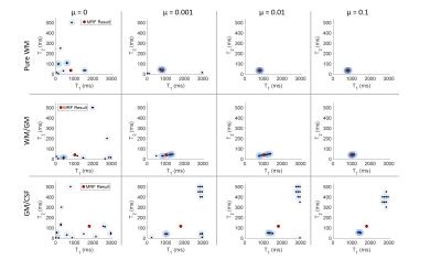
Fast fitting method for simultaneously quantifying multiple MR parameters using local optimization method with predetermined initial values
Suguru Yokosawa, Yo Taniguchi, Tomoki Amemiya, Toru Shirai, Ryota Sato, Yoshihisa Soutome, Hisaaki Ochi
A fast fitting method for quantifying multiple MR parameters using local optimization method with predetermined initial values is proposed. In the proposed method, an optimal neighborhood solution is extracted as predetermined initial values. A difference in MR parameters between the proposed method and the conventional method was less than 5 %. On the other hand, a computing time of the proposed method was approximately 15 times faster compared with the conventional method. We concluded that the proposed method can provide fast fitting process while maintaining calculation accuracy.


1442.  
Improved image texture features by Gaussian mixture models of grey-level co-occurrence matrices
Tommy Löfstedt, Patrik Brynolfsson, Tufve Nyholm, Anders Garpebring
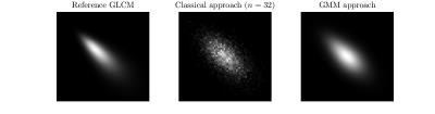
Image texture features based on gray-level co-occurence matrices (GLCMs) are useful in e.g. the analysis of MR images of tumours. However, the features can be quite sensitive to the number of grey-levels in the analysed image, in particular if the region of interest is small. In this work we propose a new method for computing the GLCM, based on Gaussian mixture models. The results show that the new method improves the estimation of the GLCM and at the same time eliminates the difficult task of selecting the number of grey-levels.


1443.   
Optimization of 2D registration using minctracc on myelin stained brain slices
Max Prihoda, Simon Hametner, Andreas Deistung, Verena Endmayr, Andrew Janke, Claude Lepage, Thomas Haider, Simon Robinson, Xiang Feng, Hans Lassmann, Jürgen Reichenbach, Evelin Haimburger, Christian Menard, Hannes Traxler, Siegfried Trattnig, Günther Grabner
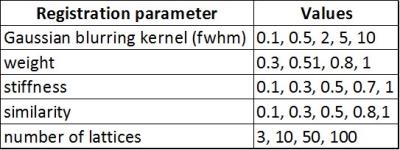
Histological analyses are important for a wide spectrum of in vivo and in vitro imaging projects. But unlike MRI or CT, histological analyses are typically performed in two dimensions. Nonlinear tissue deformation and ruptures of brain tissue are often common, making analysis in slice direction more difficult. In this work, we optimized a hierarchical, nonlinear fitting pipeline on the basis of two high resolution, myelin stained brain sections using mintracc.


1444.  
Comparison of MP2RAGE-based morphometry methods for anorexia nervosa
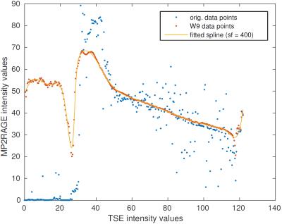
Bénédicte Maréchal, José Baiao Boto, Gkinis Georgios, Nadia Ortiz, Karl-Olof Lövblad, François Lazeyras, Maria Isabel Vargas, Alexis Roche, Tobias Kober
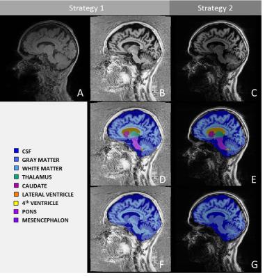
We explore the sensitivity of a morphometry tool to detect anorexia-related brain atrophy in MP2RAGE images. We compare volumetry resulting from two previously reported morphometry strategies on 16 patients with clinical suspicion of anorexia and identify both similarities and differences in brain atrophy evaluation.


1445.   
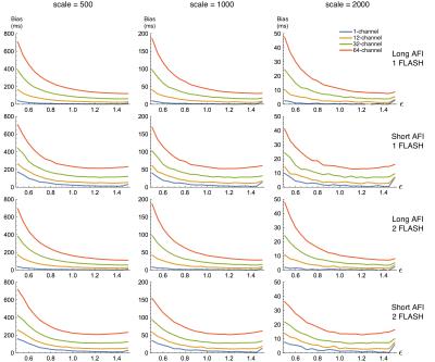
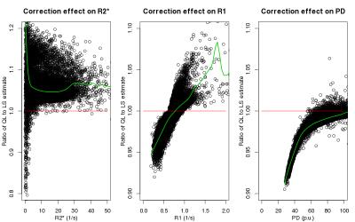
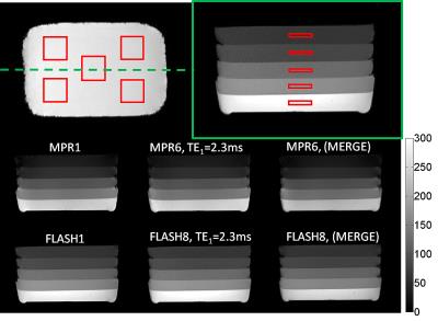
Bias and SNR of $$$T_1$$$ estimates derived from joint fitting of actual flip-angle and FLASH imaging data with variable flip angles
M. Dylan Tisdall
Previous work has suggested fitting joint AFI/FLASH data for T1 and B1+ by minimizing the 2-norm of the difference between the signal model and measurements will produce unbiased estimates of T1. We demonstrate that, contrary to previous results, the estimator has a substantial bias that varies with both the true T1 and B1+, and the receive channel count. We also demonstrate that the correct ML estimator removes the effect of channel count, and that the choice of AFI protocol has a larger impact of the quality of estimates than the addition of an extra FLASH scan.


1446.   
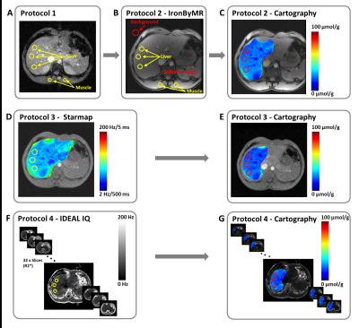
Quantification of iron liver with clinical MRI protocols
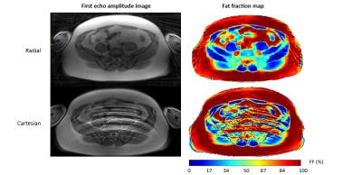
Redouane Ternifi, Philippe Pouletaut, Magalie Sasso, Véronique Miette, Fabrice Charleux, Sabine Bensamoun
Iron quantification has been assessed through the development of magnetic resonance sequences. The purpose is to improve the existing MRI iron protocols to better diagnose the degree of hemochromatosis. Five volunteers with healthy livers underwent four protocols for the quantification of iron overloads concentration (IOC). The results have demonstrated that existing clinical protocols could be improved to provide spatial distribution of iron within one slice and all over the entire liver volume. IDEAL-IQ® could be the best protocol to have IOC volume representation with a short time of acquisition and standard deviation values associated to mean IOC data. 


1447.   
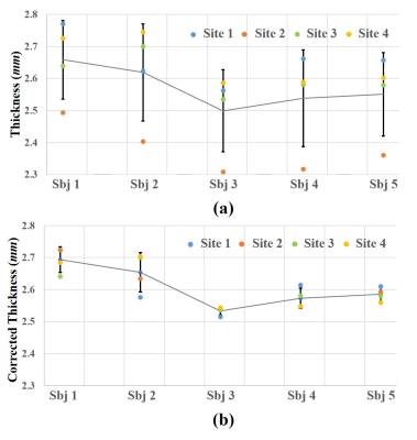
Harmonization for cortical thickness across sites in multi-center MRI study
Lin Zhao, Tuo Zhang, Xianjun Li, Chao Jin, Miaomiao Wang, Xiaocheng Wei, Hong Yin, Zengjun Zhang, Xiaoqun Yao, Xiaoling Zhang, Jian Yang
Cerebral cortex encodes crucial information of brain development, cytoarchitecture and function. However, varying data acquisition conditions at different centers could hamper group-wisely statistical analysis. This study aims to test the consistency of cortical thickness in the human brain across four sites and harmonize the deviations. Our results showed that variation of cortical thickness across sites were regionally independent, and deviation across centers could be reduced by linear regression method at a global scale, while the variations across subjects were well preserved. Those results suggest that our method has the promise in harmonizing cortical thickness measures in multi-center study.


1448.   
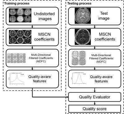
QEMDIM : Quality Evaluation using Multi-DIrectional Filter for no-reference MR image
Jinseong Jang, Kihun Bang, Hanbyeol Jang, Dosik Hwang
This paper proposes a new image quality assessment (IQA) for no-reference MRI, Quality Evaluation using Multi-DIrectional filters for MRI (QEMDIM), that is obtained from difference of statistical features between test images and numerous pre-scanned images in Mean Subtracted Contrast Normalization (MSCN) coefficient and Multi-Directional Filtered Coefficients (MDFC). the proposed method is capable of detecting various types of artifact and can be applied to clinical applications as well as being used to evaluate the performance of MRI hardware and software


1449.   
Diagnostic performance of texture analysis on MRI in differentiated degree of head and neck carcinoma
Yu Chen, Yuan Li, Yuanli Zhu, Huadan Xue, Zhuhua Zhang, Hailong Zhou, Zhengyu Jin
The aim of this study was to determine the diagnostic accuracy of pathological differentiated degree of head and neck squamous cell carcinoma (HNSCC) using MRI texture analysis.  The following texture analysis parameters were derived from the T1WI, T2WI , T2fs and Post-Gad T1WI based on different scale: entropy , mean pixel intensity, standard deviation(SD), skewness, and kurtosis. ROC curves and AUC of each parameter was determined, respectively. We conclude that the entropy at fine texture scale on Post-Gad T1WI had the best ability .


1450.  
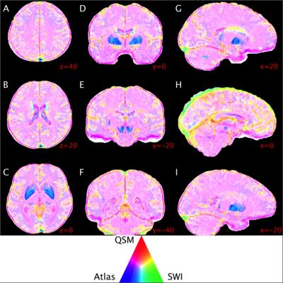
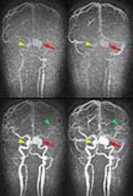
Regional variations in cerebral venous contrast using susceptibility-based MRI
Phillip Ward, Nicholas Ferris, Parnesh Raniga, Amanda Ng, David Dowe, David Barnes, Gary Egan
In this study we compared the image contrast properties of susceptibility-weighted imaging (SWI) and quantitative susceptibility mapping (QSM) for cerebral venous identification and visualisation. SWI and QSM are minimally invasive techniques to image cerebral veins with distinct contrast properties. We hypothesised that these techniques would provide complementary vein contrast in different brain regions. Contrast was measured using 1072 manually traced vein images from ten volunteers. We found regional variations in the predictive power of vein contrast and computed maps of contrast profiles that may inform which technique is best for a given application.


1451.   
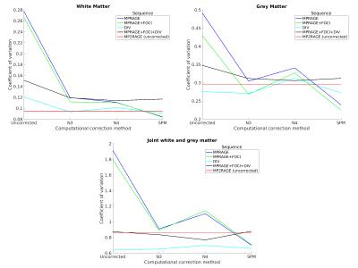
Quantitative and qualitative evaluation of bias field correction methods
Falk Luesebrink, Hendrik Mattern, Alessandro Sciarra, Oliver Speck
Bias field correction is an essential prerequisite for image analysis, especially at high field strength. In this study multiple correction methods, based on acquisition of a reference image and computational approaches with varied input parameters, are compared. The results indicate that acquisition of a conventional MPRAGE corrected by SPM yields quantitatively and qualitatively comparable results to acquiring a reference image (e.g. MP2RAGE), however, scan time is up to halfed.


1452.  
Rapid registration of EPI to high-resolution structural images
David Manners, Claudia Testa, Stefania Evangelisti, Stefano Zanigni, Caterina Tonon, Raffaele Lodi
Post-processing methods that can non-linearly register diffusion-weighted EPI data to high-resolution images are useful in the context of clinical protocols. The current research attempts to apply currently available registration methods to rapidly perform such registration. Investigations on healthy subjects show that an appropriately generated target image allows good quality registration to be performed even with freely available software. This is useful for example to provide cortical seeds for diffusion tractography.


1453.   
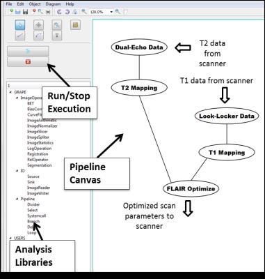
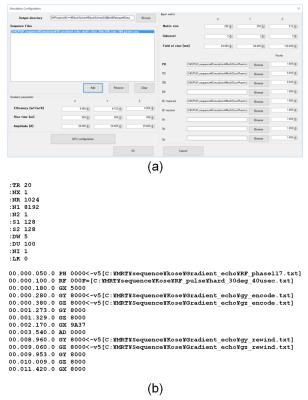
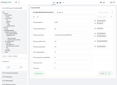
A graphical programming environment for creating and executing adaptive MRI protocols
Refaat Gabr, Getaneh Tefera, William Allen, Amol Pednekar, Ponnada Narayana
Inline processing of magnetic resonance images allows fast feedback of analysis and immediate access to quantitative information. It further allows the development of adaptive MRI protocols. Here, we present GRAPE, a development platform for graphical programming to facilitate the development of adaptive magnetic resonance imaging (MRI) protocols. This platform provides tools to enable graphical creation, execution, and debugging of image analysis algorithms integrated with the MRI scanner, all within a graphical environment. GRAPE is demonstrated with the implementation of patient-specific optimization of the scan parameters of 3D fluid-attenuated inversion recovery (FLAIR) protocol to enhance the contrast of brain lesions in multiple sclerosis, performed on a 3.0 Tesla MRI scanner. 


1454.   
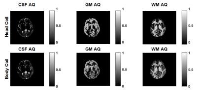
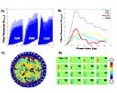
Validation of CSF based calibration for accurate and robust quantification of water content.
Zaheer Abbas, Dominik Ridder, Krzysztof Dzieciol, Nadim Jon Shah
Estimating tissue water content is challenging. Reliable quantification of the water content requires significant number of corrections and calibration to a reference. In this work, we proposed to use a region in cerebrospinal fluid for robust calibration; this is further validated in a cohort of healthy volunteers and compared to existing methods.


1455.   
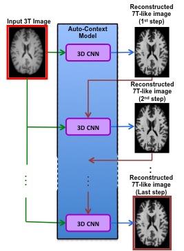
Automatic nonlinear transformation of 7T MRI brain image to Talairach stereotaxic space
Mingyi Li, Jian Lin, Katherine Koenig, Mark Lowe
Transforming MRI brain images into Talairach space will greatly facilitate the comparison of neuroimaging research results across subjects and applications of atlas to research subjects and clinical patients. We developed an automatic processing pipeline based on nonlinear registration to transform 7T MRI brain images to Talairach space. The pipeline utilized matching scores derived from brain parcellation for quality assurance (QA). The pipeline was tested on subjects including five controls, three MS patients and three ALS patients. The results showed that the method generated better results than the automatic Talairach transformation provided by AFNI. The QA scores were also comparable to those computed from 3T MRI brain images in our previous study.   


1456.  
Epilepsy Surgery Followup: Resected Tissue Analysis and Classification
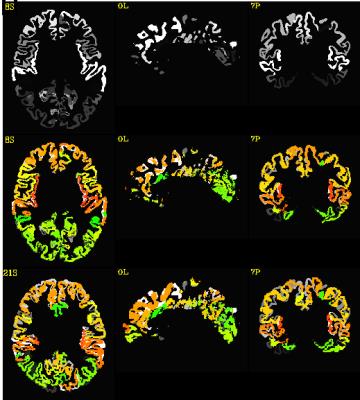
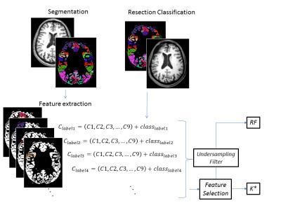
Fabrício Simozo, Tonicarlo Velasco, Luiz Murta Jr.
Focal cortical dysplasia (FCD) is one of the main causes of refractory epilepsy. There is no self-sufficient method in order to evidence the presence and location of FCD, making complete diagnosis very difficult. Although some studies have addressed FCD identification, image texture is poorly explored. This study evaluated pre and post-surgical magnetic resonance images (MRI) of epilepsy patients in order to test Machine Learning classifiers and their ability to identify dysplasia using texture features and cortical thickness. Precision and recall scores suggest the capabilities of the proposed methodology in responding to the presence of FCD tissue.


1457.  
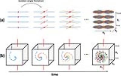
Optimization of multiple orientation QSM for building a clinically feasible protocol
Harshan Ravi, Wen-Tung Wang, Dzung Pham, John Butman
  Quantitative Susceptibility Mapping (QSM) offers unique, quantitative information about tissue magnetic susceptibility. A multi-orientation method, calculation of susceptibility through multi-orientation sampling (COSMOS), enables the dipole inversion by acquiring data at multiple orientations. In practical imaging settings, however, it is a challenge to image the subject multiple orientations.  Although small angle COSMOS  has been shown to generate reasonable QSM images, the selection of orientations within a practical acquisition protocol remains an open question. In this work, we investigated the influence of the number and direction of orientations on the outcome of small angle COSMOS for in vivo imaging.


1458.  
Differences in parameter sensitivities of GESFIDE MR signals generated on realistic angiograms and on idealized cylinders
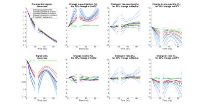
Philippe Pouliot, Louis Gagnon, David Boas, Frederic Lesage
Idealized models of cylinders for the vasculature are used in several quantitative MRI techniques such as for perfusion, CBV, vessel size and vascular MR fingerprinting. While limitations of these models are recognized, a direct comparison of the predicted MR signal between different cylinder models and those using a real vasculature as substrate has not been done to our knowledge. Here we compare the sensitivity of the MR signal for the GESFIDE sequence for 4 sets of , models of cylinders and 6 realistic angiograms from mouse somatosensory cortex. In general, simulation results are all different between the different angiograms and the different models of cylinders. This suggests that much care should be used in interpreting literature results based on models of cylinders, or as well with models on angiograms, to account for the possibility of biases in the absolute results. Correlations and differences in absolute values, for some parameters, may perhaps be less subject to bias.


1459.  
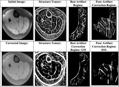
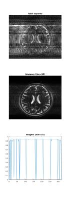
Adaptive Magnetic Resonance Image signal enhancement using squared eigenfunctions of the Schrodinger operator
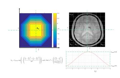
Abderrazak Chahid, Hacene Serrai, Eric Achten, Taous-Meriem Laleg-Kirati
The main of challenge of Magnetic Resonance Imaging (MRI) is dealing with high levels of noise which may corrupt the image especially since the noise is almost correlated with the image details. In this regard, we propose a new MRI enhancement method to overcome this limitation. The proposed MRI enhancement method relies on square sub-images enhancement depending on the noise level in each position using spatial adaptation of the Semi-Classical Signal Analysis (SCSA) method, where an enhancement parameter h is subject to a Gaussian distribution. The results show significant improvement in noise removal and preserving small details in the image.


1460.  
Obtaining accurate and fast unwrapped phase images
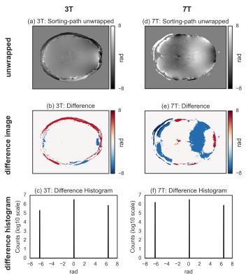
Riccardo Metere, Harald Möller
The unwrapping of phase data is a common problem in MRI. However, its solution is non-trivial for 2D or 3D images, and there has been some general research in this direction, notably in the field of optics. The two most pupular algorithms for MRI applications are: (i) a Laplacian-based method that is fast but inaccurate; (ii) a region-merging optimization method that is accurate but very slow. Here, we propose the adoption of a recently developed and freely available unwrap algorithm that significantly outperforms the other considered methods, allowing for both fast and accurate calculation of unwrapped phase images.


1461.  
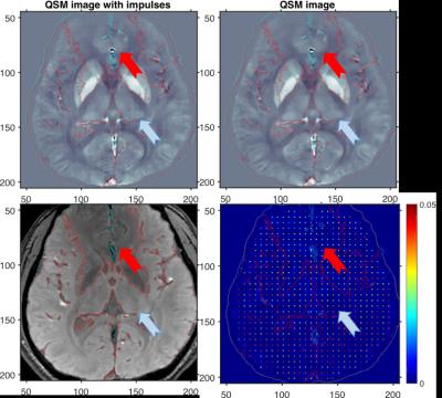
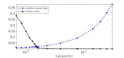
Spatial resolution properties of QSM images using MEDI algorithm
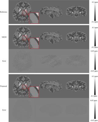
Se Young Chun
We investigated the spatial resolution of the MEDI reconstructed QSM images by deriving an analytical expression of the estimator in terms of the true QSM image. The implication of this expression is that no regularization will be applied to some part of the QSM images if the corresponding magnitude image area contains strong edges and relatively strong regularization will be applied to some part of the QSM images if the corresponding magnitude image area contains low magnitude values. Our simulations with a phantom and a in-vivo QSM image confirm this analysis; Impulse perturbations were suppressed on the area where the magnitude image has low contrasts and impulses were preserved on the area where the magnitude image has high contrast or edges.


1462.  
AWESOME-Based De-Noising of Complex-Valued fMRI Time Series
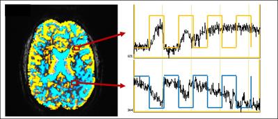
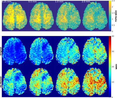
Henrik Marschner, Laurentius Huber, André Pampel, Harald Möller
In this study we investigate possible benefits of an application of ‘AWESOME’ de-noising on fMRI. The application in a high-SNR finger tapping experiment showed a reduction of the already low thermal noise contribution and therefore improvement of tSNR and reduction of false positives; no adverse effects in the form of smoothing or suppression of ‘true’ activation was observed. A second investigation of the scalability of tSNR improvement on a resting state experiment with variable slice thickness / SNR showed that thermal noise can be reliably reduced and the tSNR proportionally improved without visible reduction of detail sharpness / resolution.


1463.  
Assessment of interplatform reproducibility of T1 quantification methods used for DCE-MRI: results from a multicenter phantom study
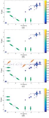
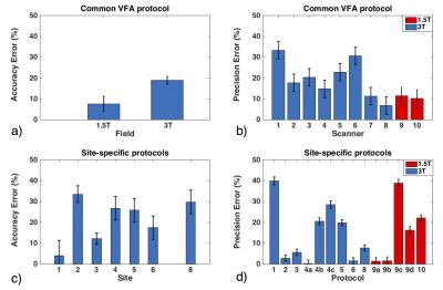
Octavia Bane, Stefanie Hectors, Mathilde Wagner, Lori Arlinghaus, Madhava Aryal, Michael Boss, Yue Cao, Thomas Chenevert, Fiona Fennessy, Wei Huang, Nola Hylton, Jayashree Kalpathy-Cramer, Kathryn Keenan, Dariya Malyarenko, Robert Mulkern, David Newitt, Karl Stupic, Lisa Wilmes, Thomas Yankeelov, Yi-Fen Yen, Stephen Russek, Bachir Taouli
Our multicenter study examined variability in T1 quantification by testing common inversion-recovery spin echo and variable flip angle (VFA) protocols, as well as T1 mapping methods used by participating sites, using a phantom with known T1 values. We found field strength dependence of the accuracy, and platform dependence of the repeatability of T1 measurements with the common VFA protocol. Accuracy for site-specific protocols was influenced by site, while repeatability, by type of protocol. Our findings suggest modified IR methods and VFA protocols with multiple flip angles and B1 correction as good methods for repeatable T1 measurement. 


1464.   
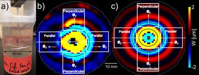
Bio-inspired optimization of technical fiber-reinforced ramifications using high-resolution MRI of Dracaena marginata branchings as concept generators
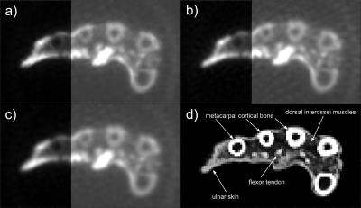
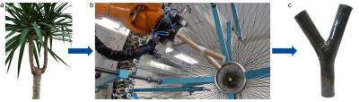
Linnea Hesse, Tom Masselter, Nils Spengler, Jan Gerrit Korvink, Jochen Leupold, Thomas Speck
MRT is still a little-known and highly underestimated imaging method within the field of functional morphology and biomechanics of plants and biomimetics. Its non-invasive and non-destructive character in combination with a large variety of applicable imaging sequences, gives this method a strong potential to shed light to various unanswered scientific questions concerning both the plant structure and function as well as on physiology. Using a Bruker Biospec 94/20 9.4T and a 3D FLASH sequence we could gain new insights into the biomechanics and development of dragon tree ramifications as a source of inspiration for the optimization of technical fiber-reinforced ramifications.


1465.   
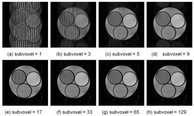
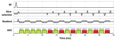
Fast Bloch-Torrey simulation of 3D RF spoiled gradient echo sequences using a number of subvoxels and molecular diffusion effect
Ryoichi Kose, Katsumi Kose
RF spoiled gradient echo sequences were studied both with experiments and  Bloch-Torrey simulation. The Bloch simulation of the 256×256×32 voxel images clarified that adequate number of subvoxels  were required for artifact-free images. The Bloch-Torrey simulation for one voxel magnetization clarified that adequate number of subvoxels were required for image intensity reproduction by diffusion effect. In conclusion, molecular diffusion effects are indispensable to reproduce the image contrast in SPGR.


1466.   
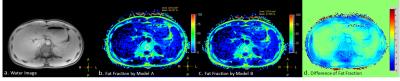
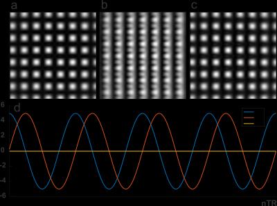
Analysis of error in Fat-Water Quantifications Originated from Models
Xiaoqi Wang, Xiaoguang Cheng, Li Xu, Li Baoqing
Fat-water separation imaging methods with multi-echo acquisition require specific fat spectrum model. The optimal spectrum model to be applied relies on the fat chemical properties as well as the acquisition scheme regarding to, for example, TR and TE. Herein we exam the consequence if inaccurate fat spectrum is used in the fat quantification processing, and analyze the related errors.


1467.   
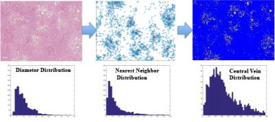
Liver Biopsy Analysis to Determine Fat Droplet Distribution
Benjamin Ratliff, Diego Hernando, Curtis Wiens, Changqing Wang, Rao Watson, Rashmi Agni, Claude Sirlin, Scott Reeder
The purpose of this work was to quantify the size and clustering of fat droplets using liver biopsy, as part of a long-term effort to characterize the relationship between tissue microstructure and quantitative MRI signals in fat-containing tissue. Three H&E stained liver core biopsies with varying fat-fractions were analyzed using segmentation software in order to generate probability density functions for fat droplet size and location. This work demonstrates that fat droplet distribution in the liver can be modeled statistically to determine the size and location distribution of fat droplets, potentially enabling characterization of the MR signal observed from fatty liver.  


1468.  
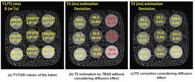
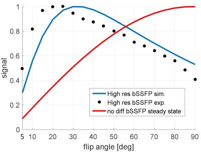
Diffusion effects on T2 relaxometry with triple echo steady-state free precession sequence
Yangzi Qiao, Chao Zou, Xin Liu, Hairong Zheng
In this study, the underestimation of T2 by TESS was revealed through simulation and phantom study. The bias becomes significant in high resolution TESS imaging with larger unbalanced gradient moment, as the diffusion effect can not be neglected. A possible correction scheme was also proposed, the results validated the diffusion effect on T2 estimation. However, the correction method relies on T1/T2/D as priori to calculate the signal change ratio through EPG algorithm. A possible solution might be simultaneous estimation on apparent diffusion coefficient and T2.


1469.   
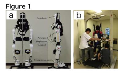
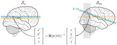
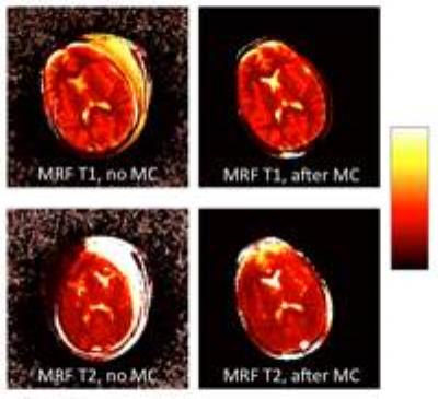
Brain activity alteration during the training period of the Hybrid Assistive Limb® (HAL) for chronic spinal cord injuries: a task-based fMRI case report
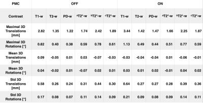
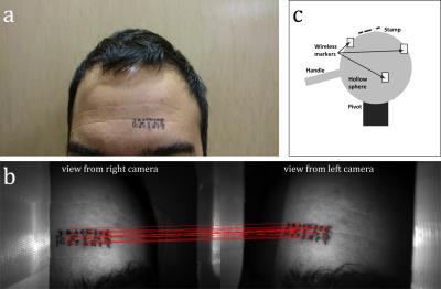
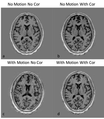
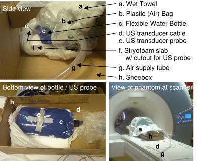
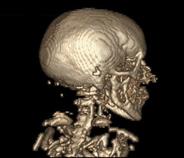
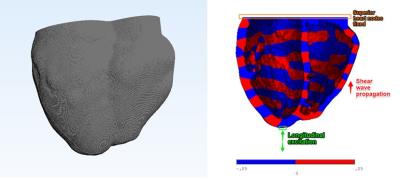
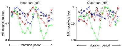
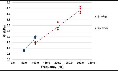
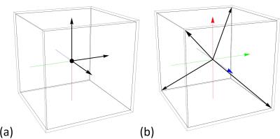
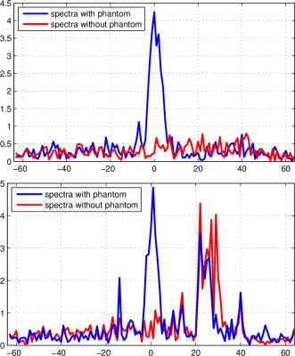
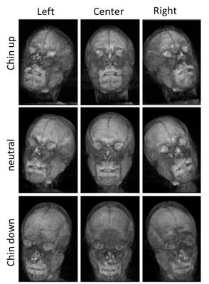
Kousaku Saotome, Akira Matsushita, Aiki Marushima, Hiroaki Kawamoto, Hideo Tsurushima, Tomohiko Masumoto, Masashi Yamazaki, Akira Matsumura, Yoshiyuki Sankai
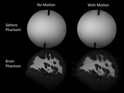
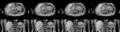
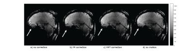
We previously developed the novel brain phantom showing image contrast and construction similar to those of in vivo MRI. This phantom has the potential to quantitatively assess the capability of the motion-corrected PROPELLER technique, which has been never approached. In the current study, we investigated the rotational frequency dependencies of the different two motion-corrected PROPELLER techniques by using our brain phantom. Our findings allow to quantitatively assess the capability of the Motion-Correction in PROPELLER.


1470.   
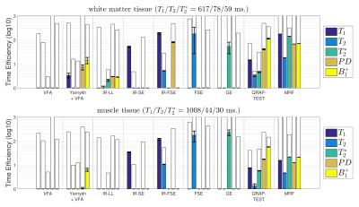
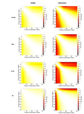
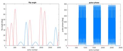
Determining the Time Efficiency of Quantitative MRI Methods using Bloch Simulations
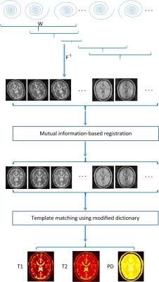
Willem van Valenberg, Frans Vos, Stefan Klein, Lucas van Vliet, Dirk Poot
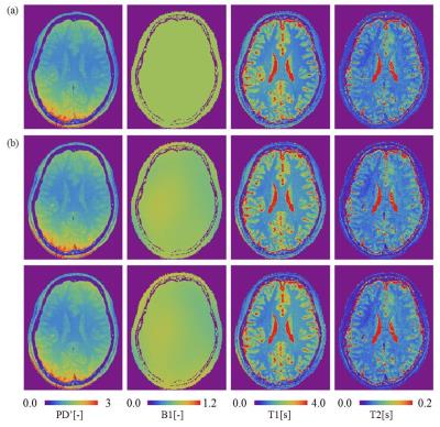
When measuring $$$T_1, T_2, T_2^*, PD$$$, or $$$B_1^+$$$, we prefer the MRI sequence that provides the best precision in the allowed scan time (i.e. having optimal time efficiency). However, experimentally determining the time efficiency is impractical when comparing many sequences, each possibly with varying settings, and multiple tissue types of interest. Here, we derive time efficiency through Bloch simulations which is applicable to any MRI sequence and tissue type. A specific strength of our framework is that it does not require an explicit fitting procedure which may not yet exist when designing novel MR sequences.


1471.  
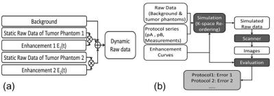
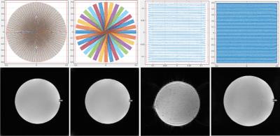
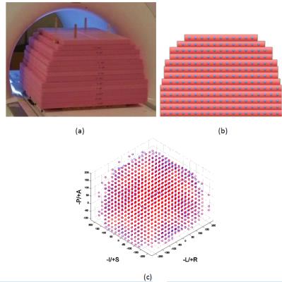
Quantitative DCE-MRI Accuracy Evaluation Using Dynamic Physical vs. Digital Phantom: a Cross-Validation
Yuan Le, Yuxiang Zhou, Eric Stinson, Stephen Riederer, Joel Felmlee
To study the image accuracy of quantitative DCE-MRI a digital phantom and computer simulation were usually used. To validate the digital phantom method, we conducted a cross-validation study comparing the image accuracy estimation from simulation with that from a dynamic physical phantom with contrast infusion. Results showed that the estimated errors were usually higher with the physical phantom, likely due to the difference in reproducibility. Consistency was found in the measurement error comparison between imaging techniques when the temporal resolution was high.


1472.   
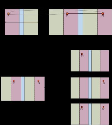
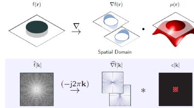
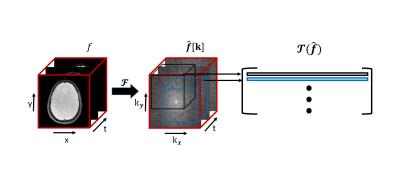
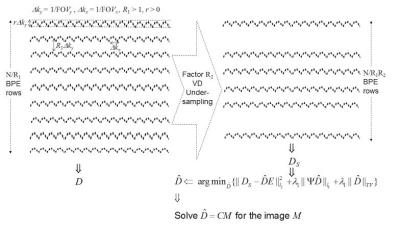
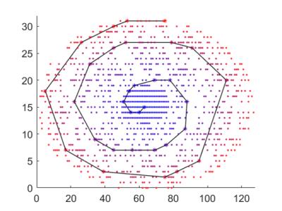
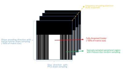
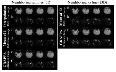
Early enhancement in breast DCE-MRI is sparse and can be imaged with a reduced FOV to increase temporal resolution
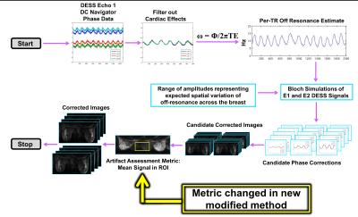
Federico Pineda, Ty Easley, Gregory Karczmar
Early enhancement in breast DCE-MRI is very sparse, if the FOV is reduced in these images and aliasing occurs, the likelihood that two significantly enhancing voxels overlap is low. We present a method for ‘unfolding’ of aliased DCE-MRI acquisitions that closely approximates fully-sampled acquisitions. This method could be used to increase the temporal resolution of DCE-MRI at very early times when enhancement is rapidly changing, allowing for the accurate measurement of early lesion kinetics.


1473.   
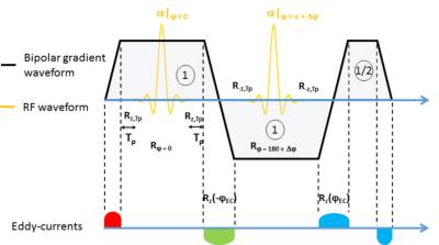
Simulation of Sound Pressure Level of MRI Scan Considering Eddy Currents
Yukari Yamamoto, Yo Taniguchi, Hisaaki Ochi, Yoshihisa Soutome
Since optimal waveforms should be chosen for each gradient pulse in order to reduce sound pressure levels (SPLs) in MRI scans, the simulation accuracy of the SPL must be improved. Assuming that the eddy current component is a cause of the disagreement between the measured and the simulated SPLs, we compared the simulation results with and without the eddy current component in this study. By including the eddy current component in the simulation, the magnitude of the SPL decreased, which reflects the decrease in the peak amplitude of the frequency component of the gradient waveform. However, the eddy current component did not affect the change trends of the SPLs depending on the change of waveforms. On the other hand, a slight change in the peak position of the frequency response functions appears to cause a significant change in the SPL, and the error of the FRF was also thought to cause disagreement between the measured and simulated SPLs.


1474.   
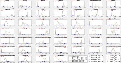
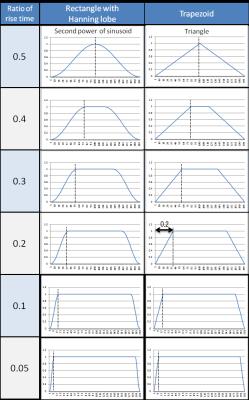
The effect of MR noise and resolution on textural features in simulated and real textures: implications for clinical practice.
Joshua Shur, Matthew Orton, Simon Doran, James D'Arcy, David Collins, Maria Bali, Martin Leach, Dow-Mu Koh
Sensitivity of textural features to acquisition parameters has important clinical implications. The aim of this study is to investigate the effect of noise and resolution on textural features. 

We compared textural features from a uniform and simulated texture, varied with noise, with experiment in a uniform phantom and organic texture.

Our data demonstrate that a uniform texture behaves as if it has inherent texture, due to presence of artefact, and this in turn will influence textural features as noise and acquisition parameters are varied.

We note that certain textural features used in clinical practice vary widely with image noise, whereas others appear to be robust.


1475.   
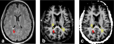
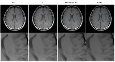
Quantification of contrast agent-induced enhancement of brain lesions in multiple sclerosis
Jung-Jiin Hsu, William Stern, Jung-Yu Hsu, Roland Henry
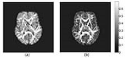
Contrast agents are routinely used in MRI to detect and evaluate tissue lesions. Conventional clinical protocols use T1-weighted sequences to visualize Gd contrast agent enhancement. Because T1-weighted MRI does not produce quantitative measurements, it is difficult to describe the lesion enhancement in quantitative terms and to infer the degree of the underlining disease activities of the lesions. A fast, whole-brain high-resolution T1 mapping method was developed to address this problem and applied to multiple sclerosis.


1476.  
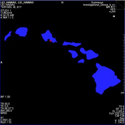
LEI-ALOHA – Magnetic Resonance Imaging in the Tropical Island Setting
Christopher Wiggins, Benedikt Poser
While there are exceptions, most major MRI research centers are located in urban areas where a sizeable population is served by a large medical infrastructure and/or university. For researchers and technical staff alike, this often precludes the possibility of combining their research program with a remote, island-based lifestyle. Here we propose a set of theoretical techniques that form a framework for a purely philosophical research program.
 
Pulse Sequences
Traditional Poster
Acquisition, Reconstruction & Analysis

 
Monday, 24 April 2017
Exhibition Hall  08:15 - 10:15


1477.   
Respiratory Self-Gating using Principal Component Analysis in 2D Golden Angle Radial Free Breathing Cine Imaging
Alexander Fyrdahl, Karen Holst, Martin Ugander, Andreas Sigfridsson
Due to respiratory motion, cardiac imaging is performed during breath holding. Breath holding can be strenuous, or even impossible, for patients with heart disease. We suggest a method for respiratory self-gating using a Golden Angle radial k-space trajectory and principal component analysis. Radial trajectories sample the middle of k-space, which corresponds to the large structure content of the image, each repetition time. The respiratory motion can be detected from the raw k-space data and the motion signal can be used to perform retrospective respiratory gating. 


1478.  
Free-Breathing T2-Weighted Abdominal Examination Using Radial 3D Fast Spin-Echo Imaging
Thomas Benkert, John Mugler III, Bjorn Stemkens, Daniel Sodickson, Hersh Chandarana, Kai Block
A 3D fast spin-echo (FSE) sequence design for free-breathing T2-weighted abdominal examination is described. Due to use of radial stack-of-stars sampling, the sequence provides pure T2 contrast and achieves high robustness to motion, enabling that patients can be scanned during shallow breathing. Using an integrated FID navigator, the sequence can additionally be combined with the self-navigated XD-GRASP reconstruction principle for patients who perform deep breathing. Furthermore, the navigator can be used for bulk-motion detection. Initial results in a free-breathing adult volunteer are shown.


1481.  
Three-dimensional T1-weighted spiral imaging of the spine
Ryan Robison, Dinghui Wang, Melvyn Ooi, James Pipe
Spine imaging requires lengthy examination times to yield sufficient SNR and to account for patient motion. Spiral MRI is a promising method for yielding high SNR acquisitions with reduced scan times and is more robust to patient motion. This work investigates the feasibility of applying spiral MRI to T1w imaging of the spine. Initial results indicate that spiral is well suited for anatomical spine imaging.


1482.  
Intra-Scan Center Frequency Drift Correction for 3D Spiral Exams
Ashley Anderson III, Ryan Robison, Dinghui Wang, Melvyn Ooi, James Pipe
High gradient demand and heating results in a changing main magnetic field and center frequency drift during minutes-long acquisitions. Center frequency drifts on the order of tens of Hz may produce visible artifacts in images acquired with long-readout spiral acquisitions. We propose a method for removing such artifacts in spiral acquisitions by demodulating individual spiral arms using a linear interpolation of center frequency measured before and after a scan.


1483.   
Slice-Accelerated Single-Shot Variable-Flip-Angle Fast Spin Echo with Very Long Echo Trains
Eun Ji Lim, Suhyung Park, Seong-Gi Kim, Jaeseok Park
A Fast spin echo (FSE) pulse sequence has been a main workhorse for clinical imaging due to its flexible contrast.  Simultaneous multi-slice (SMS) FSE in [1] was shown to be efficient for slice acceleration without much loss of signals. Nevertheless, the previous SMS FSE methods, which employ high-flip-angle, spatially-selective multi-band RF pulses for both excitation and refocusing, still remain sub-optimal due to either high energy deposition or elongated echo spacing (ESP) and thereby limited echo train length (ETL). The purpose of this work is to develop a novel, slice-accelerated single-shot variable-flip-angle (VFA) FSE with very long echo trains, in which multi-band RF pulses are used only for excitation while short hard pulses with VFA are utilized along the refocusing pulse train, enabling very short ESP and very long ETL and thus enhancing imaging efficiency. It is shown that the proposed method makes it possible to complete whole brain imaging within 30 sec without apparent artifacts and noise.


1497.   
Territorial Arterial Spin Labeling by Using Asymmetrically RF-shimmed Labeling Pulse with 4-channel RF Transmit at 3T
Kosuke Ito, Atsushi Kuratani, Nobuyuki Yoshizawa, Masahiro Takizawa
Territorial ASL (tASL) using asymmetrically RF-shimmed labeling pulse was proposed. By using 4-channel RF transmit coil, spatially asymmetric transmission was applied to pCASL pulse. Proposed method does not require longer duration of RF pulse. Also, by using knowledge of B1 map, appropriate scaling was applied to the tASL image, and labeling efficiency was consistent between nonselective ASL and tASL. Proposed method was implemented to a 3T MRI system, and demonstrated tASL in-vivo by a volunteer study. tASL of RICA, LICA, and VBA was imaged.


1484.  
SPAMM Based Dual Current Injection to Accelerate Data Acquisition in Magnetic Resonance Electrical Impedance Tomography
Nashwan Naji, Kemal Sümser, B. Murat Eyüboglu
Reducing acquisition time in magnetic resonance electrical impedance tomography (MREIT) improves signal to noise ratio and temporal resolution of measured conductivity data. On the other hand, the reconstruction accuracy of MREIT can be improved by acquiring multiple data using different current injection patterns, which in turn increases the total scan time. In this study, a novel pulse sequence is proposed to reduce the scan time in MREIT by injecting two current patterns in a single acquisition. This method is experimentally realized using a physical phantom, and its feasibility is evaluated. 


1486.   
Diffusion-weighted Echo Planar Spectroscopic Imaging in human brain at 3T
Manoj Sarma, Zohaib Iqbal, Andres Saucedo, Paul Macey, M. Albert Thomas
There have been only few attempts to obtain maps of the diffusion properties of brain metabolites with diffusion weighted spectroscopic imaging. In this study we developed an echo planar based diffusion weighted spectroscopic imaging (DW-EPSI) method which uses a pairs of bipolar diffusion gradient to measure the apparent diffusion coefficient (ADC) of metabolites across an entire slice. The sequence was tested and validated on phantom and five healthy volunteers. The ADC values from DW-EPSI  are in agreement with previous studies. The novel technique will enable a better understanding of the intracellular metabolism and water diffusivity correlates in pathologies.


1490.  
Multiple-TR neuronal resonance-MRI (NR-MRI) for detection of weak oscillating magnetic field
Ki Hwan Kim, Hyo-Im Heo, Sung-Hong Park
We propose a new approach termed neuronal resonance MRI (NR-MRI) with multiple TRs for detection of weak oscillating magnetic field. NR MRI could detect signals from oscillating magnetic fields with random frequency, phase, and on/off intervals, without synchronization between MR acquisition and neuronal oscillation. The detected signals showed higher SNR with increasing number of dynamic scans, demonstrating that temporal averaging is possible with NR-MRI. Two-TR approach showed multiple frequency components in an absolute frequency spectrum with no a priori target frequency information while suppressing the systematic noises, which warrants further investigation.


1487.  
Inversion Recovery with SMS PROPELLER
Ola Norbeck, Enrico Avventi, Henric Rydén , Stefan Skare
We show that simultaneous multi-slice (SMS) PROPELLER can be used with inversion recovery. This is accomplished by using an adiabatic multi-band RF pulse (PINS-DANTE), an in-house developed PROPELLER sequence and a reconstruction that calibrates both in-plane-GRAPPA and slice-GRAPPA weights for all PROPELLER blade angles on a single fully sampled PROPELLER blade volume.


1508.   
Sequence Design by Signal Inversion Using Extended Phase Graphs
Nicholas Dwork, John Pauly
In this work, we show that MR signal progression can be represented as an autonomous discrete linear dynamical system when the small angle approximation is made.  This formalism can be used to determine an optimal set of flip angles to yield a desired magnetic state.  We present an an example of maintaining a constant signal strength with a Fast Spin Echo sequence.


1479.   
Rapid Spinal Cord Imaging
Matthias Weigel, Oliver Bieri
For rapid spinal cord imaging, an inversion recovery prepared balanced steady state free precession (bSSFP) sequence with time-limited cine sampling was developed. It simultaneously acquires eight consecutive images of remarkable different tissue contrasts at 0.67mm in-plane resolution within a single measurement of only 51s per slice. The acquired images can be further combined to considerably improve the contrast to noise ratios (CNR) of the spinal cord tissues such as gray matter and white matter. Representative examples for images measured at different cervical spinal cord locations, various image combinations, and CNR gains are shown.


1500.   
Accelerated 3D GRASE for T2 and PD Weighted High Resolution Images
Alexandra Cristobal-Huerta, Dirk Poot, Mika Vogel, Juan Hernandez-Tamames
Parallel Imaging techniques have not been introduced for Gradient and Spin Echo sequences being a limiting factor for clinical use. Enabling PI for GRASE requires new view-ordering schemes that acquire an autocalibration region while simultaneously mitigating artifacts and obtaining the desired contrast. The purpose of this work is to present new 2D PI accelerated Cartesian view-ordering schemes with either T2 or PD contrast in multi-shot VFA 3D-GRASE, for relevant SAR and scan time reduction compared to 3D-FSE/TSE.


1488.   
Spectral-Model Based Undersampling of Multi-Phase MSI:  Application to Diffusion-Weighted Imaging Near Metal
Kevin Koch, S Kaushik
Diffusion weighted imaging around metal implants is a difficult task that requires a combination of multi-spectral MRI approaches with non-CPMG spin-echo refocusing techniques.  Current implementations of this approach have shown promising initial clinical results.  Further work on improving the acquisition efficiency of these techniques are required to achieve robust clinical viability.  Here, we present an approach that utilizes the MSI spectral domain to enable spectral undersampling of diffusion-weighted acquisitions.   The approach is also easily extendable to other multi-phase implementations of MSI, such as thermometry or relaxometry, where multiple volumes of data need to be acquired for quantitative computations.  


1480.   
Synthetic MRI of the spine using outer volume suppression and virtual coil concepts to further increase scan productivity
Suchandrima Banerjee, Ken-pin Hwang, Peng Lai, Marcel Warntjes, Ajit Shankaranarayanan
Several methods for rapid simultaneous quantification of proton density, T1 and T2 maps from a single acquisition have emerged recently, allowing for retrospective synthesis of MR images with any desired contrast weighting from these maps. This work adapts a 2D fast spin echo based mapping method to spine MRI where scans are typically long and prone to artifacts. Outer volume suppression was incorporated to be able to save time by encoding only the anatomy of interest without aliasing concerns. Interleaved k-t sampling and virtual coil methods were explored to overcome limited coil acceleration capability and to further increase scan productivity.


1489.  
MR Imaging of Magnetic Ink Patterns via Off-Resonance Sensitivity
Stephanie Perkins, Xinwei Shi, Hans Weber, Bruce Daniel, Brian Hargreaves
Printed magnetic ink creates predictable B0 field perturbations based on printed shape and magnetic susceptibility. This can be exploited for contrast in MR imaging techniques that are sensitized to off-resonance, such as fat-suppressed imaging with spectral presaturation. Magnetic ink therefore has the potential to be used in temporary tattoos for creating MR-visible skin markings of arbitrary shape and size, with applications in surgical planning, radiation therapy, tracking of joint movement, or other image registration scenarios. Here we characterize the susceptibility variations of magnetic ink and demonstrate application for MR-visible skin markings.


1491.   
Determination of the Optimum Pattern Length of MRF Sequences
Karsten Sommer, Thomas Amthor, Peter Koken, Jakob Meineke, Mariya Doneva
An important open question in magnetic resonance fingerprinting (MRF) is the optimal choice of sequence parameters, i.e. the ideal length and shape of the pattern that defines the variation of the acquisition parameters. Here we apply a Monte Carlo based measure of sequence performance to find the optimal flip angle pattern for different pattern lengths. Afterwards, the most efficient pattern is found by evaluating these optimized sequences’ actual performance in phantom experiments using various acceleration factors.


1485.   
Full-FOV, whole-brain, half-millimetre in-plane readout-segmented EPIK for high-resolution fMRI studies
Seong Dae Yun, N. Jon Shah
Since the advent of EPI, numerous approaches have been suggested to enhance its resolution for high-resolution fMRI. Recently, several methods were demonstrated for fMRI with a sub-millimetre resolution. However, none of them can achieve such resolution with a full-FOV and, at the same time, with whole-brain coverage. This work aims to develop a novel imaging method based on EPIK in combination with readout-segmentation to achieve half-millimetre resolution with a full FOV. Here, under a typical fMRI constraint (TR of 3 s), the method was shown to provide 93 slices when further combined with the multi-band technique.


1492.   
On the Influence of Intra-Voxel Dephasing in FISP-MRF with Variable Repetition Time
Sebastian Flassbeck, Simon Schmidt, Mathies Breithaupt, Peter Bachert, Mark Ladd, Sebastian Schmitter
In this work we investigate the impact of intra-voxel dephasing on standard FISP-MRF measurements and propose the use of a constant TR to improve the robustness against spatially changing magnetic fields. The results have shown that FISP MRF measurements with a variable TR are susceptible to intra-voxel dephasing, which may generate temporal signal modulations that are likely to cause quantification errors in relaxation parameters. This can be alleviated by fixing TR without loss of encoding capability. 


1495.   
Rapid background magnetic field inhomogeneity correction in MR-based oxygen imaging
Yasheng Chen, Xiaodong Zhang, Cihat Eldeniz, Dustin Ragan, Liam Comiskey, Melanie Fields, Kristin Guilliams, Jin-Moo Lee, Andria Ford, Hongyu An
Quantitative blood oxygenation level dependent (qBOLD) technique provides an invaluable and noninvasive means for mapping brain oxygen extraction fraction (OEF) and R2’ in various neurological disorders. However, background magnetic field inhomogeneity causes errors in the measurement. In this work, we propose a triple echo asymmetric spin echo sequence with imbedded Z-shimming table to measure and correct the adversary effect of the field inhomogeneity.  With both simulation and 16 patient image sets, we have demonstrated the effectiveness of the proposed technique. 


1496.  
Improving Arterial Spin Labeling Acquisition to Reduce the Effect of Delayed Arrival Time
Andre Paschoal, Renata Leoni, Antonio dos Santos, Bernd Foerster, Fernando Paiva
Arterial Spin Labeling (ASL) is a powerful technique to evaluate cerebral blood flow. To analyze hemodynamics effects with ASL, multiples acquisitions over the time are realized, which is called multiphase ASL. In conventional multiphase ASL methods, the later phases has low contrast to noise ratio, so it becomes difficult to analyze it. This study purposes a solution to this problem, through a modulation in the acquisition flip angle. With this technique, the flip angle of all phases follows a modulation equation, so that the ASL signal over the phases becomes nearly constant.


1507.  
Signal Coding for SNR Multiplying Effect and Scan Acceleration Flexibility
Yudong Zhu
In a way that augments existing encoding, signal coding promotes a multiplying effect of SNR and flexibility of scan acceleration.  Its essence is to push for noise decimation by acquiring sums of marked signals from all components where the marking can be achieved by RF, gradient or other means. Its application to multi-slice MRI opens up a regime that enjoys both a √N SNR enhancement, as analogous to that of volumetric MRI, and flexibility with scan time budget, as equal or superior to that of existing multi-slice MRI.


1493.  
An algorithm for refocusing of T2* effects in bSSFP-MRF with relaxation corrections
Mingdong Fan, Danielle Kara, Jesse Hamilton, Nicole Seiberlich, Mark Griswold, Robert Brown
It is found that bSSFP-MRF is in general subject to T2* not T2 relaxation. Like traditional bSSFP, dephasing effects of intra-voxel inhomogeneities can be refocused in bSSFP-MRF with appropriate choices in TR, TE, and FA so that parameter maps are reflective of T2 rather than T2*. An algorithm is introduced and verified in simulation for refocusing intra-voxel dephasing at TE for bSSFP-MRF with corrections to previous work for relaxation effects previously assumed as negligible. These corrections are relevant for bSSFP-MRF in which T2 is not much larger than TR to ensure that T2 maps do not contain T2* effects. 


1499.   
Tri-Fast Spin Echo: A Minimalistic Cross-Platform Multi-Spectral qMRI Pulse Sequence for Routine Clinical Use
Ning Hua, Mitchell Horn, Stephan Anderson, Hernan Jara
Purpose: To develop a simple and cross-platform pulse sequence that meets the following criteria: 1) all directly acquired images are clinically useful, 2) achieves minimalistic scan times, and 3) leads to excellent image quality as well as accurate MS-qMRI mapping. Methods: Tri-FSE consists of a single-echo-FSE sequence that is run in temporal concatenation  with a dual-echo FSE sequence that all together generate T1-, T2, and PD-weighted directly acquired images. Results: Tri-FSE was implemented at medium and high spatial resolution at 1.5T and 3.0T. Conclusion: Tri-FSE is a dual purpose simple pulse sequence that is useful for clinical and scientific purposes.


1498.   
Phase Encoding with Bloch-Siegert effect using Parallel Transmit
Yuqing Wan, Maolin Qiu, Gigi Galiana, R. Todd Constable
A gradient-free, Bloch-Siegert effect based nonlinear spatial encoding scheme was introduced for silent scanning and to eliminate gradient-induced eddy currents in conventional MRI. We implemented a phase encoding scheme with a parallel transmit system on a 7T scanner to demonstrate feasibility.  As a proof of concept, we demonstrated that B-S RF pulses can easilty produce nonlinear spatial encoding for MR imaging. The SAR limitations at 7T limit the efficiency of the encoding, but suggest the feasibility of this approach at lower fields.


1501.   
A new ultrafast 3D gradient-echo-based imaging method: RASE-II
JaeKyun Ryu, WonBeom Jung, Sun Young Chae, Geun-Ho Im, Jung Hee Lee, Seong-gi Kim, Jang-Yeon Park
One version of a new ultrafast gradient-echo-based 3D imaging technique using spatiotemporal encoding (RASE-II) is proposed which provides constant TE across all spins. RASE-II maintains most of appealing features of other spin-echo-based SPEN imaging methods such as no Nyquist ghosting and high tolerance to field inhomogeneities. RASE-II also has less sensitivity not only to SAR and B1-inhomogeneity effects because of low flip angles, but also to T2* signal modulation due to constant TE. As a promising tool for fMRI, RASE-II shows significant improvement of tSNR over GE-EPI. Its performance is demonstrated by lemon and in-vivo rat brain imaging at 9.4T.


1503.  
Blood saturation modeling in multiband multislab Time-of-Flight brain MRI
Alexis Amadon, Gaël Saïb, Nicolas Boulant, Alexandre Vignaud
Multiband multislab Time-of-Flight angiography has recently been proposed for reduced acquisition time and improved sensitivity in the human brain at 3T and 7T. However, in these previous studies, blood saturation has not been taken into account as blood traverses several slabs acquired simultaneously. Here a simple modeling of blood magnetization history is provided to take this saturation into account, which appears essential to avoid strong losses of blood to background signal ratio. Slab-dependent multiband-added VUSE pulses are simulated to counteract these losses both at 3T and 7T, from which a methodology is derived to optimize multiband TOF sequence parameters.


1502.   
Dual Echo Trajectory : Comparison to Partial Fourier Acquisition and Sequence Optimization
Jeehun Kim, Jongho Lee
In this study, we compared two acceleration methods in spin echo imaging; previously proposed Dual Echo Trajectory (DuET) and partial Fourier acquisition. Also, we further improved DuET with interleaved multislice acquisition and echo timing correction.


1504.   
Simultaneous Multi-Volume 4D Phase Contrast Flow MRI
David Feinberg, Liyong Chen
 A new technique of simultaneous 4D flow imaging acquired with simultaneous multiple 3D volumes (SMV) is presented. The velocity measurements are compared to conventional 4D flow imaging and have very good correspondence.


1505.   
Spatial Mapping Using Radio Frequencies: A Non-Linear Approach to Silent MRI
Robert Nikolov, Simona Nikolova, Clara Eng, Pierre Baldi
Recent advances for silent MRI have shown that spatial encoding can be achieved using RF rather than linearly varying static magnetic field gradients.  This has been demonstrated using homogeneous transmit (B1) fields with linearly varying phase gradients.  Similar results can be achieved with linear B1 amplitude gradients with homogeneous phase.  The efficacy of either method is limited by a maximum B1 gradient strength (phase or magnitude) per specific absorption rate. Here we demonstrate a novel approach to relieve this restriction where highly nonlinear B1 gradients can be used for combined amplitude and phase modulation with reconstruction using state-of-the-art machine learning models.


1506.  
SNR Efficiency of Combined Bipolar Gradient Echoes: Theoretical Expressions and Experimental Verification
Jean-David Jutras, Keith Wachowicz, Nicola De Zanche
Multi-echo bipolar pulse sequences are becoming increasingly popular in structural brain imaging applications that require high SNR efficiency and minimal geometrical distortions or water-fat shifts. However, various discrepant expressions for the SNR as a function of T2* and the sampling bandwidth were previously reported, making it unclear as to how multiple bipolar echoes should be combined to maximize SNR. In this study, we compare some traditional single-echo (low-bandwidth) FLASH and MPRAGE sequences with their multi-echo bipolar (high bandwidth) counterparts and validate the SNR theory via phantom measurements. Bipolar MPRAGE/FLASH yield SNR gains of ~1.3/1.6-fold, in good agreement with the theory.


1509.   
GPU optimized fast Bloch simulator for arbitrary MRI pulse sequences
Ryoichi Kose, Katsumi Kose
A GPU optimized fast Bloch simulator was developed for arbitrary pulse sequence inputs. The simulator was applied to multi-slice imaging using 256×256×512 calculation matrix. As a result, we found that the number of short pulses used to approximate the selective excitation pulse and the number of subvoxels used for imaging in the cross-sectional plane were essential to simulation speed.


1511.   
Assessing intrinsic velum height in vowels using time-resolved MRI
Martin Krämer, Melanie Weirich, Karl-Heinz Herrmann, Adrian Simpson, Jürgen Reichenbach
 Real-time MRI and synchronised audio were used to examine intrinsic velum height in German vowels. Two adult female subjects produced five repetitions of a set of sentences containing, among other target material, the point vowels /i: a: u:/ in the same phonologically non-nasalised context. Even in this small sample, the subjects exhibit variation in velum height across vowel categories, and show considerable interindividual variation in velum height during the production of the same vowel category. Collection of further data from normal subjects will be used to create a robust baseline for the assessment of abnormal velum activity.


1494.  
Ingredients for balanced SSFP Microimaging
Sebastien Bär, Thomas Oerther, Angelina Müller, Matthias Weigel, Matthias Wapler , Jochen Leupold
In this work, balanced SSFP microimaging was succesfully performed in 3D (voxel size 40μm x 40μm x 94μm) and 2D (in-plane resolution 16μm x 16μm) on a 7T small animal system and a small-bore 7T spectrometer with maximally used gradient amplitude of 1.03 T/m. Key for optimzed image quality was the choice of susceptibility matched phantom materials and the proper choice of bSSFP flip angle under consideration of diffusion attenuation due to imaging gradients.


1510.  
MRI Pulse Sequence Development Using Graphical User Interface Modules
Saulius Archipovas, Thorsten Honroth, Cristoffer Cordes, Matthias Günther, David Porter
 

We present a preliminary study on developing MRI pulse sequences using graphical user interface elements. We introduce a prototype where a sequence developer can specify a sequence structure and parameter dependencies just using the GUI. As a result, a sequence developer can concentrate on the MRI sequence design, rather than on software engineering issues. The GUI-based development framework is decoupled from the manufacturer’s development environments and promises to promote the development of basic sequence designs that are independent of the scanner platforms used.

 
Reconstruction
Traditional Poster
Acquisition, Reconstruction & Analysis

 
Monday, 24 April 2017
Exhibition Hall  08:15 - 10:15


1512.  
Strategies for Compensating for Missing k-space Data in a Novel Half-Fourier Reconstruction
Seul Lee, Gary Glover
Functional MRI (fMRI) is sensitive to off-resonance from air-tissue susceptibility interfaces. Existing half-Fourier reconstruction is vulnerable to off-resonance since it may lose most of the image energy (near k=0) with a large amount of off-resonance. In a previous study, we suggested a new half Fourier (even/odd (E/O)) reconstruction and showed it was more robust to off-resonance compared to Homodyne reconstruction. E/O reconstruction acquires every other line in k-space. Therefore, neighboring data can be used to compensate for the missing data. In this study, we suggest several strategies for compensating for missing k-space data in kx-ky as well as kz direction. 


1513.   
Reducing acquisition time while maintaining spatial resolution with extended readouts and R2* modeling
Alex Cerjanic, Giang Chau Ngo, Bradley Sutton
Single shot readouts are limited in length by field inhomogeneity and R2* relaxation. With the inclusion of a complex field map, existing field corrected reconstruction algorithms can compensate for both field inhomogeneity and R2* relaxation during extended readouts. Results for spiral acquisitions of up to 56ms in length are demonstrated on a human volunteer.


1514.   
Accelerated Regularized Image Reconstruction in Spatiotemporal MRI
Alexander Gutierrez, Di Xiao, Jarvis Haupt, Albert Jang, Steen Moeller, Michael Garwood
Interest in spatiotemporally-encoded MRI methods has increased over the last decade due mainly to their high tolerance to magnetic field inhomogeneities. However, the data acquired in spatiotemporal MRI can lead to challenging image reconstruction problems. In this abstract we propose a new framework for reconstructing images that leverages compressible structure in recent spatiotemporal encoding techniques to enable an iterative approximate inversion of the Bloch-equations for imaging. In particular, we can often obtain a visually indistinguishable reconstruction up to an order of magnitude faster than using the full inversion. 


1515.   
Fast Multi-contrast MRI Super-resolution with Similar Anatomical Structure
Hong Zheng, Zhengjian Bai, Yunsong Liu, Di Guo, Jiyang Dong, Zhong Chen, Xiaobo Qu
Since magnetic resonance imaging (MRI) can offer images of an object with different contrasts, e.g., T1-weighted or T2-weighted, the shared information between inter-contrast images can be used to benefit super-resolution. Regarding the image as a locally stationary Gaussian process and using the least square method, we found weights of a local window are to be nearly invariant to image contrasts, which can be further used to transfer the shared information from one contrast to another. We analyze this property with comprehensive mathematics and numeric experiments. The reconstructed edges are more consistent to the original high-resolution image, indicated with higher PSNR and SSIM than the compared methods.


1516.   
7T-like MR Images Synthesis from 3T MRI using Auto-Context Convolutional Neural Network
khosro bahrami, Islem Rekik, Feng Shi, Dinggang Shen
We propose a novel multi-step Convolutional Neural Network (CNN) architecture to cascade multiple CNNs, along with an Auto-Context Model (ACM), called Auto-Context CNN, to reconstruct 7T-like MR images from 3T MR images. Basically, we non-linearly map the input 3T MR images to their corresponding 7T MR images. To do so, in the training stage, we first partition the training 3T and 7T MR images into overlapping 3D patches, then we train the Auto-Context CNN to map each 3T patch to the center voxel in the corresponding 7T patch. In the testing step, we apply the trained Auto-Context CNN to generate the 7T-like MRI patch from each input 3T patch.


1517.   
Evaluation of convex programming for super-resolution MRI reconstruction using shifted slice acquisitions
Onur Afacan, Ali Gholipour, Benoit Scherrer, Simon Warfield
Super-resolution methods have recently became popular due to their ability to generate isotropic high resolution images from multiple low resolution acquisitions. In this work, we developed and evaluated a convex programming solution to the super-resolution reconstruction and applied it to combine shifted thick slice T2 images into images with isotropic resolution. With this formulation, using phantom and volunteer experiments, we show that, it is possible to generate high resolution images with better resolution and accuracy compared to the previously developed methods.


1518.  
The use of SPIRIT to reject and replace motion-corrupted data
Mark Bydder, Stanislas Rapacchi, Olivier Girard, Wafaa Zaaraoui, Jean-Philippe Ranjeva
The SPIRIT parallel imaging algorithm was evaluated for use in a data rejection and replacement scheme to reduce motion artefacts.


1519.  
A method for identifying and fixing faulty navigator corrections in system-reconstructed multi-shot 3D diffusion weighted images
Bruce Langford, Thomas Neuberger, Paul Bartell
Unusual artifacts appear in some reconstructed images of a multi-segment 3D EPI-DTI sequence with navigator correction on a 7T Bruker Biospec system running Paravision 6.1. Navigator helps to compensate for subject movement within the scanner. However, by examining the k-space of the system processed volumes and the raw navigator data, the artifacts were attributed to navigator overcompensation, which resulted in the over representation of a few lines in k-space. After zeroing the affected lines, image quality was on par with other volumes with no artifacts, eliminating the need to rescan subjects.


1520.   
Iterative reconstruction of highly undersampled multi-echo EPI – a novel dynamic contrast approach to tackle distortions
Tim Sprenger, Jonathan Sperl, Marion Menzel, Anne Menini
We propose a highly undersampled multi echo EPI sequence combined with a selfcalibrating reconstruction pipeline to adress offresonance artifacts. Ideas from the areas fieldmap, RPG and temporal subspace transform are combined yielding distortion free dynamic contrast images.


1521.  
k-t Rank Separation Reconstruction for non-Cartesian parallel fMRI
Fei Wang, Juergen Hennig, Pierre LeVan
Low-rank sparse (L-S)-reconstruction has been successfully applied to k-t-accelerated applications like cardiac cine imaging. We propose a novel way of k-t rank separation for the reconstruction of non-Cartesian parallel fMRI. Instead of reconstructing the fMRI images separately, the proposed method reconstructs images jointly. This method extracts temporal signal variation information from k-t space directly, thus exactly preserving dynamic information. The results show a higher dynamic signal recovery rate and shorter reconstruction time. 


1522.   
SQUASHER: Slice quadratic phase with HSn encoding and reconstruction
Steen Moeller, Xiaoping Wu, Noam Harel, Mike Garwood, Mehmet Akcakaya
For 3D imaging, frequency-swept excitation can be used to obtain broad bandwidth and sharp slab profiles. The quadratic phase from such RF excitation imparts spatiotemporal encoding and can be leveraged for added encoding. Here we show that, by sliding the quadratic phase across the slice-encoding direction, synergy can be achieved when combining frequency-swept excitation with Fourier encoding, which has inherent optimal noise-properties.


1523.  
Extraction of NMR Signal from a Portable Single-Sided Magnet System in a Noisy Environment
W. Hoge, Mirko Hrovat, Alan Hrovat, Mikayel Dabaghyan, Iga Muradyan, James Butler, Samuel Patz
MR is a useful method for monitoring patients with pulmonary edema.  To prevent difficult and costly transportation of patients to an MR imaging location, a portable magnet system was recently developed.The system necessarily operates in a noisy electro-magnetic environment,thus signal processing methods are needed to extract the NMR signal from a measurement system that is flooded with signal from external sources.  This work compares a traditional adaptive filter theory approach against a sub-space projection approach.  We demonstrate with phantom data measurements that these methods can improve detection of the NMR signal in anoisy environment.


1524.   
Pseudo-inverse constrained (PICO) reconstruction reduces colored noise of PROPELLER and improves the gray-white matter differentiation
Jyh-Miin Lin, Shang-Yueh Tsai, Hing-Chiu Chang, Hsiao-Wen Chung, Hsin Chia Chen, Yen-Heng Lin, Chung-Wei Lee, Ya-Fang Chen, Daniel Scoffings, Tilak Das, Jonathan Gillard, Andrew Patterson, Martin Graves
The image quality of Periodically Rotated Overlapping ParallEL Lines with Enhanced Reconstruction (PROPELLER) MRI is degraded by the “colored noise” or “blue noise”. Total variation (TV) denoising is expected to reduce colored noise and improve the overall image quality. However, no study has compared different TV algorithms for reducing colored noise. This study explores two TV denoising methods: (1) image domain denoising (IDD), and (2) Pseudo-Inverse COnstrained (PICO) reconstruction. Comparing these two TV denoising methods, PICO significantly reduces the noise level of PROPELLER and improves the gray-white matter differentiation. 


1525.  
Distortion Correction in Readout-Segmented EPI using View Angle Tilting Combined with Phase Modulated RF Pulse
Wei Liu, Kun Zhou, Fang Dong
The VAT technique has been applied in ss-EPI to eliminate the distortion along the phase encoding direction. However the long echo spacing in ss-EPI will lead to more phase errors introduced by B0 inhomogeneity and require more VAT gradient, which results an increasing image blurring and limits the spatial resolution. The rs-EPI features much shorter echo spacing compared with ss-EPI, which could be much suitable for VAT. In this study, the VAT technique is integrated into a rs-EPI sequence to further improve the distortion. In addition, phase modulated pulse is used to reduce the image blurring caused by VAT.


1526.   
An improved image-based method for field inhomogeneity map in distorted brain EPI image
Seiji Kumazawa, Takashi Yoshiura, Akihiro Kikuchi, Go Okuyama, Masataka Kitama
To correct the distortion in EPI due to field inhomogeneity, an image-based method for estimating the field map from the distorted EPI image has been proposed. However, this method suffers from long computation times. Our purpose was to improve an image-based method in terms of the computation time. Whereas the previous method synthesized EPI image in k-space requiring a lot of execution of FFT, our method synthesized EPI image in the image-domain. Our method reduced the computation time in the almost same NRMSE in previous method. Our results suggest that our improved method was able to perform a reasonable estimation of the field map.


1527.   
Selective Channel Combination of 3D Phase Offset Corrected 7T MRI Phase Images
Shaeez Usman Abdulla, David Reutens, Kieran O'Brien, Viktor Vegh
A number of MRI applications rely on accurate phase images. At ultra-high field, the limitations of various processing techniques and the absence of a volume reference coil make the combination of multi-channel signal phase challenging. We propose a method which combines phase offset corrected signal phase data across a selection of channels. We evaluated method performance at two different gradient recalled echo MRI echo times. We qualitatively and quantitatively studied the combined phase quality in distinct brain regions. We found that using a subset of channels leads to improved phase images than when all channels are used in the combination.


1528.   
Acceleration and Artifact Suppression in Selective TOF MRA with Sampling Reduction
Takashi Nishihara, Kuniharu Oka, Masahiro Takizawa, Hiroyuki Itagaki
We confirmed that 2D beam excitation presaturation-pulse (hereafter BeamSat pulse) can saturate the vessels selectively in brain. Although BeamSat can visualize hemodynamics, the scan time is long (about 5 minutes) because the based sequence is 3D TOF. The single thick slice 2D TOF was used for based sequence to reduce the scan time and the ks offset was added to saturate background brain signal. We showed that this method can accelerate the scan time to 0.6-0.8 seconds without the notable loss of vessel visualization.


1529.   
SPARKLING: Novel Non-Cartesian Sampling Schemes for Accelerated 2D Anatomical Imaging at 7T Using Compressed Sensing
Carole Lazarus, Pierre Weiss, Nicolas Chauffert, Franck Mauconduit, Michel Bottlaender, Alexandre Vignaud, Philippe Ciuciu
We present for the first time the implementation of novel non-Cartesian trajectories on a 7T scanner for 2D anatomical imaging. The proposed SPARKLING curves (Segmented Projection Algorithm for Random K-space sampLING) are a new type of non-Cartesian segmented sampling trajectories which allow fast and efficient coverage of the k-space according to a chosen variable density [1]. To demonstrate their potential, a high-resolution (0.4x0.4x3.0mm3) T2*-weighted image was acquired with an 8-fold undersampled SPARKLING trajectory. Images were reconstructed using non-linear iterative reconstructions derived from the Compressed Sensing theory.


1530.   
A Blind Deconvolution Approach to Fast MR T2 Mapping
Jingyuan Lyu, Dong Liang, Chaoyi Zhang, Ukash Nakarmi, Leslie Ying
MR parameter mapping has shown great potential but is still limited in clinical application due to the lengthy acquisition time. To accelerate the acquisition speed using multi-channel coils, we propose a novel blind deconvolution based approach to parameter mapping. The proposed method reconstructs the series of T2-weighted images, coil sensitivities of all channels, and the T2 maps simultaneously through a highly efficient, k-space based blind deconvolution approach. The experimental results show the potential of highly accelerated T2 mapping by the proposed method.


1531.   
Alternating Direction Method of Multipliers for Diffusion Basis Functions (DBF)
Odin Eufracio, Mariano Rivera, Johan Van Horebeek
We propose a new framework to solve the Diffusion Basis Functions model based on the alternating direction method of multipliers. We present an iterative, simple and efficient algorithm with closed-form updates. The proposal introduces a new regularization term to promote sparsity in the number of estimated fibers. Our experimental result shows that diffusion dictionary approaches benefit from our proposal.
 

The International Society for Magnetic Resonance in Medicine is accredited by the Accreditation Council for
Continuing Medical Education to provide continuing medical education for physicians.