ISMRM 25th Annual Meeting & Exhibition • 22-27 April 2017 • Honolulu, HI, USA

Traditional Poster Session: Musculoskeletal
1532 -1562 Cartilage, Meniscus, Tendon, Ligaments
1563 -1596 Muscle & Bone
1597 -1622 Miscellaneous MSK
 
Cartilage, Meniscus, Tendon, Ligaments
Traditional Poster
Musculoskeletal

 
Monday, 24 April 2017
Exhibition Hall  16:15 - 18:15


1532.  
Six-month follow-up of the patients with the low-grade femoral cartilage lesions using T2 mapping at 3 and 7 Tesla
Vladimir Juras, Markus Schreiner, Rahel Heule, Pavol Szomolanyi, Stefan Zbyn, Vladimir Mlynarik, Stefan Marlovits, Didier Laurent, Celeste Scotti, Joerg Goldhahn, Kubiak Ewa, Haber Harry, Ivan Frollo, Oliver Bieri, Siegfried Trattnig
Transverse relaxation time (T2) maps were assessed as a potential marker for the long-term follow-up of the patients with cartilage lesions ICRS Grade I-II in four time points (baseline, 8 days, 3 and 6 months). T2 mapping was based on a 3D triple echo steady state imaging sequence delivering high quality high-resolved T2 maps at ultra-high field MRI. The results showed opposite trends of T2 values at 3T (a decrease) and 7T (an increase) over time. The statistically significant difference was found in case of deep zone of the cartilage lesion at 3T. T2 mapping could be used in the future as a good alternative to cartilage biopsies in clinical trials on new therapies aimed at cartilage regeneration.


1533.   
Automated Segmentation of Knee Cartilage from MR Images using Sequential Multi-atlas Registration and Shape-constrained Locally-weighted Voting for Quantitative Knee Joint Assessment
Han Sang Lee, Helen Hong, Young Cheol Yoon, Junmo Kim
We propose a multi-atlas segmentation method for the knee cartilage in T2 PD MR images using sequential multi-atlas registrations and locally-weighted voting (LWV). To select training atlases similar to the test image, a 2D projection image-based atlas selection method is proposed. Then, to extract a bone model to be used as registration target in cartilage segmentation, the bone is segmented by sequential multi-atlas registrations and LWV. Finally, to segment a cartilage without leakage into low-contrast surroundings, the cartilage is segmented by bone-mask-based cartilage registration and shape-constrained LWV with distance and structure similarity weights, as well as atlas similarity weight.


1534.  
MR Microscopy of early changes in osteoarthritis – Ex-vivo study of the osteochondral degenerative process at 7T
Benedikt Hager, Sonja Walzer, Vladimir Juras, Martin Zalaudek, Xeni Deligianni, Oliver Bieri, Andreas Berg, Joachim Friske, Reinhard Windhager, Siegfried Trattnig
Visualizing the early changes in osteoarthritis is still challenging the musculoskeletal imaging community. Especially structures of the osteochondral junction cannot be depicted by conventional MRI due to their thinness and short T2/T2* relaxation times. A variable echo time sequence in combination with high field 7T and a MR microscopy setup, allows microscopic resolution and echo times in the sub-millisecond range and can be used for visualizing short T2/T2* structures of the cartilage-bone interface such as calcified cartilage. In this study we focused on imaging early changes in osteoarthritis as approach for bringing biological information closer to clinic related applications. 


1536.  
The effect of loading on ultrashort echo time magnetization transfer (UTE-MT) imaging and modelling parameters in human articular cartilage: a feasibility study
Saeed Jerban, Ma Yajun, Michael Carl, Xin Cheng, Eric Chang, Jiang Du
UTE_MRI is assumed to be a great non-invasive technique to assess articular cartilage (AC) as a unique viscoelastic tissue, with a very short T2. Early stage of cartilage osteoarthrosis, is hypothesized to affect the mechanical properties of AC, sooner and quicker than its morphology. This study, focused on the application of the UTE_MT modelling and MT ratio variations in femorotibial AC in loaded knee. Compressing the cadaveric human intact knee with different loads, using an MRI-compatible device, resulted in significant increases macromolecule fraction. The local MTR maxima in the AC were interestingly shifted towards the contact point after loading.


1537.  
Accelerated Musculoskeletal MRI using Region of Interest Compressed Sensing
Amaresha Konar, Nithin Vajuvalli, Shivaprasad Chikop, Sairam Geethanath
MusculoSkeletal (MSK) MRI is used for analyzing injuries, and the knee injury is one of the common injuries reported in sports. Current work uses Compressed Sensing (CS) based reconstruction technique called Region Of Interest Compressed Sensing (ROICS). The proposed method has been demonstrated on five datasets at chosen acceleration factors. The reconstructed images are compared with the reference images using line intensity profile and NRMSE graph to demonstrate the utility of the proposed ROICS method on MSK MRI. The qualitative and quantitative result shows that ROICS performs better than CS. Current and future work involves the prospective implementation of ROICS on MSK MRI.


1538.  
Algorithm for Semi-Automatic Segmentation of Hip Acetabular Cartilage Applied to Patients with Femoroacetabular Impingement
Casey Johnson, Joost Mulders, Douglas Martin, Patrick Morgan, Jutta Ellermann
A new algorithm to segment the acetabular cartilage based on a single 3D DESS data set is presented. This development was motivated by a need to simplify visualization of 3D quantitative maps of acetabular cartilage damage for surgical planning and arthroscopic correlation. A rapid segmentation algorithm is an important element of this framework, which will be used to guide reparative arthroscopic surgery of patients with femoroacetabular impingement.


1539.   
Comparison of T1? quantification, SNR, and reproducibility in 3D Magnetization-Prepared Angle-Modulated Partitioned k-space Spoiled Gradient Echo Snapshots (3D MAPSS) and 3D Fast Spin Echo (CubeQuant) techniques.
Matthew Tanaka, Valentina Pedoia, Dharshan Chandramohan, Weitian Chen, Xiaojuan Li
Though T2 cartilage mapping has been shown to be sequence dependent, few studies have looked at the inter-sequence variation of cartilage T1ρ quantification. This study compares T1ρ quantification, SNR, and reproducibility of the MAPSS and CubeQuant T1ρ sequences. Four healthy controls received unilateral knee scans. Each patient was scanned twice and was removed from the scanner between scans. Significant differences were found in T1ρ quantification with comparable SNR and reproducibility. This study highlights the importance of using same sequences for quantitative cartilage imaging in multicenter studies.


1540.  
Comparative T2 and T1? Mapping of Patellofemoral Cartilage under in situ Mechanical Loading with Prospective Motion Correction
Thomas Lange, Benjamin Knowles, Michael Herbst, Kaywan Izadpanah, Maxim Zaitsev
Robust comparative T2 and T mapping of patellofemoral cartilage under in situ loading is demonstrated with prospective motion correction and T2 and T changes in response to loading are evaluated quantitatively, focusing on the load-bearing lateral patellar facet. T2 and T mapping were performed with equal spatial resolution and field of view for different loading conditions (0/20/40 kg). While no significant T2 differences for the three loading conditions were observed, T in superficial patellar cartilage was significantly reduced under loading. The quantitative findings suggest that T is a more reliable and sensitive marker for load-induced changes than T2.


1541.  
Correlation time mapping of articular cartilage degeneration in equine model
Abdul Wahed Kajabi, Mikko J. Nissi, Juuso Ketola, Jaakko K. Sarin, Victor Casula, Juha Töyräs, René Weeren, Harold Brommer, Jos Malda, Irina A.D. Mancini, Matti Hanni, Miika T. Nieminen
Correlation time mapping was utilized to study structural alterations in articular cartilage of the femoral trochlea harvested from skeletally mature equines. Samples originated from a study in which surgically created lesions in the lateral trochlear ridge of the femur were filled with different materials and were left to heal for 12 months. Correlation time maps of the cartilage or repaired tissues were obtained from fitting T relaxation dispersion data. The correlation times of the repair tissue from the lesion site and tissue next to the lesion site were significantly shorter than the correlation times in a location distant from the lesion, indicating gradual degenerative changes observed in early osteoarthritis. 


1542.  
Multiparametric MRI assessment reveals early cartilage degeneration at 2 and 8 weeks after anterior cruciate ligament transection in a rabbit model
Victor Casula, Abdul Wahed Kajabi, Simo Ojanen, Mikko Finnilä, Rami Korhonen, Walter Herzog, Simo Saarakkala, Mikko Nissi, Miika Nieminen
The sensitivity of several quantitative MRI parameters to early degenerative changes in articular cartilage was investigated in an experimentally induced anterior cruciate ligament transection (ACLT) rabbit model. Namely T1, continuous wave T (CWT), adiabatic T (AdT) and T (AdT), T2 and TRAFF (relaxation along a fictitious field) were measured at 9.4 T. All parameters showed significant elongation within two weeks of ACLT, except TRAFF. Rotating frame parameters showed some evidence of superior sensitivity to cartilage alterations. A multiparametric approach may enhance the discrimination power of quantitative MRI for early cartilage degeneration.


1543.  
T2-mapping evaluation of talar cartilage for chronic lateral ankle instability (LAI) with isolated anterior talofibular ligament (ATFL) tear, and combined ATFL and calcaneofibular ligament (CFL) tear
Hongyue Tao, Yang Qiao, Yiwen Hu, Xu Yan, Kui Ma, Yinghui Hua, Shuang Chen
The study aimed to use T2-mapping to quantitatively evaluate talus cartilage for chronic lateral ankle instability (LAI) with isolated anterior talofibular ligament (ATFL) tear, and combined ATFL and calcaneofibular ligament (CFL) tear. Seventeen patients with ATFL tear, 10 with ATFL+CFL tear, and 21 healthy subjects were recruited. All participants underwent T2-mapping scan, and patients completed American-Orthopedic-Foot-and-Ankle-Society (AOFAS) scoring. The results indicated that chronic LAI with ATFL+ CFL tear may result in much larger and more severe cartilage degeneration than isolated ATFL tear, and medial anterior of talus could be the main cartilage compartment affecting patients’ clinical symptom and prognosis.


1544.   
T1? acquired at higher Spin Lock Frequency and Composite R2 - R1? are better predictors of Patient-Reported Outcome in Osteoarthritis
Taylor Leong, Valentina Pedoia, Colin Russell, Sharmila Majumdar
Synopsis:  The agreement and associations of T1p values with different spin lock frequencies were evaluated for the detection of radiographic knee OA and association with patient-reported outcomes. Analyses were performed in vivo at 3T MRI. Significant differences were identified between FSL 500 Hz, 3 TSL and T2 in all knee compartments and between FSL 700 Hz, 3 TSL and FSL 500 Hz, 3 TSL. While similar behavior was observed in distinguishing radiographic OA, T1ρ computed at higher FLS (700 Hz) and R2-R1ρ 700Hz   demonstrated a better association with patient-reported outcomes.


1545.   
Knee dGEMRIC at 7 T: Validation against 1.5 T and comparison of T1 mapping methods
Pernilla Peterson, Emma Olsson, Lars Olsson, Carl Johan Tiderius, Jonas Svensson
dGEMRIC (delayed Gadolinium Enhanced Magnetic Resonance Imaging of Cartilage) is a well-established technique for cartilage quality assessment in osteoarthritis at 1.5 T. The aim of this study was to establish dGEMRIC at 7 T by validation against 1.5 T and comparison of three T1 mapping methods: inversion recovery, variable flip angle and look locker. Both healthy volunteers and patients with early signs of osteoarthritis were scanned at both field strengths postcontrast. We conclude that dGEMRIC is feasible at 7 T and that inversion recovery is the preferred T1 mapping approach at 7 T.


1546.   
T1? assessment of cartilage in a large animal model of traumatic joint injury
Daniel Thedens, David Heckelsmiller, Barbara Laughlin, Mothana Saad Eldine, Douglas Pedersen, Douglas Fredericks, Jessica Goetz
MRI offers the opportunity for early characterization of cartilage in injured joints with emerging contrast mechanisms such as T1ρ. The purpose of this work was to investigate the ability of T1ρ to detect early cartilage changes in a large animal (goat) model of PTOA. T1ρ imaging of injured joints showed increased relaxation times six months after injury compared to uninjured contralateral joints in five animals. T1ρ may help better elucidate the time course and mechanisms of cartilage degeneration in PTOA. 


1547.   
Biomechanical T2-mapping in osteoarthritis with 3D-TESS at 7T MRI
Sebastian Roehrich, Pavol Szomolanyi, Markus Schreiner, Vladimir Juras, Rahel Heule, Oliver Bieri, Siegfried Trattnig
Biochemical MRI of hyaline cartilage shows promising results for the evaluation of OA. In this study we implemented in-vivo compression and a fast 3D-TESS sequence at 7T for cartilage transversal relaxation time (T2)-mapping of healthy volunteers and patients with risk factors for the development of early osteoarthritis of the knee (OA). Results show a location-dependent and differing behavior of T2-values under compression between the two groups, further increasing the value of T2-values as possible biomarker for OA.


1548.   
T2 -mapping and dGEMRIC of the patellar cartilage - long term follow-up after patellar stabilizing surgery in childhood
Eva Bengtsson Moström, Eveliina Lammentausta, Thröstur Finnbogason, Lars Weidenhielm, Per-Mats Janarv, Carl Johan Tiderius
Recurrent patellar dislocation in childhood often require surgical stabilization, but the effects on cartilage quality after surgery is unknown. 17 patients were examined with T2 and dGEMRIC ≥5 years after surgery. dGEMRIC was shorter centrally, whereas T2 was longer most medially in the patellar cartilage of the operated patella (p<0.05).  The short dGEMRIC indicates loss of glycosaminoglycans in the patella of the operated knee. Longer T2 may be an early sign of joint pathology. These findings may indicate an imbalance in the synthesis of matrix molecules, a sign of early cartilage degeneration. 


1549.  
Cartilage T2 Relaxation Times in Both Knees of Healthy Individuals
James Yoder, Uchechukwuka Monu, Garry Gold
The ACL-injured population is predisposed to developing radiographic Osteoarthritis post injury. Quantitative biomarkers such as T2 relaxation times evaluate the macromolecular composition of the cartilage matrix. Assessing the variation in this quantitative measure between knees of healthy individuals can help us identify degenerative difference brought on by injury. Using projection maps and pre-defined compartments, we measured the T2 relaxation times and quantified intrinsic differences between the knees of a group of healthy volunteers using coefficient of variation. This study demonstrates that there are similar T2 relaxation times for the left and right knees of healthy individuals.


1550.   
A prospective natural history study on cartilage composition changes in patients with low-grade cartilage injury
Didier Laurent, Vladimir Juras, Vladimir Mlynarik, Markus Schreiner, Pavol Szomolanyi, Stefan Zbyn, Celeste Scotti, Joerg Goldhahn, Harry Haber, Ewa Kubiak, Ronenn Roubenoff, Stefan Marlovits, Siegfried Trattnig
Changes in the macromolecular structure of articular cartilage were monitored in patients with a low-grade cartilage defect by using a comprehensive, compositional MRI approach over a period of 6 months. Preliminary results showed that, while no large change in glycosaminoglycan contents was observed, the bi-layer structure bound to the collagen fiber organization may have been further deteriorated over time.


1551.  
The Effect of Fat Saturation on Calculated T2 and T2* Relaxation Times in Articular Cartilage
Mary Hall, Feliks Kogan, Garry Gold
This study aimed at identifying differences in T2 and T2* relaxation times in knee cartilage with and without the use of fat suppression.  Four volunteers were scanned with 3D cones, 2D multi-echo FSE, and qDESS sequences with and without fat sat.  For cones and qDESS, T2* and T2 values increased with use of fat sat.  For 2D FSE, values did not change.  The results show that use of fat sat can have an effect on quantitative MRI parameters.  


1552.  
Simulated reorientational correlation times of collagen-associated water
Jouni Karjalainen, Matti Hanni, Mikko Nissi, Miika Nieminen
Correlation times of molecular motions are fundamentally related to magnetic relaxation. We use molecular dynamics simulations to compute the reorientational correlation times of water molecules associated with a model collagen molecule. Our aim is to provide a reference independent of measurements of correlation times of water in articular cartilage. The results suggest that although the reorientation slows down close to the protein surface, the diffusion between the different water pools appears to render the average correlation times short as compared to a water molecule trapped close to the macromolecule.


1553.  
T1? and T2 of Articular Cartilage in the Thumb Carpometacarpal Joint
Matthew Koff, Parina Shah, Ryan Breighner, Darryl Sneag, Ogonna Nwawka, Hollis Potter
Radiography is commonly used to evaluate osteoarthritis at the thumb carpometacarpal (CMC) joint, but newer quantitative magnetic resonance imaging (MRI) techniques provide additional information regarding biochemical composition of trapezial (TM ) and 1st metacarpal (MC) articular cartilage. Morphologic MRI and quantitative T1ρ and T2 mapping of the thumb CMC joint was performed. T1ρ and T2 values of the TM and MC were similar, and full ligamentous tears were not found. This pilot study showed that quantitative T1ρ and T2 mapping is feasible at the human thumb CMC joint.


1554.   
3D modeling of cartilage surfaces at the knee using clinical magnetic resonance imaging data
James MacKay, Josh Kaggie, Tom Turmezei, Graham Treece, Martin Graves, Andrew McCaskie, Fiona Gilbert
We present a surface-based method for analyzing articular cartilage at the knee using clinical MRI data. This method offers several advantages over existing approaches, including the ability to visualize the spatial distribution of cartilage thickness and compositional parameters, and perform surface-based comparisons between individuals and within individuals over time using statistical parametric mapping. We outline the steps involved in the analysis pipeline and describe initial results in 5 human cadavers and 2 human subjects in vivo, demonstrating the promise of this technique.


1555.   
Quantitative susceptibility mapping of articular cartilage at different orientations to investigate susceptibility anisotropy
Olli Nykänen, Juha Töyräs, Ville Kolehmainen, Lassi Rieppo, Simo Saarakkala, Karin Shmueli, Mikko Nissi
In this study we examined ex-vivo bovine articular cartilage using quantitative susceptibility mapping (QSM). Our purpose was to find a reliable QSM measurement and estimation protocol and then study how different enzymatic and chemical degradations affect the susceptibility of articular cartilage, in order to establish the feasibility of QSM for the assessment of articular cartilage. Treatments by trypsin (to degrade proteoglycans) and EDTA (to remove calcifications) were found to have minimal effects on the susceptibility. However, a significant depth-wise anisotropy of susceptibility in cartilage was observed. Further studies are warranted to investigate the susceptibility changes in cartilage.


1556.  
In Vivo Multicomponent T1? and T2 Relaxation Mapping of Human Knee Cartilage
Azadeh Sharafi, Ding Xia, Gregory Chang , Ravinder Regatte
In this study, we demonstrated the feasibility of in-vivo biexponential T1ρ and T2 relaxation measurement of knee articular cartilage using 3T MRI in clinically feasible scan times in eight healthy volunteers. Our preliminary results demonstrate  that the biexponential model better represents the relaxation behavior in articular cartilage and can be used to distinguish different water compartments associated with macromolecules (tightly bound and loosely bound water)  in the cartilage.


1557.   
Compressed Sensing Microscopic MRI in Articular Cartilage at 7T: Quantitative Determination of GAG Concentration using dGEMRIC Method
Nian Wang, Farid Badar, Yang Xia
To evaluate the compressed sensing feasibility of quantification GAG concentration using dGEMRIC method, high resolution T1 mapping of articular cartilage was accelerated using various acceleration factors. It demonstrates that using CS doesn’t introduce major deviations in quantification of GAG concentration even with acceleration factor of 16, which holds great promise in making quantitative imaging techniques more accessible for clinical applications.


1558.   
Quantitative multiparametric relaxation time mapping of experimental model of osteoarthritis in the equine
Mikko Nissi, Abdul Wahed Kajabi, Juuso Ketola, Jaakko Sarin, Victor Casula, Irina Mancini, Harold Brommer, René van Weeren, Jos Malda, Juha Töyräs, Miika Nieminen
In this study, sensitivity of several quantitative MRI (qMRI) parameters, namely T1, T2, continuous wave (CW) T, adiabatic T, adiabatic T and TRAFF were investigated for their sensitivity to secondary degenerative changes in equine articular cartilage specimens (n=13+6 lesions and controls, respectively) due to presence of surgically induced lesions in the adjacent tissue. Significant differences in relaxation times between ROIs in the secondary affected, lesion repair tissue and adjacent controls area or control animals were detected. The findings suggest that properties of cartilage are altered due to the presence of a lesion in adjacent tissue.


1559.  
Zonal differences of T1rho and T2 relaxation times of the meniscus: comparison study between normal and osteoarthritis knees
Shigeo Hagiwara, Shoichiro Takao, Hon Yu, Yasuhito Kaneko, Taiki Nozaki, Ran Schwarzkopf, Hiroshi Yoshioka
 We compared the zonal differences of T1rho and T2 relaxation times of the meniscus in normal and osteoarthritis knees. Our study indicates that T1rho and T2 relaxation times in degenerative meniscus decrease and demonstrate more homogenous values throughout the meniscus.


1535.  
The effect of loading on T2* and MT ratio in tendons: A feasibility study
Saeed Jerban, Amin Nazaran, Michael Carl, Xin Cheng, Eric Chang, Jiang Du
UTE_MRI is assumed to be a great non-invasive technique to assess tendons as a unique viscoelastic tissue, with a very short T2. Early stage of tendon diseases, is hypothesized to affect the mechanical properties of tendon sooner and quicker than its morphology. This study, focused on the UTE-based T2* and MTR variations of the tendon samples under mechanical loading. Stretching the tendon samples with different loads using an MRI-compatible device resulted in significant reductions of T2* and increases in MTR. The MTR regional variations and shifts, obtainable by presented approach will help for assessing tendon quality and health level.


1560.   
 
A Comparison of T2 Measurement Changes on MRI and Clinical Correlation in a Pilot Randomised Controlled Trial Comparing Meniscal Allograft Transplantation to Physiotherapy
Nick Smith, Charles Hutchinson, Victoria Sherwood, David Wright, Peter Thompson, Andy Metcalfe, Matthew Costa, Tim Spalding
This pilot study shows the T2 changes that occur within cartilage in patients with meniscal transplantation. A full trial to prove whether meniscal allograph translation is justied 


1561.   
Regionally Dependent T2* Values of the Patellar Tendon in Collegiate Basketball Players
Erin Argentieri, Parina Shah, Ogonna Nwawka, Matthew Koff
Patellar tendinosis is a significant debilitation in collegiate and professional athletes that frequently leads to reduced performance. The quantitative magnetic resonance imaging (MRI) technique of T2* mapping is well suited to evaluate patellar tendinosis. This study evaluated regional differences of T2* values in collegiate basketball players pre-season. Short and long T2* values were both prolonged proximally, and no correlations were found with corresponding morphologic imaging. Continued longitudinal imaging will permit the evaluation of the development of tendinosis or micro-tears.


1562.   
Spiral Fingerprinting of Articular Knee Cartilage and Bone
Joshua Kaggie, Guido Buonincontri, Michela Tosetti, James MacKay, Rolf Schulte, Ferdia Gallagher, Martin Graves
Magnetic resonance fingerprinting (MRF) seeks to acquire MRI parameter maps at shorter overall scan duration than achievable through separate image acquisitions.  This work demonstrates multi-slice MRF at 3T for T1, T2, and proton-density mapping of a healthy human knee at a high in-plane spatial resolution (0.7x0.7mm$$$^2$$$) with an eight-channel transmit/receive coil at 3T.  The spiral trajectories enabled quantification of tissues with ultrashort echo-times, such as trabecular bone.
 
Muscle & Bone
Traditional Poster
Musculoskeletal

 
Monday, 24 April 2017
Exhibition Hall  16:15 - 18:15


1563.   
Quantitative Evaluation of T2 Signal Intensity for the Assessment of Muscle Denervation
Parina Shah, Erin Argentieri, Matthew Koff, Darryl Sneag
Presence and severity of muscle denervation due to peripheral neuropathy are conventionally evaluated using needle electromyography (EMG); the results of which are critical in the diagnosis of nerve injury and prognosticating nerve recovery. Routine MRI can confirm the presence of denervation but is unable to quantify severity and relies on qualitative detection of diffuse T2-weighted signal hyperintensity of the muscle and fatty infiltration (if chronic). This pilot study explores the role of T2 mapping in the diagnosis denervation and for quantification of severity. T2 mapping may be an important complement to EMG results, particularly given the drawbacks associated with EMG.  


1564.   
Noninvasive MRI Biomarkers for Muscular Dystrophy Progression in Young Muscle
Joshua Park, Ravneet Vohra, Thomas Klussmann, Niclas Bengtsson, Jeffrey Chamberlain, Donghoon Lee
Muscular dystrophy is a family of inherited diseases characterized by progressive muscle weakness that leads to muscle damage and wasting, and in the case of Duchenne muscular dystrophy (DMD), is fatal. Clinical measures of muscular dystrophy rely on surgical biopsy, which is invasive and provides a limited overview of the disease’s progression. Magnetic resonance imaging (MRI) may provide valuable information pertaining to tissue characteristics of this disease. We performed multi-parametric MRI to assess the changes in young dystrophic mice. The changes observed in skeletal muscles demonstrate MRI parameters may be used to track disease progression and future treatment options.


1565.   
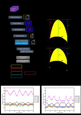
Semi-automated analysis of diaphragmatic motion with cine MRI in controls and non-ambulant Duchenne Muscular Dystrophy (DMD) patients
Courtney Bishop, Rexford Newbould, Valeria Ricotti, Christopher Sinclair, Jordan Butler, RB Matt Evans, Jasper Morrow, Mike Hanna, Paul Matthews, Tarek Yousry, John Thornton, Francesco Muntoni, Robert Janiczek
This study presents both an analysis pipeline for measuring diaphragmatic motion from cine MRI data, and the application of this image processing technique to investigate exploratory MRI endpoints of respiratory function in both healthy controls and non-ambulant DMD boys.  Cine-derived metrics of diaphragm motility and contractility correlated with sitting spirometry-derived forced vital capacity, and showed relationships with disease progression surrogates of age and months non-ambulatory, as well as a longitudinal change over 12 months.  Longitudinal changes were not seen in spirometry measures.


1566.   
Potential of Stimulated Echo Diffusion-weighted Imaging as Disease Marker in Duchenne Muscular Dystrophy
Bauke Kogelman, Maaike Putten, Matt Hall, Chris Clark, Andrew Blamire, Paola Porcari, Louise Weerd
We used stimulated echo diffusion weighted imaging (STE-DWI) in a mouse model of Duchenne muscular dystrophy (mdx). From the data in mdx or wild type animals we observe that although muscle fibre size is smaller in the mdx mouse, the diffusion data show increased diffusibility – opposite to the hypothesised effect if fibre size is the main determinant of restricted water diffusion. Muscle fibre permeability is significantly greater in the mdx mouse, suggesting that the overall system permeability has a countering effect on diffusion restriction. Additional modelling is required to capture these two opposing effects.


1567.   
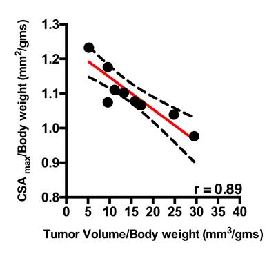
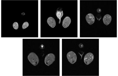
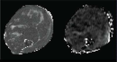
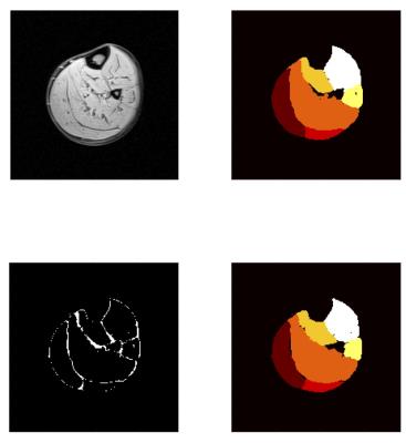
Magnetic resonance biomarkers for cachexia in a mouse model of pancreatic ductal adenocarcinoma.
Ravneet Vohra, Matthew Campbell, Joshua Park, Kayla Gravelle, Stella wHang, Yak-Nam Wang, Joo-Ha Hwang, David Marcinek, Donghoon Lee
Cancer induced cachexia is prevalent with many cancers and is characterized by loss of muscle and fat mass. In pancreatic cancer the syndrome affects approximately 80% of patients. Non-invasive multi-parametric MRI was used to monitor the progression of cachexia in a genetically engineered mouse model of pancreatic ductal adenocarcinoma (PDAC). Using pixel-by-pixel comparison, we demonstrate a significant decrease in skeletal muscle T2 and increase in magnetization transfer ratio (MTR). Additionally, we found significant difference in diffusion parameters.


1568.   
Fast delineation of calf muscles for quantitative MRI applications
Pierre-Yves Baudin, Morin Beyeler, Pierre Carlier, Olivier Scheidegger
One typical obstacle in quantitative MRI lies in the delineation of the regions of interest. When not absolutely necessary, per-muscle analyses are often abandoned, resulting in the loss of a wealth information. A software dedicated to accelerated segmentation of muscle images was recently introduced. It was tested here on the calf muscles of healthy volunteers and Duchenne patients. Results showed an important gain in average speed and were close to the manual segmentation used as reference. Thus, the tested software proved to be a valuable tool for quantitative analysis of skeletal muscle NMR images.


1569.   
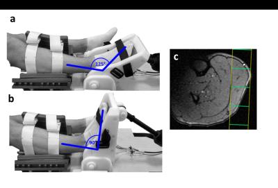
Influence of muscle fiber tension on 31P MRS recovery parameter after intense exercise
Kevin Moll, Alexander Gussew, Jürgen Reichenbach
When performing exercise induced 31P MRS motion and activities from the everyday life and the positioning of the investigated extremities within the MR scanner should be considered, since this could induce partial ischemia effects like muscular cramps. The aim of this study was to prove the effect of muscle tension on metabolic parameter measured by 31P MRS after an intense exercise. PCr recovery as well as pH kinetic was significantly slowed by a partial ischemia preventing a quantitative evaluation. 


1570.   
Multi-parametric characterization of highly fat infiltrated muscular dystrophy patients: results of a multi-variate analysis.
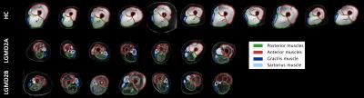
Alberto De Luca, Alessandra Bertoldo, Denis Peruzzo, Maria Grazia D'Angelo, Martijn Froeling, Filippo Arrigoni
Limb Girdle Muscular Dystrophies (LGMD) are a family of myopathies characterized by progressive degeneration and fat infiltration of muscular tissue. In the context of a study that includes Dixon, T2quantification and diffusion MRI (dMRI), we investigated a multi-variate analysis to remove the effect of fat from concurrent measures and correlate them with clinical indexes of strength. In our dataset of highly infiltrated patients, T2 and dMRI metrics were strongly biased by fat. Our results show that it is possible to mitigate the bias by multi-variate modeling of the FF effect while retaining disease specific effects. 


1571.   
Changes in muscle morphometry, composition and performance in patients with sporadic inclusion body myositis
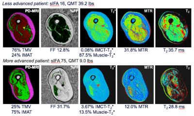
Didier Laurent, Jonathan Riek, Christopher DJ Sinclair, John S. Thornton, Ronenn Roubenoff, Dimitris A. Papanicolaou, Parul Houston, Tarek Yousry, Pedro M. Machado, Michael G. Hanna
A comprehensive MRI approach was used to characterize changes in muscle structure and composition in patients with sporadic inclusion body myositis (sIBM) over 1-year. Results showed progressive deterioration in muscle quality, e.g. increased connective tissue volume in the most affected muscles, and this appeared to be associated with a functional impact.


1572.   
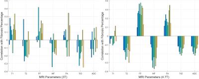
Correlating MRI and Histological Parameters in GRMD Muscles: A Comparison Between 3T and 4.7T Acquisitions
Aydin Eresen, Stephen McConnell, Sharla Birch, Wade Friedeck, Jay Griffin, Joe Kornegay, Jim Ji
The relationship between localized histological truth and various MRI quantitative measures were investigated using canine models. In this study, GRMD pectineus muscle samples were imaged on a 3T clinical scanner and a 4.7T small-bore scanner. Trichrome stained histology slice was registered to the 3D MRI volume. Localized quantitative MRI parameters are correlated with histology analysis (muscle, fibrosis and interstitial tissue percentage). Parameters derived from the 4.7T MRI data show consistent correlations, while those from 3T MRI data do not show consistent correlation.


1573.   
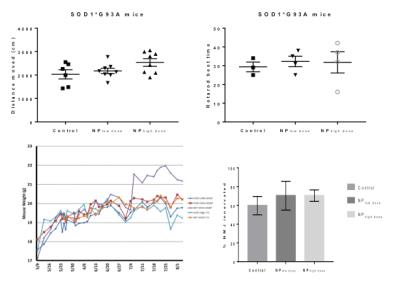
Targeted modulation of retinoid signaling using polymeric nanoparticles in a mouse model of Amyotrophic Lateral Sclerosis
David Medina, Eugene Chung, Ricki Ceton, Robert Bowser, Rachael Sirianni, Gregory Turner
Retinoid signaling activity in the CNS, mediated by RARβ, directly influences ALS pathology development and progression in vivo and CNS targeted delivery of RARβ agonists can reduce ALS pathology in vivo when delivered systemically via polymeric nanoparticles.  In this study the therapeutic affect of adapalene, a RARβ agonist, loaded nanoparticles were examined by measuring quadriceps volume in a SOD1G93A mouse model of ALS.


1574.  
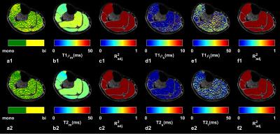
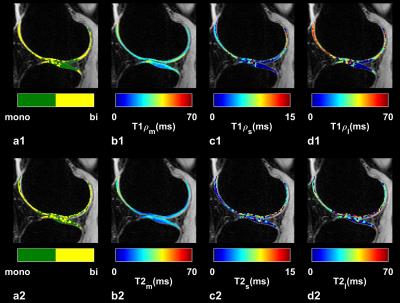
Multicomponent 3D-T1? and T2 Relaxation Mapping of Skeletal Muscle: In-vivo Feasibility
Azadeh Sharafi, Gregory Chang , Ravinder Regatte
In this study, we investigated the in-vivo feasibility of multicomponent 3D-T1ρ and T2 relaxation mapping in calf muscle using 3T MRI in clinically feasible scan times on eight healthy volunteers. Our preliminary results demonstrate that the biexponential model better characterized the relaxation behavior in calf muscle and can be used to differentiate between different water compartments associated with macromolecules (collagen and contractile proteins) and extracellular/vascular water in calf muscle.


1575.   
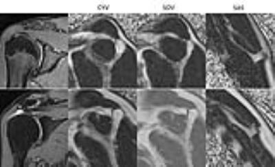
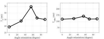
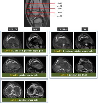
Fat fraction and muscular volume of entire supraspinatus muscle with/without tendon tear using IDEAL-IQ sequence
Taiki Nozaki, Yasuyo Teramura, Takeshi Hara, Saya Horiuchi, Atsushi Tasaki, Yasuyuki Kurihara, Hiroshi Yoshioka
Muscular atrophy and fatty degeneration of the supraspinatus muscle occurs after rotator cuff tear. It is often necessary to evaluate for these muscle abnormalities following tears to determine surgical candidacy. We usually evaluate this on a single slice. However, it is not well known whether this slice is representative of fatty degeneration and muscular atrophy overall. Furthermore, it is also not known how fatty degeneration and muscular atrophy will progress after tear. The purpose of this study was to make a standardized fat fraction and muscular volume model in patients with or without supraspinatus tendon tear using IDEAL-IQ sequence. 


1576.   
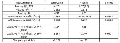
Perturbed muscle mitochondrial function in Sarcopenia
Jamie Ho, Stacey Tay, Subhasis Banerji , Mary Stephenson
In this study we use 31P MRS in combination with Near Infra-red Spectroscopy to assess muscle mitochondrial function and changes in oxygenation in patients with Sarcopenia as to date, the etiology and molecular mechanisms of sarcopenia remain poorly understood. 10 males diagnosed with sarcopenia and 10 healthy controls were scanned where 31P MRS: A fully relaxed 31P MR spectrum data  and exercise paradigm were acquired. MVC were significantly different between the sarcopenic and healthy groups (p=0.0044).  Despite this, there was a tendency for increased ATP turnover in patients compared with healthy controls (44% and 66% MVC (ATP turnover p= 0.5642 44%MVC, p= 0.6128 66%MVC). Patients with sarcopenia showed significantly higher oxidative ATP synthesis ATP at 66%MVC where p= 0.0477. These results indicate that at least some of this increased requirement can be met oxidatively, with no contribution from breakdown of PCr at the end of exercise.


1577.   
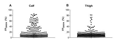
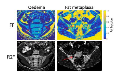
Disease progression in skeletal muscles of Myotonic Dystrophy Type 1 evaluated using quantitative MRI
Linda Heskamp, Marlies van Nimwegen, Guillaume Bassez, Marieke Ploegmakers, Jean-Francois Deux, Baziel van Engelen, Arend Heerschap
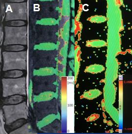
We studied the occurrence and progression of fatty infiltration, atrophy and edema-like processes in calf and thigh muscles of Myotonic Dystrophy Type 1 (DM1) patients. Fat fraction (FF) and muscle volume (MV) were obtained by a DIXON-sequence and T2 of muscle water (T2water) was calculated by a bi-component extended-phase-graph model. Calf and thigh muscles show fatty infiltration and increased T2water in non-fat infiltrated and fat infiltrated muscles. Atrophy is observed in four calf muscles and one thigh muscles. FF significantly increases in 10 months in fat infiltrated and non-fat infiltrated muscles, but MV only decreases in fat infiltrated thigh muscles.


1578.   
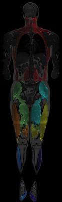
Body Composition Analysis Combined with Individual Muscle Measurements using Dixon-MRI
Janne West, Thobias Romu, Sofia Thorell, Hanna Lindblom, Emilia Berin, Anna-Clara Spetz Holm, Lotta Lindh Åstrand, Magnus Borga, Mats Hammar, Olof Dahlqvist Leinhard
Body composition analysis is increasingly important for diagnosis and follow-up in many patient groups and medical conditions. The combined fat and muscle quantification on global and regional level is not commonly reported. In this study a Dixon-MRI based acquisition and body composition analysis was extended to quantify individual muscles. Test-retest reliability was established in a clinically relevant group of 36 postmenopausal women. This method enables advanced phenotyping combined with measurements of specific muscles to target clinical questions.


1579.  
A Potential Pathophysiological Link Between Generalized and Localized Muscle Fat Infiltration in Chronic Whiplash Patients
Anette Karlsson, Anneli Peolsson, James Elliott, Thobias Romu, Helena Ljunggren, Olof Dahlqvist Leinhard
Muscle fat infiltration (MFI) in the cervical multifidi muscles and whole-body skeletal muscle tissue was measured in participants with chronic whiplash associated disorders (WAD) using whole-body fat-water separated MRI to investigate potential interaction between deep neck muscle and generalized MFI. Thirty participants with chronic WAD and 30 matched controls were included. MFI in multifidi was strongly associated to whole-body MFI as well as to severity of WAD. The strong association between Multifidi MFI and whole-body MFI indicates that both generalized factors and localized effects related to the trauma may be important for understanding the pathophysiology of chronic WAD.


1580.  
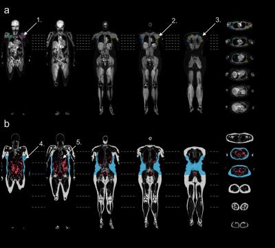
Whole-body MRI in facioscapulohumeral muscular dystrophy: Preliminary results
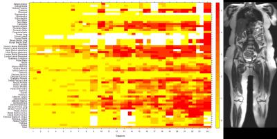
Doris Leung, Li Pan, John Carrino, Kathryn Wagner, Michael Jacobs
Facioscapulohumeral muscular dystrophy (FSHD) is a hereditary disorder that causes progressive muscle wasting. Whole-body MRI (WBMRI) was used to scan 24 adults with genetically-confirmed type 1 FSHD. Muscles were scored for fat infiltration and edema-like changes. Fat infiltration scores were compared to muscle strength and function measurements. Our analysis reveals a distinctive pattern of muscle involvement and sparing in FSHD. Averaged fat infiltration scores for muscle groups in the legs were statistically significantly associated with quantitative muscle strength and 10-meter walk time. We conclude that WBMRI offers a promising disease biomarker in FSHD and other muscular dystrophies.


1581.  
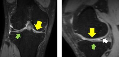
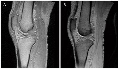
DTI imaging application of vastus medialis oblique muscle in recurrent patellar dislocation
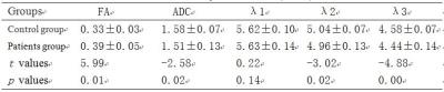
Lisi Liu, Zhuozhao Zheng, Huishu Yuan, Lizhi Xie
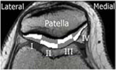
The osseous-related factors that influence patellofemoral joint instability have been well-studied in plenty of the previous reported literatures. However, the muscle-related factors that affect patellofemoral joint instability have not been fully revealed. MRI is a noninvasive imaging method, including conventional MRI scans and diffusion tensor imaging (DTI) sequence, which can respectively reflect the macroscopic and microscopic structures of the muscle fibers.1,2 In the current study, the cross-sectional area were measure with DTI parameters of FA, ADC, and λ1, λ2, λ3 of vastus medialis oblique (VMO). Thereafter, these parameters were compared between the recurrent patellar dislocation patients and the healthy volunteers. 


1582.  
Diffusion Tensor imaging in the Injuries of the Levator Ani Muscle in Female Pelvic Floor Dysfunction
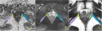
Huici Zhu, Jianyu Liu, Yan Zhou, Lizhi Xie
To find the most vulnerable component of levator muscle, this research compared the levator ani muscle which contains pubovisceral, puborectal and iliococcygeal muscles between the control group and the patient group by using DTI. 49 female PFD (pelvic floor dysfunction) patients and 51 female healthy subjects underwent DTI imaging on a 3.0T MR system. A significant difference was found in the FA value of the right pubovisceral muscle between the two groups, and therefore, this part are more likely to be damaged comparing with other compartments of levator ani muscle. DTI with fiber tractography permits the evaluation of the injuries of levator ani muscle, which has a certain effect in revealing the pathogenesis of the female pelvic floor dysfunction.


1583.   
Analysis of T2 Relaxation Times in Vastus Lateralis Muscle after Anterior Cruciate Ligament Injury
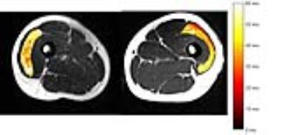
Richard Lawless, Peter Hardy, Anders Andersen, Brian Noehren
Anterior cruciate ligament (ACL) injuries are associated with a persistent decrease in quadriceps muscle strength despite rehabilitation. Previous studies have shown that ACL injury results in muscle disorganization, which could account for the loss in strength. We sought to use dual-spin echo and multi-spin echo sequences to estimate T2 of the vastus lateralis in injured and uninjured limbs of twenty-two ACL injured subjects. T2 of the injured limb was significantly longer than the uninjured limb for both pulse sequences. Our results suggest that T2 relaxation may provide clinicians a means to quantitatively monitor muscle recovery after ACL injury.    


1584.   
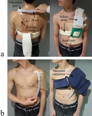
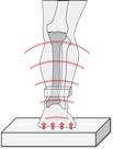
Optimal excitation location for magnetic resonance elastography of the supraspinatus muscle
Daiki Ito, Tomokazu Numano, Kazuyuki Mizuhara, Koichi Takamoto, Takaaki Onishi, Hisao Nishijo
It is difficult to palpate the supraspinatus muscle since it is difficult to determine their anatomic location superficially. Magnetic resonance elastography (MRE) can measure tissue stiffness quantitatively only if vibrations reach these tissues. To vibrate the tissues efficiently, it is necessary to determine the best excitation location. We investigated the excitation location suitable for MRE of the supraspinatus muscle. When the excitation location was placed on the trapezius muscle, the wave images represented clear wave propagation compared with those on the head of humerus. Therefore, optimal excitation location may be the trapezius muscle in the MRE of the supraspinatus muscle.


1585.   
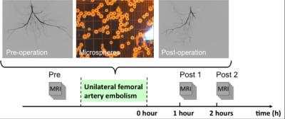
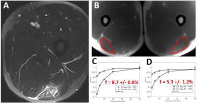
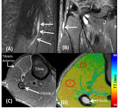
Simultaneous measurement of muscle transverse relaxation rates of R2, R2' and R2*: application in unilateral femoral artery embolization
Chengyan Wang, Rui Zhang, Bihui Zhang, Haochen Wang, Xiaodong Zhang, Min Yang, Jue Zhang, Xiaoying Wang, Jing Fang
Diagnosis of the ischemic lesion is challenging and often requires the use of pharmaco-mechanical methods. This study demonstrates the feasibility of using a susceptibility-based MRI technique with the multi-echo gradient and spin echo (MEGSE) sequence to assess the skeletal muscle oxygenation alternations in animal model of unilateral femoral artery embolization. The results shown that the skeletal muscle R2 decreases obviously from after femoral artery embolization, while R2’ increases correspondingly. MEGSE seems to be an appropriate method for the evaluation of skeletal muscle ischemia after embolization.


1586.  
Feasibility of Quantifying Bone Metabolism in the Femoral Neck in Human Subjects using Multi-Modality Imaging
Christian McHugh, William Raynor, Tom Werner, Abass Alavi, Chamith Rajapakse
18F-sodium fluoride (NaF) is a readily available radiotracer that has shown great potential to study bone metabolism associated with osseous diseases such as osteoporosis. We analyzed PET/CT and MRI data to study the effects of bone volume fraction and marrow distribution on the uptake of NaF in the proximal femur. Our data showed that the mean standardized uptake (SUVmean) decreased with age and became more significant when local adjustments for bone and bone marrow were taken into account. We foresee the importance of high-resolution microstructural bone MRI for the partial volume correction of PET data for accurate quantification bone metabolism.


1587.  
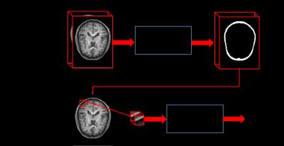
Segmentation of CMF Bones from MRI with A Cascade Deep Learning Framework
Dong Nie, Li Wang, Jianfu Li, Daeseung Kim, James Xia, Dinggang Shen
Accurate segmentation of CMF bones from MRI is one of the most important fundamental steps in clinical applications, and it can also be used in other areas, such as character animation and assistive robotics. In this paper, we propose a cascade framework based on the recently well-received and prominent deep learning methods. Specifically, we first propose a 3D fully convolutional network architecture for a coarse segmentation of the bone tissue. Further, we propose to utilize CNN for fine-grained level segmentation around the predicted bone tissue area. The conducted experiments show that our proposed 3D deep learning model could achieve good performance in terms of segmentation accuracy.


1588.   
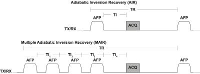
Robust Pore Water Suppression in Cortical Bone with Multiple Adiabatic Inversion Recovery
Kevin Harkins, Sasidhar Uppuganti, Jeffry Nyman, Mark Does
Measurement of bound water concentration in cortical bone with MRI is a promising method for evaluating bone fracture risk. One approach to measure bound water involves suppression of pore water signal with an adiabatic inversion-recovery pulse sequence. However, this approach requires a priori knowledge of pore water T1 which itself is expected to vary with bone porosity. We propose to minimize the effect of subject-dependent pore water T1 variation by in bound water imaging using a multiple adiabatic inversion recovery preparation optimized to suppress pore water over a broad T1 domain. 


1589.  
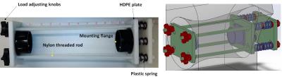
MRI Assessment of the Effect of Low-Magnitude Mechanical Stimulation on Postmenopausal Bone Loss
Chamith Rajapakse, Wenli Sun, Christian McHugh, Ben Newman, Mona Al Mukaddam, Peter Snyder, Felix Wehrli
Low-magnitude mechanical stimulation (LMMS) has shown great potential as a non-pharmacological intervention for improving bone quality in animal models. However, human trials have yielded less compelling evidence, possibly related to difficulties in maintaining adherence and use of conventional imaging techniques not being able to detect subtle longitudinal changes. Here we investigated the use of high-resolution structural bone MRI in monitoring treatment efficacy of LMMS in postmenopausal women. The data show that baseline bone volume fraction at the distal tibia is associated with the changes observed over one year of active LMMS or placebo treatment.


1590.   
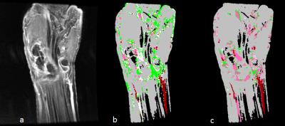
Potential of High Resolution Isotropic Microstructural MRI of the Proximal Femur
Austin Alecxih, Elizabeth Kobe, Alyssa Johncola, Marissa Evans, Shivali Patel, Sun Kim, Benjamin Newman, Gregory Chang, Christian McHugh, Chamith Rajapakse
Microstructural MRI based finite element modelling is an area of current research in regards to the role that this technology can play as an efficient and affordable metric for quantifying bone integrity and for identifying fracture risk.  This study was conducted in an attempt to unveil the extent to which MRI resolution impacts the quantitative output generated by finite element models pertaining to the parameter of bone stiffness of the proximal femur in vitro. Conclusions reveal a statistically significant variance relative to the values generated for two differing MRI resolution classes.  


1591.   
Simplified approach to quantification for hand synovitis in rheumatoid arthritis using dynamic contrast enhanced MRI: pixel-by-pixel time intensity curve shape analysis
Yuto Kobayashi, Tamotsu Kamishima, Taro Sakashita, Hiroyuki Sugimori, Shota Ichikawa, Atsushi Noguchi, Michihito Kono, Toshitake Iiyama, Tatsuya Atsumi
Quantification for synovitis using time intensity curve (TIC) shape analysis of rheumatoid arthritis has been believed to require long acquisition time up to 6-7 minutes to observe washout phase. In this study, we found that wash out phase does not contribute to accurate depiction of synovitis in the hand and simplified TIC shape analysis could significantly decrease acquisition time from about 6 minutes to 3 minutes.


1592.   
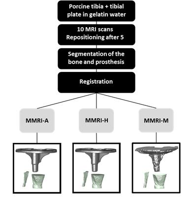
A novel approach to measure tibial component migration by low field markerless magnetic resonance imaging
Femke Schroder, Frank Simonis, Dean Pakvis, Rianne Huis in't Veld, Bennie ten Haken
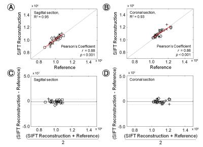
This study evaluated if low field MRI is a practical alternative for Roentgen stereophotogrammetric-analysis (RSA) to measure prosthetic migration. This also included determining the optimal registration method for this purpose. The detection of migration on low field MRI was sufficient for clinical use in two of the translation directions and all three rotational directions. Manual registration proved to be the most accurate method for markerless MRI (MMRI) estimation of the migration. 


1593.  
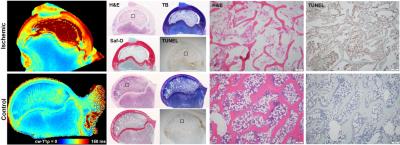
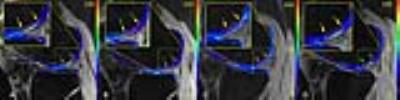
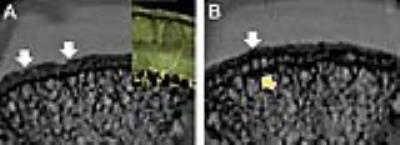
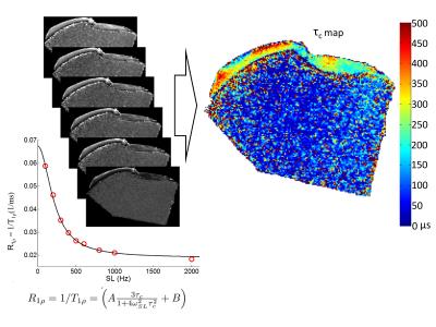
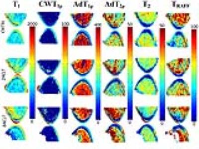
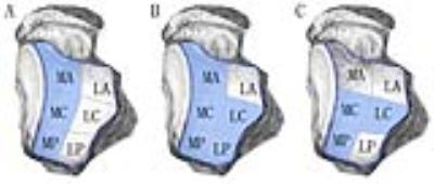
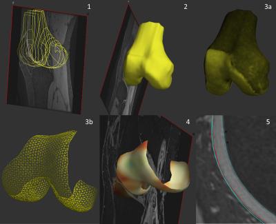
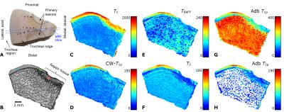
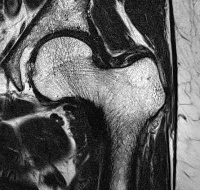
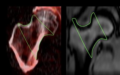
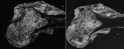
Quantitative Mapping of the Ischemic Femoral Head in a Piglet Model of Legg-Calve-Perthes Disease
Casey Johnson, Luning Wang, Ferenc Toth, Cathy Carlson, Harry Kim, Jutta Ellermann
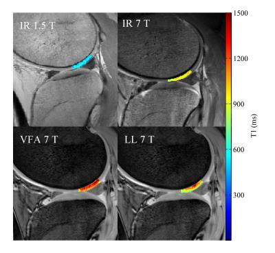
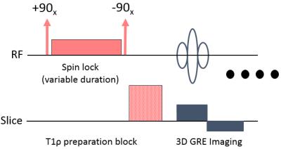
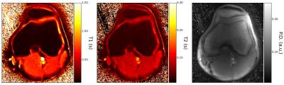
This study investigated the sensitivities of T1, T2, continuous-wave T1ρ, adiabatic T1ρ, and RAFF relaxation times to ischemia-induced necrosis and subsequent repair of the developing femoral head in a piglet model of Legg-Calve-Perthes disease (LCPD), a disabling childhood hip disorder. Quantitative maps of ischemic and control femoral heads acquired ex vivo at 9.4T MRI were compared numerically and validated with histology. Our findings reveal that the relaxation times provide complementary information on the status of the pathological hip, which can potentially address a clinical need for diagnostic imaging tools to assess the early stages of LCPD.


1594.  
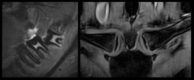
Age estimation using MR imaging of the third molar teeth and the medial clavicular epiphysis: Validation of a multifactorial approach
Thomas Widek, Pia Baumann, Heiko Merkens, Thomas Ehammer, Andreas Petrovic, Isabella Klasinc, Martin Urschler, Eva Scheurer
High migration rates in the last years put forensic age estimation of living people at the forefront of forensic research. The established standard for age estimation uses images acquired with ionizing radiation, therefore radiation-free alternatives such as MRI are currently of high interest. In this study a CT based multifactorial approach that combines wisdom teeth and clavicles was validated with MRI data. The sensitivity to estimate subjects under 18 years of age as minors with MRI lies above 93%. Our results showed that MR could replace the CT based multifactorial Approach.


1595.   
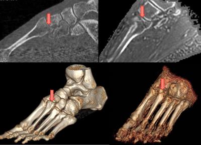
Three-dimensional fracture visualization with Zero Echo Time MRI
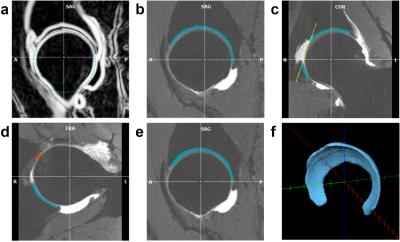
Gaspar Delso, Geoffrey Warnock, Caroline Zellweger, Felipe de Galiza, Irene Burger, Martin Huellner, Patrick Veit-Haibach
The present study is aimed at assessing the performance of proton density-weighted zero-echo time (ZTE) acquisition for the visualization of bone fractures.


1596.   
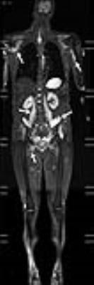
An Efficacy Analysis of Whole-Body Magnetic Resonance Imaging in the Diagnosis and Follow-Up of Polymyositis and Dermatomyositis
Zhen-guo Huang, Min-xing Yang, Bao-xiang Gao, Xiao-liang Chen, He Chen, Kai-ning Shi
To evaluate the value of whole-body magnetic resonance imaging (WBMRI) in diagnosing muscular and extramuscular lesions in patients with polymyositis (PM) and dermatomyositis (DM). A retrospective analysis of WBMRI data was performed on PM / DM patients who met the Bohan and Peter diagnostic criteria. WBMRI comprehensively displays the muscular involvement in PM / DM patients, and has the ability to diagnose other associated extramuscular diseases, such as ILD and systemic malignancy. WBMRI can also help screen for multifocal steroid-induced osteonecrosis.
 
Miscellaneous MSK
Traditional Poster
Musculoskeletal

 
Monday, 24 April 2017
Exhibition Hall  16:15 - 18:15


1597.   
Deformable Registration of In Vivo Human Spine Magnetic Resonance Imaging Using Scale Invariant Feature Transform (SIFT) Algorithm
Dong-Hoon Lee, Do-Wan Lee, Yong Hyun Chung, Bong-Soo Han
Deformable registration process for in vivo human spine MR images can provide the crucial information to investigate the diagnostic performance and treatment effects of pathologies. Here, based on the scale invariant feature transform (SIFT) algorithm, we attempted to evaluate the deformable registration process for spine images, and compared with commercially mounted algorithm in MRI system. The results qualitatively and quantitatively showed fine results, and clearly showed the reproducibility of the use of SIFT algorithm compared with commercially mounted stitching algorithm on MRI system. Our approach can be helpful for the extension of other medical imaging modalities for image deformable registration.


1598.  
Perfusion and Fat Content of Lumbar Vertebral Body: Global and Regional Analysis
Hou-Ting Yang, Yi-Jui Liu, Hing-Chiu Chang, Xiang-Wei Xie, Ming-Fun Lin, Wing P. Chan
Vertebral blood perfusion has been reported that there was a decrease trend in normal aging people and post-menopause women and those with arteriosclerosis, fat marrow increased and osteoporosis. Although DCE-MRI and MRS have shown potential probing bone marrow content and blood perfusion characteristics of vertebra body in previous studies, all of these were ROI analysis only and lack regional information for bone marrow content and blood perfusion. Therefore, The purposes of this study were to investigate the regional perfusion and bone marrow content in vertebra body with use of DCE-MRI and six-point Dixon IDEAL technique by pixel-by-pixel analysis.


1599.  
Non-Gaussian water diffusion kurtosis imaging and T2* mapping of Intervertebral Disc Degeneration at 3T MR
Feifei Zeng, Yun fei Zha, Dong Xing, Liang Li, Hui Lin
To assess the feasibility of detecting early biochemical changes of nucleus pulposus (NP) in intervertebral disk degeneration (IVDD) by diffusion kurtosis imaging (DKI), and compare mean kurtosis (MK) and T2* mapping in IVDD according to Pfirrmann grades. Twenty individuals underwent lumbar sagittal T2-weighted image, DKI and T2* mapping. DKI related parameters—mean kurtosis (MK) and T2* values were measured. The differences between MK and T2* value in NP were highly significant. Pfirrmann grades were inversely significantly correlated with T2* values in the NP, and positively correlated with MK values, especially between Pfirrmann grade II ,III and ?.


1600.  
Bone marrow iron assessment using quantitative susceptibility mapping in iron overload rabbit model
Yoonho Nam, Eo-Jin Hwang, Joon-Yong Jung
The aim of our study was to assess the feasibility of quantitative susceptibility mapping (QSM) as a tool to quantify bone marrow iron level non-invasively. Nine rabbits were scanned before and after iron accumulations, and bone marrow susceptibility values of these rabbits were evaluated from the reconstructed QSM images. The results showed a linear correlation between the measured susceptibility changes and the injected iron doses.


1601.   
Quantitative study of longitudinal distribution of fat fraction in the lumbar spine
Huiying Chen, Huishu Yuan, Lizhi Xie, Ning Lang
The aim of this study was to explore the fat fraction(FF) variation of inter and intra vertebral bodies in the lumbar spine, by using water-fat imaging. In lumbar spine, L1 and L2 vertebras demonstrated lower FF level but with more common ‘margin higher’ FF distribution pattern comparing to other vertebras, which might be related to stress and activities of the vertebra. We suggested that FF distribution pattern indicate certain skeletal remodeling that affects bone strength, which may be a promising indicator in assessing prevalent fracture besides the increased average FF of the whole vertebral body.


1602.   
Intervertebral disc heterogeneity decoded with MRI histogram analysis
Christian Waldenberg, Hanna Hebelka, Helena Brisby, Kerstin Lagerstrand
Low back pain has major consequences for both the individual as well as for the society which often results in sick leave from work. Unfortunately, health care today lacks diagnostic techniques and procedures necessary to select and treat patients who would most benefit from treatment. Histogram analysis based on MRI imaging has the potential to display disc heterogeneity and possibly depict painful discs. A total of 98 histograms of 49 intervertebral discs were generated based on three mid-sagittal slices of high quality T2W MRI images and T2-maps. Using MATLAB, a two component Gaussian mixture distribution model was fitted to the histogram data in order to retrieve disc heterogeneity measures. Mann-Whitney U-test displayed a significant difference in disc heterogeneity measures with increased Pfirrmann grade. 


1603.  
Synthetic contrast optimization of bone metastases
Catharina Petersen, Marcel Warntjes
To achieve optimal contrast when imaging bone metastases, the patient would have to imaged multiple times at various contrast settings. This is not possible in practice, resulting in the use of standard contrasts that are often sub-optimal due to patient-specific variations in both healthy and metastatic tissue.

Using synthetic MRI, it is possible to synthesize a multitude of contrasts from a single scan. This makes it possible to determine optimal contrast weighing post-scan. Studying bone metastases, synthetic images where used to develop a mathematical model of perceived contrast, taking both tissue properties and the human visual system into account.


1604.   
The presence of newly defined classical features of intraneural perineurioma may obviate the need for fascicular biopsy and pathologic diagnosis
Thomas Wilson, Shelby Stewart, B. Matthew Howe, Robert Spinner, Kimberly Amrami
Intraneural perineurioma is typically diagnosed by targeted fascicular biopsy and histopathologic analysis.  Through review of clinical history and magnetic resonance imaging of consecutive patients undergoing fascicular biopsy, we define the classical features of intraneural perineurioma as no cancer history, unifocal disease, moderate-severe T2 hyperintensity, moderate-severe contrast enhancement, homogeneous contrast enhancement, fusiform shape, and enlargement of involved nerves.  This combination of features has a high specificity and moderate sensitivity for the diagnosis of perineurioma.  When the classical features of perineurioma defined in this study are present, the high specificity of this combination of features may obviate the need for fascicular biopsy.  


1605.  
The value of dynamic contrast-enhanced MRI quantitative parameters in evaluating the efficacy of neoadjuvant chemotherapy for osteosarcoma
ma huan, li kun, cao peng
Osteosarcoma is the most common bone sarcoma in children and adolescents 1. Response of osteosarcoma to NAC should be assessed preoperatively, but conventional imaging has limit, and histopathological examination cannot dynamically observe the response of tumor to treatment. DCE-MRI can reflect the pathophysiology of tumor at microvascular and cellular level. Currently, researchers have used DCE-MRI qualitative and semi-quantitative method to analyze the response of osteosarcoma to NAC, but studies on DCE-MRI quantitative analysis are rarely reported. Therefore, we analyzed the quantitative parameter change of DCE-MRI in 17 patients of osteosarcoma before and after NAC, and compared good response and poor response to chemotherapy in order to explore the value of DCE-MRI quantitative parameters in assessing the response of osteosarcoma to NAC. 


1606.  
Fat Fraction Histogram Analysis: Insights into the Biology of Lipomatous Tumors
Mikael Skorpil, Henric Rydén, Elisabet Lidbrink, Otte Brosjö, Johan Berglund
Fat content evaluation in soft-tissue tumors is based on visual grading of T1-weighted images with uncertain accuracy and inter-rater variability. We used a multi-point Dixon technique to quantitatively measure the distribution of proton density fat fraction in different lipomatous tumors.  The effect of radiotherapy on fat content in a myxoid liposarcoma was also assessed, with histograms revealing a distinct alteration. Our data give supporting evidence that maturation of tumor cells is the cause for the lipoma-like areas seen after radiotherapy of myxoid liposarcomas. On a voxel level (2×2×2 mm3) various lipomatous tumors exhibited different fat fraction histograms.


1607.   
Characterization of Through-Plane and In-Plane Artifacts using a 3D-Printed Grid Phantom with an Embedded Metal Hip Implant
Gregory Hong, Matthew Teeter, Jaques Milner, Steven Pollmann, Maria Drangova, David Holdsworth
Metal Artifact Reduction (MAR) is required for orthopedic imaging near implants. Novel MAR techniques have been developed in the past decade, creating a need for quantitative evaluation of the effectiveness of geometric distortion correction. We have developed a 3D-printed modular conformal grid phantom, consisting of a grid of regularly spaced spherical markers.  This phantom provides a measure of inherent field inhomogeneity, and contains a conformal cavity in which a metal object can be embedded. This approach provides a means to characterize through-plane and in-plane artifacts across a 3D volume, facilitating testing and validation of novel MAR techniques during development. 


1608.  
Cartesian UTE with echo time below 0.5 millisecond
Franciszek Hennel, Markus Weiger, Manuela Rösler, Roger Luechinger, Bertram Wilm, Klaas Pruessmann
MRI with sub-millisecond TE typically uses non-Cartesian sampling strategies such as radial UTE leading to increased sensitivity to eddy currents and a high computational burden. We report that TE values in the range so far reserved for radial UTE can be reached with Cartesian sampling using a gradient coil dedicated for muscoskeletal applications in humans.


1609.   
Iterative Denoising of Undersampled PROPELLER Diffusion-Weighted Imaging near Metal Implants
Sampada Bhave, S. Shivram Kaushik, Kevin Koch
Diffusion weighted imaging is widely used in musculoskeletal MR imaging. Current implementations of diffusion-weighted imaging include the use of PROPELLER DUO imaging sequence and multi-spectral MRI imaging techniques. However, these implementations are restricted by long acquisition times. In this work we propose to an approach to undersample the blades in a uniform fashion and denoise the reconstructed image using total variation based denoising approach. This approach provides acceptable results up to an acceleration factor of 3 and reduces the scan time. This reduction in scan time can be used to acquire multiple slices within clinically feasible scan times. 


1610.   
Ultrashort Echo Time Chemical Shift Quantitative Susceptibility Mapping (UTE-CS-QSM) for Hemosiderin Assessment in Hemophilic Arthropathy
Xing Lu, Annette Drygalski, Alexey Dimov, Lena Volland, Zhe Liu, Yi Wang, Jiang Du, Eric Chang
Hemosiderin deposition in hypertrophic synovium is a hallmark of progressive hemophilic arthropathy and accumulates with joint bleeding. . Conventional quantification of hemosiderin uses semi-quantitative MRI scores, assigning grades of small, moderate, or large to assess the burden of hemosiderin. To date, there is no known method to assess or quantify hemosiderin concentration within synovium due to the extremely short T2 values of this tissue. In this pilot study, we demonstrated that the clinically compatible IDEAL UTE-CS-QSM technique can be used to quantify hemosiderin depositions. We have shown that synovium in joints with hemophilic arthropathy demonstrates various susceptibility values, likely corresponding to different concentrations of hemosiderin.


1611.   
Chemical Shift T1 Quantification with Three-dimensional Ultrashort Echo Time Cones Imaging and IDEAL (3D UTE-Cones-IDEAL) Processing
Xing Lu, Michael Carl, Yajun Ma, Yanchun Zhu, Yinghua Zhao, Wenhui Yang, Eric Chang, Jiang Du
Conventional T1 measurements are subject to errors due to fat contamination and invisibility of the short T2 components. In this study, we aimed to develop 3D UTE with Cones sampling and IDEAL processing (3D UTE-Cones-IDEAL) with variable TRs for robust fat/water separation and more accurate T1 estimation of joint tissues ex vivo and in vivo.  The results show that this technique can robustly separate water and fat signal and lead to longer T1s for water-rich tissues (such as muscle, tendon and peripheral nerve), and shorter T1s for fat-rich tissues.


1612.   
On the Source of MRI Off-Resonance Observables in Arthroplasty-Induced Metallosis
Kevin Koch, Matthew Koff, Thomas Bauer, Hollis Potter
The deposition of metal particles near total joint replacements has substantial impact on patient morbidity.   Current standards of clinical management of patients with symptomatic total hip replacements are highly leveraged on the presence of wear debris in periprosthetic tissues.  Our group has recently developed methods to perform off-resonance based identification of metal particle deposits.  Here, we provide analysis on the source of tissue-based metal particle Larmor frequency offsets.  The results of this analysis provide new insight on the role of off-resonance observables in the progression of symptomatic failed total hip replacements.   


1613.  
Fat Fraction and R2* as Measures of Bone Marrow Composition and Structure: Validation in Fat-Water-Bone Phantoms
Timothy Bray, Alan Bainbridge, Shonit Punwani, Yiannis Ioannou, Margaret Hall-Craggs
MRI is becoming central to clinical decision-making in diseases involving the bone marrow, including spondyloarthritis and multiple myeloma. Both diseases cause alterations in marrow composition and structure, but there is currently no reliable method for quantifying these changes. Here, we describe a series of fat-water-bone phantoms enabling validation of proton density fat-fraction (PDFF) and R2* as measures of marrow composition and trabecular bone mineral density (BMD) respectively. Our data suggest that PDFF measurements can be achieved with excellent accuracy. Furthermore, there was a strong relationship between R2* and BMD, suggesting that R2* could be used to monitor BMD in vivo. 


1615.   
Application of lower extremity oxygenation imaging in animal model of microsphere-induced femoral artery embolization
Chengyan Wang, Haochen Wang, Bihui Zhang, Rui Zhang, Xiaodong Zhang, Min Yang, Jue Zhang, Xiaoying Wang, Jing Fang
Embolization following percutaneous intervention is a universal problem occurring in all vascular beds, especially in lower extremities. This study assesses the performance of a noninvasive MRI based oxygenation imaging, in characterizing muscle oxygen extraction fraction (OEF) level after microsphere-induced lower extremity embolization in animal models. By comparison of the calculated muscle OEFs and R2’ before and after surgery, significantly increase could be found. The results show promise in the evaluation of early muscle ischemia in many clinical diseases such as acute lower limb embolism or peripheral artery disease.


1616.  
Age estimation of soft tissue hematomas using 3.0 T MRI: A feasible approach
Kathrin Ogris, Thomas Widek, Eva Hassler, Andreas Petrovic, Thorsten Schwark, Peter Grabuschnigg, Eva Scheurer
Forensic imaging requires reliable analysis of hematomas and their resolution as they can have significant medico-legal consequences. In 30 healthy volunteers autologous blood was injected into the subcutaneous fatty tissue of the thigh and repetitive MRI scans of the artificial hematoma were performed on a 3T scanner. Due to the contrast behavior three relevant age ranges were defined and validated. The generated approach correctly classified all tested hematomas with an age less or equal 24 hours. The development of a reliable and objective MR-based age estimation method of hematomas is in the interest of justice. 


1618.   
2D Adiabatic UTE T1rho Imaging: Reduced Sensitivity to the Magic Angle Effect
Yajun Ma, Yanchun Zhun, Jiang Du, Eric Chang
Spin lattice relaxation in the rotating frame (T1rho) has been proposed as a biomarker to probe biochemical changes in cartilage. It reflects the slow interactions between motion-restricted water molecules and their local macromolecular environment, and provides unique biochemical information in the low frequency region ranging from a few hundred hertz to a few kilohertz. Changes to the extracellular matrix (ECM), such as proteoglycan (PG) loss, may be reflected in measurements of T1rho and T1rho dispersion. However, T1rho based on conventional continuous wave (CW) spin-locking preparation pulses demonstrate strong magic angle effects. Studies have shown an increase of over 100% in measured T1rho when the fibers were oriented from parallel to 54° relative to the B0 field. Adiabatic T1rho has been proposed to reduce the magic angle effect. However, many joint tissues such as the deep layers of articular cartilage, menisci, ligaments and tendons have short T2 relaxation and cannot be reliably imaged and quantified using the adiabatic T1rho sequence. In this study we aimed to develop a novel 2D adiabatic ultrashort echo time (UTE) T1rho sequence for magic angle insensitive imaging of both short and long T2 tissues in the joint. 


1619.   
Efficacy of double inversion recovery MR imaging in evaluation of the synovium without contrast enhancement
Wook Jin, YeNa Son, Geon-Ho Jahng, Jang Gyu Cha, Seong Jong Yun, Kyung Nam Ryu
A double-inversion recovery (DIR) sequence simultaneously voids two sources of signals from two different tissues by applying two 180 degree inversion pulses before a signal acquisition. To investigate the efficacy of the DIR image in visualization of the knee synovium without contrast enhancement,  contrast-enhanced T1-weighted fat suppressed images (CET1FS) were compared with DIR images in knee MR. Our results showed DIR images were well correlated with CET1FS images.


1620.   
The use of quantitative perfusion metrics in evaluating the relationship between perfusion level and intervertebral disc degeneration
jiao wang, yun fei zha, dong xing, bing wu, hui lin
To explore the relationship between perfusion level and intervertebral disc degeneration.18 individuals underwent MRI exams including DCE-MRI, T2*mapping as well as conventional lumbar imaging. The cranial and caudal VSB and CEP perfusion parameters (Ktrans, Kep, Ve) as well as T2* values of discs were measured. it was found that T2* showed a negative correlation with the Pfirrmann grades, while showed no correlation with the perfusion parameters.


1621.  
A Prospective, Longitudinal Assessment of Adverse Local Tissue Reactions in Resurfacing Hip Arthroplasty Versus Primary Total Hip Arthroplasty
Matthew Koff, Owen Drinkwater, Danyal Nawabi, Edwin Su, Douglas Padgett, Hollis Potter
Total hip arthroplasty devices are successful achieving pain reduction, but adverse local tissue reactions (ALTRs) frequently occur for metal-on-metal hip resurfacing arthroplasty (HRA) patients. This longitudinal study evaluated if HRA patients have a greater prevalence of ALTRs as compared to ceramic-on-poly (COP) patients. Images were acquired at 2 time points, with a 1 year interval, and evaluated for synovitis and ALTR. ALTR prevalence and synovial thickness was greater in HRA than COP subjects at both time points, with significant increases at the second time point. This study will permit a better understanding of the natural history in the development of ALTR near arthroplasty.


1617.   
Assessment of the Myotendinous Junction After Injury in Elite Athletes Using 3D Ultrashort Echo Time Magnetization Transfer (UTE-MT) Imaging and Modeling
Yajun Ma, Heinz Hoenecke, Douglas Chang, Jiang Du, Eric Chang
Myotendinous injuries are very common in sports, most often affecting the lower extremities. Both clinical examination and MRI have been widely used for the diagnosis and prognosis of acute myotendinous injuries. However, current paradigms remain insufficient as re-injury rates are as high as 34%. For elite athletes, the balance between rehabilitation time and risk of re-injury after return to play can be extremely challenging. Using conventional MRI techniques, clinical interpretation focuses on the evaluation of edema, fluid, and hemorrhage rather than the assessment of the injured/healing components of the myotendinous junction. Furthermore, immature and mature fibrous scar tissue as well as native tendon have very short T2 values and are “invisible” with clinical MRI, thus precluding their distinction and assessment. A technique to quantitatively characterize macromolecules in injured/healing myotendinous junctions could be particularly useful in recovering athletes.


1614.  
Simultaneous Quantification of Fat Fraction and R2* as measures of Bone Oedema/Adiposity and Structure in Spondyloarthritis
Timothy Bray, Alan Bainbridge, Corinne Fisher, Shonit Punwani, Yiannis Ioannou, Margaret Hall-Craggs
Treatment decisions in patients with spondyloarthritis increasingly involve the use of MRI to assess the extent, severity and type of inflammation surrounding the joints. However, conventional spin echo imaging requires subjective interpretation and gives little information about trabecular structure, despite the fact that new bone formation and bone destruction may both occur in spondylorthritis. Here, we describe the use of chemical shift-encoded MRI as a quantitative method for assessing inflammation of the sacroiliac joints. Specifically, we demonstrate the use of proton-density fat fraction and R2* as measures of bone marrow composition and structure, respectively, in both active and chronic inflammation.


1622.  
In vivo assessment of trabecular bone in the proximal femur
Maria Kalimeri, Christiani Jeyakumar Henry, Su Xiao Di, Marlena Kruger, John James Totman
Osteoporosis is a bone disease that predominately affects postmenopausal women. Routine screening of osteoporosis is performed by means of Dual X-ray Absorptiometry (DXA) which is limited by its inability to differentiate between cortical and trabecular bone, reduced accuracy due to overlying tissue and the use of ionising radiation. In this abstract, we investigate the use of MRI as an alternative, in a cohort of 95 Chinese-Singaporean postmenopausal women. Strong correlations with DXA measurements were observed, in addition to significant differences between the normal and osteopenic, normal and osteoporotic groups.
 

The International Society for Magnetic Resonance in Medicine is accredited by the Accreditation Council for
Continuing Medical Education to provide continuing medical education for physicians.