|
Applications of Microstructural Imaging in Disease
Combined Educational & Scientific Session
ORGANIZERS: Noam Shemesh, Fernando Calamante, Jennifer McNab
Monday, 18 June 2018
| N01 |
13:45 - 15:45 |
Moderators: Fernando Calamante, Noam Shemesh |
Skill Level: Intermediate to Advanced
Session Number: M-06
Overview
Microstructural changes in diseases typically occur relatively early on, making them interesting biomarkers for disease onset and staging. This CES aims to first describe the state-of-the-art in methods designed to probe different aspects of microstructure and their application in disease, followed by an overview of applications in cancer.
Target Audience
M.D.s and Ph.D.s interested in the applications of MRI methods sensitive towards microstructure to investigate disease.
Educational Objectives
As a result of attending this course, participants should be able to:
-Describe diffusion- and relaxation-based methods and how they impart sensitivity towards microstructure;
-Identify which microstructural method is more appropriate for investigating different types of microstructural changes upon disease; and
-Describe the state-of-the-art in microstructural imaging.
13:45
|
 |
 Microstructure/Diffusion-Mediated MRI Signals Microstructure/Diffusion-Mediated MRI Signals
Jens Jensen
Diffusion MRI is highly sensitive to the microstructural properties of biological tissues, such as cellularity and membrane permeability. However, the connections between standard diffusion measures and specific microstructural properties are complex and subtle, making the biological interpretation of changes in diffusion measures associated with disease very challenging. Microstructural modeling has frequently been combined with diffusion MRI to improve interpretability, but the reliability of model predictions is often limited by uncertainties in their underlying assumptions. Here we review these considerations by examining several examples of how microstructure affects commonly employed diffusion measures.
|
14:15
|
|
Application of Microstructure/Diffusion-Mediated Signals to Study Disease
Matthew Budde
This talk will summarize state-of-the-art techniques to probe tissue microstructure and describe ongoing efforts to understand how the biology of nervous system tissue relates to DWI signal and models.
|
14:45
|
0132.
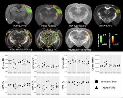 |
Do Non-Gaussian diffusion MRI methods improve the detection or specification of cellular alterations following traumatic brain injury?
Elizabeth Hutchinson, Sarah King, Alexandru Avram, M Irfanoglu, Michal Komlosh, Susan Schwerin, Eli Shindell, Sharon Juliano, Carlo Pierpaoli
Following traumatic brain injury (TBI), numerous microscale cellular alterations appear and evolve with a range of consequences for adverse outcomes and recovery. Diffusion tensor MRI (DTI) has been identified as a potentially sensitive tool for characterizing these changes, but is notably limited in providing specific information about particular cellular alterations and more advanced non-Gaussian frameworks have been developed that may address these limitations. To assess the utility of non-Gaussian modeling for improved detection and specification of TBI-related cellular alterations, we compared DTI, DKI and MAP-MRI in mouse brains following mild TBI and their correspondence to histopathology in the same tissue.
|
14:57
 |
0133.
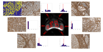 |
Histological Validation of in-vivo VERDICT MRI for Prostate using 3D Personalised Moulds
Elisenda Bonet-Carne, Maira Tariq, Hayley Pye, Mrishta Appayya, Aiman Haider, Colleen Bayley, Joseph Jacobs, Alexander Freeman, David Hawkes, David Atkinson, Greg Shaw, Hayley Whitaker, Daniel Alexander, Shonit Punwani, Eleftheria Panagiotaki
VERDICT analysis can successfully distinguish benign from malignant prostate tissue in-vivo showing promising results as a cancer diagnostic tool. However, the accuracy with which model parameters reflect the underlying tissue characteristics is unknown. In this study, we quantitatively compare the intracellular, extracellular-extravascular and vascular volume fractions derived from in-vivo VERDICT MRI against histological measurements from prostatectomies. We use 3D-printed personalised moulds designed from in-vivo MRI that help preserve the orientation and location of the gland and aid histological alignment. Results from the first samples using the 3D mould pipeline show good agreement between in-vivo VERDICT estimates and histology.
|
15:09
|
0134.
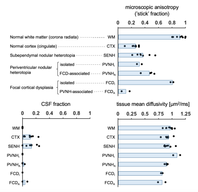 |
Microscopic diffusion anisotropy reveals microstructural heterogeneity of malformations of cortical development associated with epilepsy: A b-tensor encoding study at 7T
Björn Lampinen, Ariadni Zampeli, Filip Szczepankiewicz, Maria Compagno Strandberg, Kristina Källén, Isabella Björkman-Burtscher, Markus Nilsson
Malformations of cortical development are macro- or microscopic abnormalities of the cerebral cortex. Here, we investigated such malformations associated with epilepsy using b-tensor encoding, which is a recently developed technique that permits estimation of microscopic anisotropy also in regions where diffusion is isotropic on the voxel level. Results show a large heterogeneity in microscopic anisotropy between lesions, which we hypothesize represents different levels of axonal content. The characteristics of some types of lesions depended strongly on whether they were associated to other lesions, which could be clinically helpful for indicating hidden sources of epileptic seizures.
|
15:21
 |
0135.
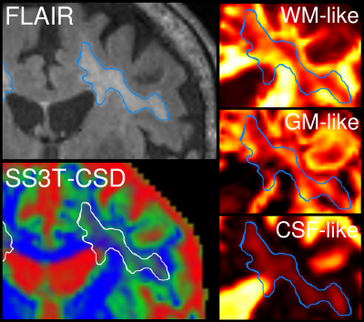 |
Investigating microstructural heterogeneity of white matter hyperintensities in Alzheimer’s disease using single-shell 3-tissue constrained spherical deconvolution
Remika Mito, Thijs Dhollander, David Raffelt, Ying Xia, Olivier Salvado, Amy Brodtmann, Christopher Rowe, Victor Villemagne, Alan Connelly
White matter hyperintensities (WMH) observed on FLAIR MRI are highly prevalent in Alzheimer’s disease. Although often associated with cognitive decline, such associations are highly variable, likely due to the underlying pathological heterogeneity within these lesions. Here, we explore this potential heterogeneity in vivo in an Alzheimer’s disease cohort, by investigating relative tissue fractions obtained using single-shell 3-tissue constrained spherical deconvolution (SS3T-CSD). We show distinguishable tissue profiles of lesions based on classification as periventricular or deep, and additionally show heterogeneity within lesions, thus highlighting the pitfalls of binary classification of WMH, and the value of investigating their underlying diffusional properties.
|
15:33
|
0136.
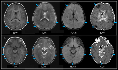 |
Neurite Orientation Dispersion and Density Imaging: Added Value in the Detection of Tubers in Patients With Tuberous Sclerosis Complex
Xiali Shao, Xuewei Zhang, Wenrui Xu, Hua Guo, Zhe Zhang, Jieying Zhang, Tao Jiang, Weihong Zhang
The aim of this study was to evaluate the performance of Neurite Orientation Dispersion and Density Imaging (NODDI) in depicting cortical tubers in patients with tuberous sclerosis complex (TSC). By comparing with conventional MRI and DTI, the intracellular volume fraction (ICVF) derived from NODDI showed privilege over both techniques with higher sensitivity and better contrast ratio. Our result has revealed that NODDI was better at detecting microstructural disruption than DTI and conventional MRI sequences with a more reasonable model assumption, and may somehow shed light on the management of epilepsy in TSC patients.
|
15:45
|
|
Adjournment & Meet the Teachers |
|
