Zhe Liu
By Pinar Ozbay
It is our pleasure to present one of the Editor’s picks for July, Preconditioned Total Field Inversion (TFI) Method for Quantitative Susceptibility Mapping (QSM), from Cornell University. In this work Zhe Liu, Pascal Spincemaille and colleagues proposed an algorithm which allows mapping of tissue magnetic susceptibility in regions with large dynamic susceptibility ranges, such as cavities, bones, and hemorrhages in the head. There are two main steps in QSM algorithm which are removal of background fields to calculate the local field, and solving the local field-to-susceptibility problem. The latter is an ill-posed problem by nature, hence is mainly referred to as the step of inverse problem in the literature of QSM. Their method calculates susceptibility maps via ‘total field inversion’, which generalizes those two steps as one optimization problem, and further employs preconditioning to achieve fast convergence.
Pinar: Could you tell us about your backgrounds, and you got into the field of MRI?
Zhe: I came from the major Automatic Control, a program which is a combination of computer science and electrical engineering for my undergraduate. In my senior year, I started working on motion correction in MRI in CBIR research center at Tsinghua University. I found MRI very interesting, because I could use the knowledge I learnt from signal processing and optimization courses.
Pascal: I am a physicist from Belgium, and I used to work on theoretical physics. Then I decided to look for something more practical and thought I would do MRI for a while, travel around the world and get back to Belgium, but I never got back. I am really happy I ended up in MRI, which is a broad field that makes a difference in people’s lives.
Pinar: This will be the 3rd QSM-related paper in MRM Highlights, and each time we ask the authors to briefly introduce QSM. What is exciting about QSM?
Zhe: For me QSM is interesting in understanding the nature of the inverse problem. It has two major advantages which make it widely useful: In comparison to other phase-contrast methods like susceptibility-weighted imaging (SWI), it has less blooming artifacts, and it is more accurate for identifying areas which are rich in iron or calcium, such as hemorrhage and MS lesions. The second advantage is it reflects the tissue magnetic properties independently of imaging parameters, so it is ideal for quantification, such as measuring oxygen consumption levels.
Pascal: Susceptibility imaging got started by treating susceptibility as an artifact, and trying to find the source of that artifacts. That’s an interesting problem from a mathematical, physical and as well clinical point of view. In order to do QSM, we had to learn a lot about optimization algorithms, and treat those solvers less as a black box. On the side of the applications, there are two important ones that are implemented at Cornell, one of which is preoperative planning for Deep Brain Stimulation. Namely, one day, a surgeon we collaborate with walked into the scanner control room and saw the subthalamic nucleus (STN) on a QSM image that happened to be displayed on the viewer. He thought it was the ideal image for deciding on electrode placement, because he could see the STN very well due to high iron content. The other application comes from the MS research group at Cornell. As QSM could be used to determine the MS lesion stage, they are looking into using it as Gd replacement for follow-ups.
Pinar: Could you describe the proposed method in your work, i.e. Preconditioned Total Field
Inversion?
Pascal: QSM turns the images you acquired with MRI into a susceptibility map. The physics tells you that the field inhomogeneity is generated by the convolution of the susceptibility distribution with the unit dipole kernel. And the field you calculate from the complex data has a background and a local component. So, when you first remove the background field, you are left with the local field, which should be generated only by the tissue inside the brain (that’s your hope). Once you have the local field, it is easier to do the second step, which is deconvolution (solving the inverse problem). The contrast you expect to see is on the order of 0.1 to 0.3ppm. If you don’t do enough iterations it appears as it doesn’t work, but it eventually converges, and if you have a smaller dynamic susceptibility range, your algorithm would run faster. So, what we thought is, why don’t you solve those two in one step?
Zhe: The problem with the two steps is that the error of the first step (background field removal) might propagate and cause artifacts in the second step (inverse problem). Because these two steps can be explained by the same physical model we tried to fuse them in one equation, and we observed that we can even use the same iterative solver (conjugate gradient). The only thing is we need more iterations to reach a solution given large dynamic range of susceptibility, as mentioned by Pascal. In this work, we think of ways to accelerate the speed of the TFI. And for that, we use a preconditioner, which reflects contrast with strong external sources and weak tissue sources, and utilizes additional information such as R2* to extract hemorrhage site.
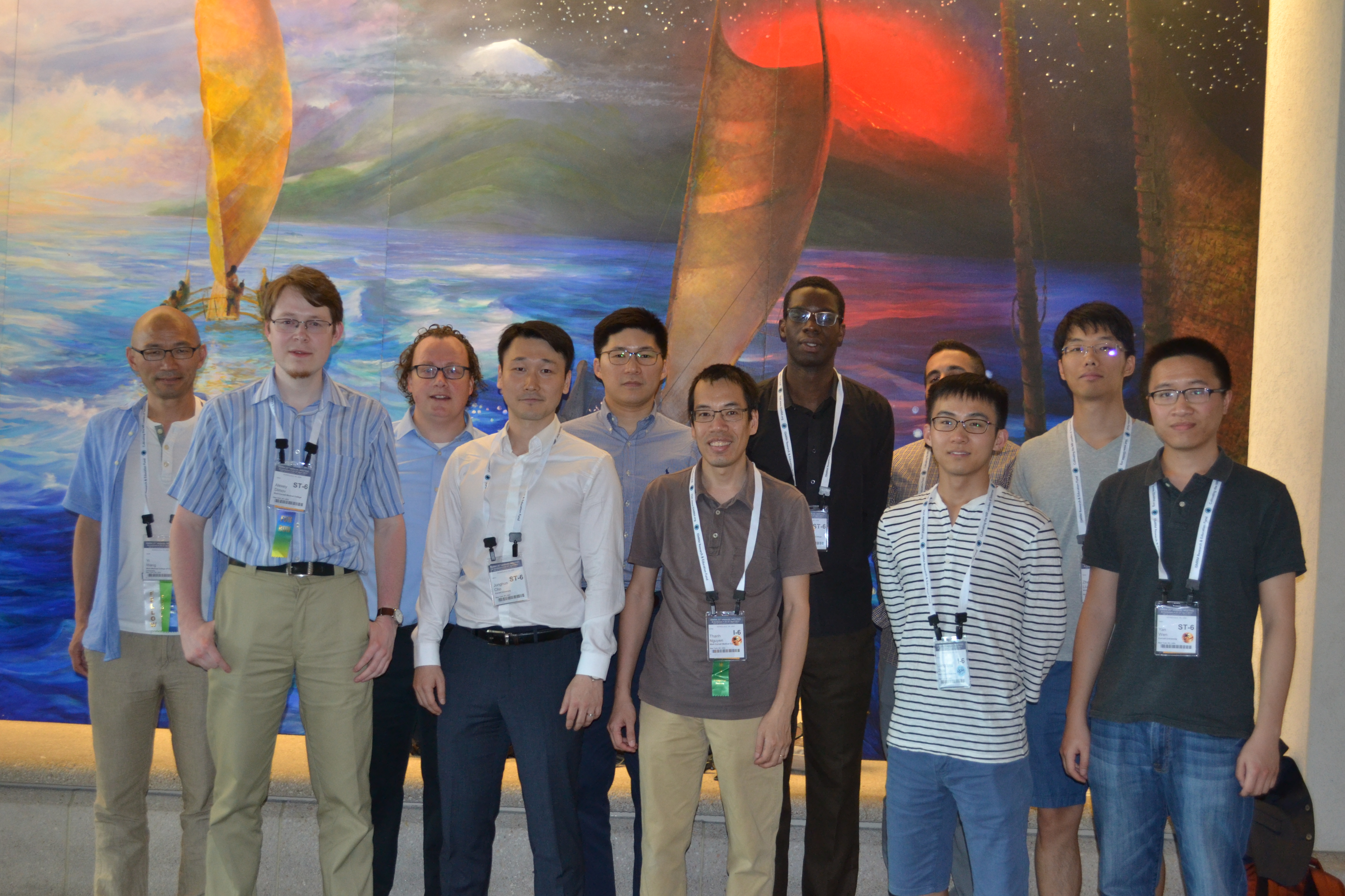
Cornell MRI Research Lab: Yi Wang, Alexey Dimov, Pascal Spincemaille, Junghun Cho, Youngwook Kee, Thanh Nguyen, Kofi Deh, Ramin Jafari, Liangdong Zhou, Zhe Liu and Yan Wen (From left to Right)
Pinar: Could you give us a summary of your results?
Zhe: Our results show that in healthy subjects we can improve the homogeneity of QSM compared to local field inversion technique. Compared to the other TFI methods which are based on Laplacian operation, we could preserve the cortical structure of the brain. In the end, we showed that it is possible to do QSM using preconditioned TFI in the whole head and to measure susceptibility in skull. In the patients with hemorrhage, we are able to reduce the artifacts around the hemorrhage.
Pascal: For brain, QSM techniques are well-developed, as the brain only has susceptibility contrast, no fat and other interferences. Outside of the brain (as for liver or cardiac QSM), we need to do water-fat separation first. In those cases, we then almost always use preconditioned TFI, and we believe that’s the next frontier for making QSM work in the body.
Pinar: Do you see QSM as a ‘push button’ method in the scanners soon?
Zhe: It is currently a fully automated process at Cornell and collaborated sites, with a dedicated server for QSM reconstruction. The entire process (scanner –> QSM server -> reconstructed maps -> scanner) takes 5-7 minutes, so the results are always ready by the end of the scan session.
Pascal: Other research sites that are interested to use this method can contact us, and we will help them install the system on their sites. Matlab QSM code from our lab is also available, and we are working on incorporating TFI into it. Below is our MATLAB code (http://weill.cornell.edu/mri/pages/qsm.html) and the automated and compiled mode is available upon request.