|
|
||
|
OCTOBER 2013 • Vol. 2, Issue 4 |
||
|
|
||
|
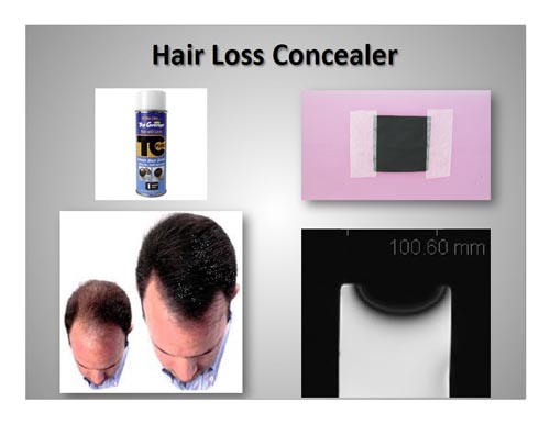
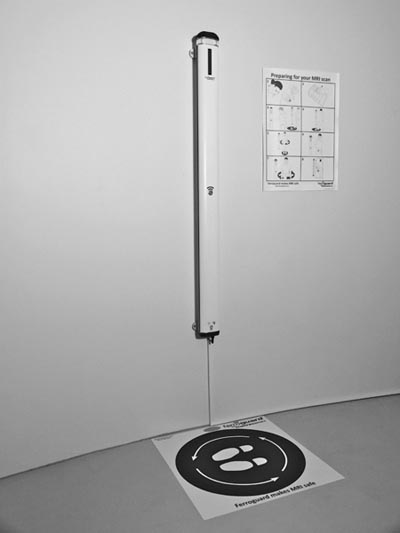
Hair Loss Concealer: Unusual MRI Artifacts and Positive Alarm on Ferromagnetic Detection System |
||
|
||
| (This article represents the views of its author only and does not reflect those of the International Society for Magnetic Resonance in Medicine and are not made with its authority or approval) | ||
|
Cosmetics,
including eye makeup, nail polishes, body lotions,
hair loss concealers and others are frequently used
to improve or enhance the appearance of the human
body. Several reports have indicated that certain
cosmetics, especially those containing iron oxide or
“heavy metal particles”, can produce unwanted
artifacts on magnetic resonance (MR) images (1-5).
This information has mostly been limited to eye
makeup (e.g., mascara, eye shadow, and eye liner)
and restricted to MR systems operating at 1.5-Tesla
or less. Many other types of cosmetics may contain ingredients that cause artifacts on MR images and it is well known that artifacts are inherently larger in association with 3-Tesla MR systems (1, 6, 7). Artifacts may create issues if the area of interest is the same as where the cosmetic was applied or if its presence was unknown to the radiologist, thus, potentially causing it to be interpreted as pathology (1-5). A recent study conducted by Escher, et al. (1) involved investigating artifacts at 3-Tesla for thirty-eight different types of cosmetics. The findings indicated that, of the thirty-eight cosmetics studied, the following exhibited artifacts on MRI: two nail polishes; five eyeliners; three mascaras; three eye shadows; and one hair loss concealer. The artifact size ranged from small (eye shadow) to very large (hair loss concealer) and tended to be associated with the presence of iron oxide or other metal-based ingredient. Therefore, it was concluded that many commonly used cosmetics cause artifacts that could create issues for MRI procedures. As part of the pre-MRI screening procedure, consideration should be given to patient management with regard to this information and a policy should be in place to prevent problems related to cosmetics. The policy may include advising patients before they arrive for MRI exams to thoroughly remove all cosmetics. Alternatively, it may be necessary to have makeup removal items available at the MRI facility to take off different types of cosmetics (e.g., nail polish remover). In the case of the hair loss concealer (Top Coverage
Plus Bald Spot Eraser, TC Plus While it would be beneficial to identify a patient using hair loss concealer before performing MRI of the brain, the patient may be unwilling to divulge the use of this product and surely not aware of the artifact issue. Accordingly, if an MRI technologist or radiologist observes a large signal void mostly related to the surface of the scalp in a patient that does not have a neurological metallic implant, the possibility of hair loss concealer as being responsible for the artifact should be considered. |
||
|
||
|
|
||
|
||
|
REFERENCES (1) Escher KB, Shellock FG. Evaluation of MRI artifacts at 3-Tesla for 38 commonly used cosmetics. Magnetic Resonance Imaging 2013;31:778-782. (2) Smith FW, Crosher GA. Mascara--an unsuspected cause of magnetic resonance imaging artifact. Magn Reson Imaging. 1985;3:287-9. (3) Weiss RA, Saint-Louis LA, Haik BG, McCord CD, Taveras JL. Mascara and eye-lining tattoos: MRI artifacts. Ann Ophthalmology 1989;21:129-131. (4) Wright RM, Swietek PA, Simmons ML. Eye artifacts from mascara in MRI. AJNR Am J Neuroradiol. 1985;6:652. (5) Sacco D, Steiger DA, Bellon EM, Coleman PE, Haacke EM. Artifacts caused by cosmetics in MR imaging of the head. Am J Roentgenol 1987;148:1001-1004. (6) Hargreaves BA, Worters PW, Pauly KB, Pauly JM, Kock KM, Gold GE. Metal-induced artifacts in MRI. AJR Am J Roentgenol. 2011;197:547-55. (7) Shellock FG. Reference Manual for Magnetic Resonance Safety, Implants, and Devices: 2013 Edition. Biomedical Research Publishing Group, Los Angeles, 2013. (8) Shellock FG, Karacozoff AM. Detection of implants and other objects using a ferromagnetic detection system: implications for patient screening prior to MRI. American Journal of Roentgenology 2013;201:720-725. |
||
| Dr. Shellock announces his new book "MRI Bioeffects, Safety, and Patient Management" which is a fundraiser for the Lupus Research Institute. Please go to: http://www.mrisafetybook.com/ for more information. | ||