ISMRM 24th Annual Meeting & Exhibition • 07-13 May 2016 • Singapore

Electronic Poster Session: Cardiovascular 1

2664 -2671 Atherosclerosis Imaging
2672 -2695 MRA
2696 -2719 Velocity & Flow Quantification

Exhibition Hall 

10:45 - 11:45

    Computer #

2664.   
31 The Ability to Detect Intracranial Arterial Calcifications:  Evaluation of 3D TOF MRA and SNAP
Mahmud Mossa-Basha1, Haining Liu1, Dan S. Hippe1, Niranjan Balu1, Jie Sun1, Dean Shibata1, and Chun Yuan1
1Radiology, University of Washington Medical Center, Seattle, WA, United States
Intracranial arterial calcification evaluation has shown increasing importance in the literature based on associations with current and future stroke events, dementia and cognitive decline.  We hypothesize that the proton-density image, SNAP Ref, that is generated with Simultaneous Non-contrast Angiography and intraPlaque hemorrhage (SNAP) can more adequately assess calcifications compared to 3D TOF MRA relative to the reference standard, thin slice CTA.


2665.   
32 Simultaneous multi-slice carotid vessel wall MRI with DIR-FSE
Sagar Mandava1, Mahesh Bharath Keerthivasan1, Kevin Johnson2, Diego R. Martin3, Ali Bilgin1,3,4, and Maria I. Altbach3
1Electrical and Computer Engineering, University of Arizona, Tucson, AZ, United States, 2Siemens Healthcare, Tucson, AZ, United States, 3Medical Imaging, University of Arizona, Tucson, AZ, United States, 4Biomedical Engineering, University of Arizona, Tucson, AZ, United States
The DIR-FSE sequence is popularly used to assess plaque build up but is known to be a single slice technique due to a non-selective inversion pulse used in the DIR module. In this work we present a technique to improve the SNR efficiency of DIR-FSE sequences by multi-band excitation. The proposed technique can generate multiple slices at the exact null point of blood and the acquired data can be used to create upto 16 TE images and T2 maps for all the acquired slices. 


2666.   
33 Post-Contrast T1w Black Blood Images of Atherosclerotic Plaque using 3D DANTE prepared Stack of Stars (3D DANTE-SOS) Sequence and ciné Reconstruction Method using Retrospective Ordering and Compressed Sensing (ciné-ROCS)
Seong-Eun Kim1, John A Roberts1, J Scott McNally1, Bradley D Bolster, Jr.2, Gerald S Treimam3,4, and Dennis L Parker 1
1Department of Radiology, UCAIR, University of Utah, Salt Lake City, UT, United States, 2Siemens Healthcare, Salt Lake City, UT, United States, 3Department of Surgery, University of Utah, Salt Lake City, UT, United States, 4Department of Veterans Affairs, VASLCHCS, Salt Lake City, UT, United States
Contrast enhanced T1 imaging has been used to aid in the detection of the fibrous cap overlying the necrotic core. We have implemented 3D SOS with retrospective cardiac cycle gated compressed sensing reconstruction to minimize, characterize artifacts in post-contrast imaging. Radial based k-space trajectory may offer reduced motion sensitivity and more robust ciné-ROCS reconstructions due to its inherent oversampling of central k-space. Ciné-ROCS reconstruction from 3D SOS acquisition demonstrates improved demonstration of fine plaque structures and vessel wall movement due to cardiac motion to the regular reconstruction and may help provide information on symptomatic plaque development and response to treatment.


2667.   
34 Motion insensitive high resolution in vivo DWI of Carotid Artery Wall Imaging using 3D Diffusion Weighted Driven Equilibrium Stack of Stars (3D DW-DE SOS) sequence
Seong-Eun Kim1, J Scott McNally1, Bradley D Bolster, Jr.2, Gerald S Treimam3,4, and Dennis L Parker 1
1Department of Radiology, UCAIR, University of Utah, Salt Lake City, UT, United States, 2Siemens Healthcare, Salt Lake City, UT, United States, 3Department of Surgery, University of Utah, Salt Lake City, UT, United States, 4Department of Veterans Affairs, VASLCHCS, Salt Lake City, UT, United States
DWI might provide a tool for discriminating intraplaque hemorrhage and lipid core from other components. Motion insensitive 3D DW-DE SOS has been developed to acquire high resolution DWI to improve the accuracy of ADC measurements. This technique was able to yield high resolution ADC that could provide clear ROI selection for important plaque components. Increased spatial resolution in motion insensitive 3D DW-DE SOS can improve the sensitivity of ADC maps in plaque component identification. The results obtained indicate that an ADC map may be of substantial value in identifying lipid and hemorrhage within overall plaque burden.


2668.   
35 Motion Insensitive 3D technique for simultaneous measurement of in-vivo ADC and T2* in Atherosclerotic plaque using a 3D Multiple Echo Diffusion Weighted Driven Equilibrium Stack of Stars (3D ME-DW-DE SOS) Sequence.
Seong-Eun Kim1, J Scott McNally1, Bradley D Bolster, Jr.2, Gerald S Treimam3,4, and Dennis L Parker 1
1Department of Radiology, UCAIR, University of Utah, Salt Lake City, UT, United States, 2Siemens Healthcare, Salt Lake City, UT, United States, 3Department of Surgery, University of Utah, Salt Lake City, UT, United States, 4Department of Veterans Affairs, VASLCHCS, Salt Lake City, UT, United States
DWI has the potential to provide complementary information that will allow better discrimination of plaque components such as lipid core. Iron has consistently been found in higher concentrations in atherosclerotic plaque compared to vessel tissue. In previous studies, intraplaque T2* distinguished symptomatic from asymptomatic plaques in patients with carotid atherosclerosis. In this work we introduce simultaneous measurement of ADC and T2* using a motion insensitive high resolution 3D multiple echo diffusion weighted driven equilibrium Stack of Stars sequence. This technique can provide high resolution T2* and ADC values simultaneously, which may provide important clinical information to detect plaque progression.


2669.   
36 Characterization of carotid plaque using sampling perfection with application-optimized contrast using different flip angle evolution (SPACE) sequence - Permission Withheld
Yoshimitsu Ohgiya1, Nobuyuki Ohike 2, Naomi Yagi 1, Hiroto Sasamori1, Jiro Munechika 1, Masanori Hirose 1, and Takehiko Gokan 1
1Showa University School of Medicine, Tokyo, Japan, 2Department of Pathology, Showa University Fujigaoka Hospital, Yokohama-shi, Japan
The purpose of this study was to investigate whether 3D turbo spin-echo (TSE) SPACE sequence can evaluate plaque characteristics of the carotid artery. Mann-Whitney test was used to examine differences in the contrast ratio (CR) between soft plaques and fibrous plaques on T1- and T2-weighted images. The mean CR of the soft plaques (1.54 ± 0.25) was significantly higher than that of the fibrous plaques (1.17 ± 0.13) (p < 0.001) on T1-weighted images. In conclusion, T1-weighted TSE SPACE sequence can evaluate main components of plaques in the carotid artery with high sensitivity and specificity.


2670.   
37 Selective reacquisition for motion artifact reduction in quantitative T2 mapping of carotid artery vessel wall
Robert Frost1, Aaron T. Hess2, Linqing Li3, Matthew D. Robson2, Luca Biasiolli2, and Peter Jezzard1
1FMRIB Centre, Nuffield Department of Clinical Neurosciences, University of Oxford, Oxford, United Kingdom, 2Oxford Centre for Clinical Magnetic Resonance Research, Division of Cardiovascular Medicine, University of Oxford, Oxford, United Kingdom, 3Section on Magnetic Resonance Spectroscopy, National Institute of Mental Health, Bethesda, MD, United States
Ghosting and blurring artifacts caused by swallowing or coughing can be a significant problem in quantitative T2 mapping of atherosclerotic plaque in the carotid artery. The method is based on a multi-slice multiple spin-echo sequence which acquires k-space lines sequentially with a 2 s gap between lines. A navigator echo was added at the end of the echo-train to identify and reacquire data corrupted by motion. The selective reacquisition reduced ghosting and blurring artifacts in healthy volunteer scans with intentional swallowing motion.


2671.   
38 Feasibility of Vessel Wall Imaging of the Superficial Palmar Arch using 7T and 3T MRI
Alison N. Pruzan1,2, Audrey Kaufman1,2, Claudia Calcagno1,2, Yu Zhou1,2, Zahi A. Fayad1,2, and Venkatesh Mani1,2
1Department of Radiology, Icahn School of Medicine at Mount Sinai, New York, NY, United States, 2Translational and Molecular Imaging Institute, Icahn School of Medicine at Mount Sinai, New York, NY, United States
Evaluation of atherosclerosis in smaller arteries in the hand may be clinically useful in certain disease conditions such as Diabetes. We sought to demonstrate feasibility of vessel wall imaging of the superficial palmar arch using 7T and 3T MRI in comparison with very high frequency micro ultrasound. Results indicated that 7T imaging of the palmar arch was feasible and subjective image quality analysis was better than 3T and ultrasound.
Exhibition Hall 

10:45 - 11:45

    Computer #

2672.   
49 Four-Dimensional, Multiphase, Steady-State Imaging with Contrast Enhancement (MUSIC) with Improved Functional Assessment
Ziwu Zhou1, Fei Han1, Takegawa Yoshida1, Kim-Lien Nguyen1, Paul Finn1, and Peng Hu1
1Radiological Sciences, University of California, Los Angeles, Los Angeles, CA, United States
A recent proposed technique called four dimensional, multiphase, steady-state imaging with contrast enhancement (MUSIC) enables detailed anatomical assessment of cardiovascular system. However, limited available cardiac phase prevents accurate functional assessment of the heart. In this study, we compared original MUSIC with an improved MUSIC technique that generates double cardiac phases. Initial results suggest a more accurate left ventricular volume measurement and better appreciation of cardiac wall motion can be achieved with more cardiac phases.


2673.   
50 Effect of Gadolinium-Induced Susceptibility on First-Pass Single-echo Dixon CE-MRA and Methods for Correction
Eric G. Stinson1, Joshua D. Trzasko1, and Stephen J. Riederer1
1Radiology, Mayo Clinic, Rochester, MN, United States
Single-echo Dixon imaging for contrast-enhanced MR angiography (CE-MRA) can provide the advantages of multi-echo Dixon without the tradeoff of longer acquisitions and reduced temporal resolution. Single-echo Dixon imaging assumes that both the water and fat signals are real and have known initial phase and phase due to field inhomogeneities. However, when a paramagnetic Gadolinium-based contrast agent is injected for CE-MRA, the field may be perturbed and render the a priori phase estimates invalid. The purpose of this study is to demonstrate the effect of Gd-induced field perturbations on first-pass single-echo Dixon CE-MRA and describe strategies to avoid or correct artifacts.


2674.   
51 Preliminary evaluation of Respiratory Self Navigated Whole-Heart Angiography in Combination with Ultra-small Super-paramagnetic Iron Oxide Particles
Davide Piccini1,2, Peter J. Weale3, Saeed Mirsadraee4, Rachel O. Forsythe5, Annette S. Cooper6, and Scott Semple5,6
1Advanced Clinical Imaging Technology, Siemens Healthcare, Lausanne, Switzerland, 2Department of Radiology, University Hospital (CHUV) and University of Lausanne (UNIL), Lausanne, Switzerland,3Siemens Healthcare Ltd, Camberley, United Kingdom, 4NHS Lothian & Clinical Research Imaging Centre Queen's Medical Research Institute, University of Edinburgh, Edinburgh, United Kingdom, 5Centre for Cardiovascular Science, University of Edinburgh, Edinburgh, United Kingdom, 6Clinical Research Imaging Centre, University of Edinburgh, Edinburgh, United Kingdom
Respiratory self-navigated 3D-radial MRA is an efficient and robust method for the depiction of coronary luminal anatomy at 1.5T. Higher magnetic fields are more challenging, due to off-resonance artifacts. Contrast-enhanced imaging is usually beneficial. However, variable contrast during prolonged scans can deteriorate the self-navigation signal. Here we investigate the use of an ultra-small super-paramagnetic iron oxide particles (USPIO) contrast agent with longer half-life in combination with self-navigation at 3T. Systolic and diastolic datasets from 10 volunteers were acquired and analyzed. We show that USPIO contrast-enhanced self-navigated coronary MRA is feasible at 3T with good image quality and reliable motion correction.


2675.   
52 Feasibility of Subtractionless Three-Station First-Pass Peripheral MRA at 3 T
Holger Eggers1, Bernhard Schnackenburg2, Marc Kouwenhoven3, Alan Huang3, Tim Leiner4, Rolf Gebker5, and Sebastian Kelle5
1Philips Research, Hamburg, Germany, 2Philips Healthcare, Hamburg, Germany, 3Philips Healthcare, Best, Netherlands, 4Department of Radiology, University Medical Center Utrecht, Utrecht, Netherlands, 5German Heart Institute Berlin, Berlin, Germany
Chemical shift encoding-based water-fat imaging, or Dixon imaging, has recently been demonstrated to permit first-pass peripheral MRA without subtraction. While previous work focused on an evaluation at 1.5 T, this work explores the feasibility of this subtractionless method at 3 T. Results on the first six patients are presented and are compared with the established subtraction method. Substantial improvements in image quality are seen, and the use of three stations is, despite particular challenges at 3 T, preliminarily found to be practicable.


2676.   
53 International, Multicenter Phase 3 Blinded Study of the Structural Visualization, Diagnostic Efficacy and Safety of Gadobutrol (Gadavist) Enhanced MRA of the Renal Arteries compared to Time-of-Flight MRA using CTA as the standard of reference (SoR)
Jacob Agris1, Neda Rastegar2, Kelly Fabrega-Foster2, Sheela Agarwal1,3, Daniel Haverstock4, and Ihab Kamel2
1Radiology, Bayer, Whippany, NJ, United States, 2Radiology, Johns Hopkins, Baltimore, MD, United States, 3Radiology, Massachusetts General Hospital, Boston, MA, United States, 4Statistics, Bayer, Whippany, NJ, United States
International, multicenter phase 3 blinded study comparing the performance of CE-MRA with Gadobutrol, a high relaxivity and highly stable macrocyclic contrast agent, to 2D Time-of-flight MRA (ToF) using CTA as the standard of reference (SoR). 317 patients suspected of renal artery disease were enrolled. There was almost no error in the Gadobutrol MRA vessel measurements (0.0mm Gadobutrol vs 0.5mm ToF for stenosis measurements) and superior assessability as well as superior specificity was demonstrated reducing the need for additional imaging studies by 50%. Gadobutrol enhanced MRA of the renal arteries has superior visualization, more accurate measurements and is a valuable alternative to CTA without any ionizing radiation.


2677.   
54 Lung Perfusion: MRI vs SPECT for screening in suspected Chronic Thromboembolic Pulmonary Hypertension
Christopher S Johns1, Smitha Rajaram2, David Capener1, David G Kiely3, Andrew J Swift1,4, and Jim M Wild1
1Academic Unit of Radiology, University of Sheffield, Sheffield, United Kingdom, 2Radiology Department, Sheffield Teaching Hospitals, Sheffield, United Kingdom, 3Sheffield Pulmonary Vascular Clinic, Sheffield Teaching Hospitals, Sheffield, United Kingdom, 4Institute of Insilico Medicine, The University of Sheffield, Sheffield, United Kingdom
A comparison of perfusion SPECT and MRI in screening for chronic thromboembolic disease. To assess the role of MRI perfusion in the clinical imaging pathway in this patient group. 


2678.   
55 Estimation of Error in Volume Measurement for 100 Intracranial Aneurysms Imaged Serially with CE-MRA at 1.5T and 3T
Farshid Faraji1, Alastair Martin1, and David Saloner1
1Department of Radiology, UCSF, San Francisco, CA, United States
Intracranial aneurysms are localized dilations in blood vessels occurring in 1-6% of the population which can have devastating consequences in the event of rupture. Many aneurysms are asymptomatic, found incidentally, and fall below the surgical size threshold. For this reason, physicians choose to follow aneurysms with imaging rather than opting for surgical or endo-vascular intervention. Here we further our analysis of a previously presented image processing technique using contrast-enhanced MRA to follow intracranial aneurysms longitudinally. We investigate the measurement error of this post-processing technique in 100 intracranial aneurysms, and evaluate the differential effects of imaging at 1.5T and 3T.


2679.   
56 Intra-individual quantitative and qualitative comparison of 4D-MRA- and dynamic CTA-bolus-time-curves of gadopentetate dimeglumine and gadobutrol in minipigs - Permission Withheld
Dariusch Reza Hadizadeh1, Vera Catharina Keil1, Gregor Jost2, Hubertus Pietsch2, Martin Weibrecht3, Bishr Agha1, Christian Marx1, Michael Perkuhn3, Hans Heinz Schild1, and Winfried Albert Willinek4
1Radiology, University of Bonn, Bonn, Germany, 2MR and CT Contrast Media Research, Bayer Healthcare, Berlin, Germany, 3Innovative Technologies, Research Laboratories, Philips Technologie GmbH, Aachen, Germany, 4Department of Radiology, Neuroradiology, Sonography and Nuclear Medicine, Brüderkrankenhaus Trier, Trier, Germany
In an animal model bolus kinetics and image quality after injection of 1M gadobutrol (standard and half-dose) and 0.5M gadopentetate dimeglumine (standard-dose) had been investigated in 4D-MRA at 3T and dynamic CTA. The first pass arterial peak Gd-concentrations (quantified by CTA) were higher for standard-dose compared to half-dose gadobutrol. In 4D-MRA the first pass arterial peak enhancement was comparable for both gadobutrol doses and gadopentetate dimeglumine due to peak cut-off effects at high vascular Gd concentrations. Markedly higher venous bolus peaks were found for standard-dose gadobutrol. Image quality of 4D-MRA was rated significantly higher for both doses of gadobutrol.


2680.   
57 Evaluate Relative Helicity of Aortic Flow for Marfan Syndrome by 4D Flow Phase Contrast MRI
Pin-Chen Chen1, Hsin-Hui Chiu2, Wen-Yih Isaac Tseng3, and Hsu-Hsia Peng4
1Institute of Systems Neuroscience, Hsinchu, Taiwan, 2Department of Pediatrics, Taipei, Taiwan, 3Center for Optoelectronic Biomedicine, Taipei, Taiwan, 4Department of Biomedical Engineering and Environmental Sciences, Hsinchu, Taiwan
We aim to evaluate relative helicity of aortic flow for patients with Marfan syndrome (MFS) by 4D flow phase-contrast MRI. An individual helicityAED mapping was composed to provide the information of the spatiotemporal distribution of relative helicity. The E sign represents the relative helicity cores shown in the early diastole whereas D sign represents the relative helicity core shown time-delayed and downstream. The A sign represents an additional relative helicity core other than D, E, or the normal single cores. MFS patients showed abnormal relative helicity core in helicityAED mapping, which provided promising approaches for patient managements in the future.


2681.   
58 Evaluation of Dual Agent Relaxation Contrast MR Lymphangiography (DARC-MRL) for Pre-surgical Identification of Lymphatic Channels
Beth Ripley1, Gregory J Wilson1, and Jeffrey H Maki1
1Department of Radiology, University of Washington Medical Center, Seattle, WA, United States
A new method of lymphatic channel mapping was evaluated in a retrospective review of pre-surgical lymphedema MR exams. Dual Agent Relaxation Contrast MR Lymphangiography (DARC-MRL) uses intracutaneous injection of a Gd contrast agent for enhancement of lymphatic channels and concomitant intravenous injection of USPIO (ferumoxytol) for suppression of (otherwise lymphatic-obscuring) veins. 43 MR exams using DARC-MRL were compared to 43 matched exams using conventional (no USPIO) MR lymphangiography. Exams were graded for degree of venous contamination, and DARC-MRL exams exhibited dramatically less obscuring venous contamination than the conventional method.


2682.   
59 Cardiac and Respiratory Motion-Resolved Free-Running Whole-Heart Coronary MRA of Patients Using 5D XD-GRASP Reconstruction.
Giulia Ginami1, Simone Coppo1, Li Feng2, Davide Piccini1,3, Tobias Rutz4, Ricardo Otazo2, Daniel Sodickson2, Matthias Stuber1,5, and Jerome Yerly1,5
1University Hospital (CHUV) and University of Lausanne (UNIL), Lausanne, Switzerland, 2Center for Advanced Imaging Innovation and Research (CAI2R), Department of Radiology, New York University School of Medicine, New York, NY, United States, 3Advanced Clinical Imaging Technology, Siemens Healthcare, Lausanne, Switzerland, 4Division of Cardiology and Cardiac MR Center, University Hospital of Lausanne (CHUV), Lausanne, Switzerland, 5Center for Biomedical Imaging (CIBM), Lausanne, Switzerland
Free-running self-navigated techniques have been introduced in order to allow for time resolved three-dimensional whole-heart MR acquisitions. A more recently proposed free-running 5D (x-y-z-cardiac-respiration) XD-GRASP (eXtra-Dimensional Golden-angle RAdial Sparse Parallel MRI) approach enables acquisition and reconstruction of cardiac- and respiratory-motion resolved 3D volumes. In this study, we investigated the potential of 5D XD-GRASP in a clinical setting.


2683.   
60 MRI of peripheral vascular calcifications using stack-of-stars 3D FLASH
Marcos Paulo Botelho1, Shivraman Giri2, Ioannis Koktzoglou1,3, Alto Stemmer4, and Robert R. Edelman1,5
1Radiology, NorthShore University HealthSystem, Evanston, IL, United States, 2Siemens Healthcare, Chicago, IL, United States, 3Radiology, University of Chicago Pritzker School of Medicine, Chicago, IL, United States, 4Siemens Healthcare GmbH, Erlangen, Germany, 5Radiology, Feinberg School of Medicine, Northwestern Univesity, Chicago, IL, United States
We evaluated a novel approach for visualizing and quantifying peripheral arterial calcifications using a stack-of-stars 3D FLASH pulse sequence.  The technique permitted isotropic 1mm3 spatial resolution and displayed dark calcifications against a relatively uniform bright background.  Banding artifacts relating to chemical shift were minimized by the use of radial in-plane spatial encoding and an in-phase echo time, while vascular signal was enhanced by the use of a low flip angle near the Ernst angle of blood. In patients with peripheral arterial disease, there was excellent correlation using CT angiography as the standard of reference.  


2684.   
61 Non-Contrast Gate-Free Aortic MR Angiography with Robust Fat Suppression
Nobuyuki Toyonari1, Masami Yoneyama2, Seiichiro Noda1, Yukari Horino1, and Kazuhiro Katahira1
1Kumamoto Chuo Hospital, Kumamoto, Japan, 2Philips Electronics Japan, Tokyo, Japan
Non-contrast MR angiography (MRA) is a promising method to diagnose and follow-up of vascular diseases such as dissecting aortic aneurysm. Conventionally, 3D gated balanced steady-state free precession (bSSFP) is used for the aorta, but it has several limitations. To overcome bSSFP’s limitations , we propose a new technique based on gradient echo DIXON sequence with flow-independent relaxation-enhanced non-contrast MRA technique (Relaxation-Enhanced Angiography without Contrast and Triggering: REACT). We showed that REACT could provide more robust and stable MRA without any artifacts and failed fat suppression compared with conventional bSSFP. 


2685.   
62 Time-of-flight unlimited: Novel self-gated unenhanced peripheral MR angiography with continuous table movement in less than 7 minutes - Permission Withheld
Michael O. Zenge1, Justin Ream2, Ankur Doshi2, Mary Bruno2, Christopher Stroehlein3, Peter Speier3, Hersh Chandarana2, and Harald H. Quick4,5
1Siemens Healthcare, Malvern, PA, United States, 2Radiology, NYU Langone Medical Center, New York, NY, United States, 3Siemens Healthcare, Erlangen, Germany, 4Erwin L. Hahn Institute for MR Imaging, University Duisburg-Essen, Essen, Germany, 5High Field and Hybrid MR Imaging, University Hospital Essen, Essen, Germany
Although unenhanced MR angiography eliminates the need for contrast injection, it would use ECG triggering which adds extra time and complexity to patient preparation. In the current work, time-of-flight (TOF) unlimited introduces self-gated rapid, radial MR imaging with continuous table movement for seamless coverage of the peripheral vasculature. In-vivo experiments in five healthy volunteers and two patients were performed. Initial results are more than promising and TOF unlimited demonstrates similar image quality compared to conventional ECG triggered TOF despite sevenfold accelerated data acquisition. Thus, novel TOF unlimited may be a time efficient screening tool for peripheral arterial disease.


2686.   
63 Reduction of image artifacts in non-contrast-enhanced velocity-selective peripheral MRA at 3T
Taehoon Shin1, Qin Qin2, Jang-Yeon Park3,4, Robert S. Crawford5, and Sanjay Rajagopalan6
1Diagnostic radioloy and nuclear medicine, University of Maryland, Baltimore, MD, United States, 2Radiology, Johns Hopkins University, Baltimore, MD, United States, 3Biomedical Engineering, Sungkyunkwan University, Suwon, Korea, Republic of, 4Center for neuroscience imaging research, Insititute for Basic Science, Suwon, Korea, Republic of, 5Vascular and endovascular surgery, University of Maryland, Baltimore, MD, United States, 6Cardiovascular Medicine, University of Maryland, Baltimore, MD, United States
Velocity-selective (VS) magnetization-prepared non-contrast-enhanced MR angiography has advantages of large 3D FOV, arbitrary 3D spatial resolution and need for single acquisition only. Peripheral VS-MRA has shown great potential at 1.5T but might be challenging at 3T due to large B0 and B1 inhomogeneity in the pelvis and legs. In this study, we show that the effects of B0 and B1 offsets are manifested as arterial signal loss, stripe artifact and background signal variation. We develop multiple-refocused VS excitation pulses and propose successive applications of two VS preparation pulses with shifted excitation profiles to suppress these artifacts.


2687.   
64 Dual-phase coronary MR angiography using image based respiratory navigation - Permission Withheld
Markus Henningsson1 and Rene Botnar1
1Imaging Sciences and Biomedical Engineering, King's College London, London, United Kingdom
Image-based navigation (iNAV) is a promising respiratory motion correction approach for coronary MR angiography (CMRA). However, for dual-phase CMRA the technique may fail due to the different cardiac motion states of the systolic and diastolic acquisition. Here we propose the use of separate independent systolic and diastolic iNAV acquisitions to address this issue. We compared iNAV to the conventional diaphragmatic one-dimensional navigator (1D NAV) in 8 healthy subjects. The proposed technique achieves similar or improved coronary vessel sharpness compared to 1D NAV while reducing dual-phase CMRA acquisition time.


2688.   
65 FSE-based Non-Contrast-enhanced Magnetic Resonance Venography for evaluation of the upper extremity veins compared with Contrast-enhanced MRV and Ultrasound
Ruth P Lim1,2,3, Emma Hornsey1, Dinesh Ranatunga1,2, Huming Hao4, Lucy McKenna1, Julie Smith1, Tim Spelman5, Jason Chuen3,4, and Mark Goodwin1,2
1Radiology, Austin Health, Melbourne, Australia, 2Radiology, The University of Melbourne, Melbourne, Australia, 3Surgery, The University of Melbourne, Melbourne, Australia, 4Surgery, Austin Health, Melbourne, Australia, 5Centre for Population Health, Burnet Institute, Melbourne, Australia
Venous mapping is important in end stage renal disease patients requiring vascular access for hemodialysis. We compare image quality (IQ) and measured vessel caliber of FSE based non-contrast MRV  (NC-MRV) to contrast-enhanced MRV (CE-MRV) and US in available segments in 10 healthy volunteers.  Central and arm vein IQ was diagnostic, but forearm vein IQ was suboptimal, inferior to CE-MRV. No difference in vessel caliber between sequences was demonstrated for most segments between NC-MRV and CE-MRV, but both MRV techniques yielded significantly larger caliber measurements than US, raising concern regarding application of MRV-derived measurements to clinical practice.


2689.   
66 Imaging Deep Inferior Epigastric Perforators with Phase Contrast Magnetic Resonance Angiography: A Feasibility Demonstration
Xiangyu Yang1, Michael Miller2, and Michael V Knopp1
1Radiology, The Ohio State University, Columbus, OH, United States, 2Plastic Surgery, The Ohio State University, Columbus, OH, United States

Currently, MRA is not considered the optimal technique for preoperative imaging of perforators before flap reconstructive plastic surgeries. In this prospective study, we demonstrate that the quality of perforator MRA can be greatly enhanced by using the phase contrast technique. Perforator imaging with PC-MRA not only generates images with better quality and contrast than X-ray and iodinated contrast based CTA, the current clinical gold-standard, but also offers unique features, such as the capability to visualize venous flow and differentiate perforator arteries and veins, that are highly relevant to the design, planning, and execution of DIEP flap reconstructive surgery.  


2690.   
67 Vessel-selective time-resolved cerebral angiograms in less than one minute
Eleanor S K Berry1, Peter Jezzard1, and Thomas W Okell1
1FMRIB Centre, Nuffield Department of Clinical Neurosciences, University of Oxford, Oxford, United Kingdom
Vessel-encoded pseudo-continuous arterial spin labeling (VEPCASL) can be used to obtain vessel-selective time-resolved angiograms. Here we compare accelerated Cartesian and radial readouts to demonstrate the feasibility of speeding up their acquisition, enabling better prospects for use of the method in a clinical setting. It was possible to acquire 2D vessel-selective angiograms in less than one minute. Accelerated (undersampled) radial acquisition consistently led to angiograms with higher signal-to-noise ratio and better quality peripheral artery imaging versus accelerated Cartesian imaging.


2691.   
68 Clinically-Acceptable Non-Contrast Thoracic MRA using 3D Radial k-space Sampling and Compressed Sensing
Marc D Lindley1,2, Daniel Kim2, Kristi Carlston2, Leif Jensen2, Daniel Sommers2, Ganesh Adluru2, Edward VR DiBella2, Christopher J Hanrahan2, and Vivian S Lee2
1Physics, University of Utah, Salt Lake City, UT, United States, 2Radiology, UCAIR, University of Utah, Salt Lake City, UT, United States
As an alternative to contrast-enhanced (CE) MRA, we developed an accelerated non-contrast MRA of thoracic aorta using a combination of T2-prepared and fat saturation preparations, b-SSFP readout, 3D radial stack of stars sampling with tiny golden angles, and compressed sensing. This NC-MRA was compared with standard ECG-gated CE-MRA in 8 patients. Normalized signal difference and aortic diameters were not significantly different between CE- and NC-MRA methods.


2692.   
69 Non-Contrast MR Angiography with Arterial Spin Labeling: Initial Experience in Pediatric Patients With a 3D Spiral pCASL and CINEMA Protocol
Amber Pokorney1, Niccolo Stefani2, Zhiqiang Li3, Jonathan M. Chia2, John Condie4, Houchun Harry Hu1, and Jeffrey H. Miller1
1Radiology, Phoenix Children's Hospital, Phoenix, AZ, United States, 2Philips, North America, Cleveland, OH, United States, 3Barrow Neurological Institute, Phoenix, AZ, United States, 4Neurology, Phoenix Children's Hospital, Phoenix, AZ, United States
This pilot clinical study evaluates the diagnostic utility of a 3D spiral pCASL approach and a 3D dynamic PASL pulse sequence (i.e., CINEMA) in evaluating the neurovasculature in pediatric patients.  With increasing recent concerns over the possible deposition of Gadolinium in the brain, a viable clinical protocol that can supplant traditional contrast-enhanced MR angiography is particularly relevant to the pediatric population.  Our clinical results in patients ranging from 1 year of age to adolescents demonstrate that both 3D spiral pCASL and dynamic PASL are robust approaches and yield diagnostically useful information that supports clinical findings from conventional TOF angiography.      


2693.   
70 Pseudocontinuous spin-labeled quiescent-interval slice-selective (QISS) magnetic resonance angiography
Ioannis Koktzoglou1,2, Marcos P Botelho1,3, Shivraman Giri4, Amit Pursnani5, and Robert R Edelman1,3
1Radiology, NorthShore University HealthSystem, Evanston, IL, United States, 2University of Chicago Pritzker School of Medicine, Chicago, IL, United States, 3Northwestern University Feinberg School of Medicine, Chicago, IL, United States, 4Siemens Healthcare, Chicago, IL, United States, 5Medicine, NorthShore University HealthSystem, Evanston, IL, United States
To describe a pseudocontinuous arterial spin-labeled quiescent-interval slice-selective (pCASL QISS) pulse sequence for vessel-selective nonenhanced MR angiography. 


2694.   
71 Clinically-Feasible Non-Contrast Abdominopelvic MRA using 3D Radial Stack-of-Stars k-space Sampling and Compressed Sensing
Marc D Lindley1,2, Daniel Kim2, Kristi Carlston2, Leif Jensen2, Daniel Sommers2, Ganesh Adluru2, Edward VR DiBella2, Christopher J Hanrahan2, and Vivian S Lee2
1Physics, University of Utah, Salt Lake City, UT, United States, 2Radiology, UCAIR, University of Utah, Salt Lake City, UT, United States
Quadruple inversion-recovery (QIR), non-contrast (NC) MRA was developed as an alternative to contrast-enhanced MRA, and it performance was evaluated in patients with peripheral arterial disease.  The scan time for QIR with respiratory gating, however, is on the order of 10-15 minutes. We sought to accelerate QIR using a combination of 3D radial stack-of-stars sampling with tiny golden angles and compressed sensing (CS). This study shows that 5.3-fold accelerated QIR with radial k-space sampling and CS produces images that are comparable to those produced by original QIR (e.g., 2.7-fold acceleration using GRAPPA). In 10 human subjects, normalized signal difference and vessel dimensions were not significantly different between original QIR and 5.3-fold accelerated QIR with radial k-space sampling and CS.


2695.   
72 Pulmonary Magnetic Resonance Angiography: The direct and indirect findings of pulmonary embolism and their mimics
John Bisges1, Scott K. Nagle1, Christopher J. François1, Peter Bannas2, Michael D. Hope3, J. Paul Finn4, Karl Vigen1, Thomas M. Grist1, Scott B. Reeder1, and Mark L. Schiebler1
1Radiology, UW-Madison, Madison, WI, United States, 2Radiology, University of Hamburg-Eppendorf, Hamburg, Germany, 3Radiology, University of California at San Francisco, San Francisco, CA, United States, 4Radiology, University of California at Los Angeles, Los Angeles, CA, United States
The use of pulmonary magnetic resonance angiography (MRA) is playing an increasingly important role for the primary diagnosis of pulmonary  embolism (PE) and other causes of acute chest pain. We will define appropriate imaging  scenarios  for the clinical use of this test.  Then, using a pictorial essay approach, we will demonstrate the various imaging features that: (A)  directly indicates  the presence of PE; (2) indirectly suggests the presence of PE; (3) findings that directly show right heart strain; (4) indirect findings suggesting elevated central venous pressure and most importantly; (5) those findings that can mimic PE. After review of these teaching cases, imaging  physicians will be able to confidently make the diagnosis of PE on pulmonary MRA examinations.
Exhibition Hall 

10:45 - 11:45

    Computer #

2696.   
73 Performance of Self-Calibrated Phase Contrast Correction in Pediatric and Congenital Cardiovascular MRI
Ana Beatriz Solana1, Erin A. Paul2, Ek Tsoon Tan3, Amee M. Shah2, Wyman W. Lai2, Christopher J. Hardy3, and Anjali Chelliah2
1GE Global Research, Garching bei Muenchen, Germany, 2Dept of Pediatrics, New York-Presbyterian Morgan Stanley Children's Hospital of New York, New York, NY, United States, 3GE Global Research, Niskayuna, NY, United States
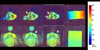
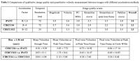
Phase contrast (PC) MR flow measurements are affected by multiple sources of error, including background phase offsets.  The gold-standard approach to correct these offsets involves repeating PC measurements on a static phantom, prolonging each CMR study and impeding exam workflow. Here, we compared the performance of a self-calibrated correction to static-phantom corrected PC data obtained from a pediatric and congenital heart disease population. Self-calibrated correction results showed strong agreement with phantom-corrected data for all vessel types and differed from static-phantom correction by a mean difference in Qp/Qs values of only 0.069. 


2697.   
74 Analysis and correction of eddy current induced artifacts in spiral phase contrast MRI using Point RESolved Spectroscopy
Rene Bastkowski1, Kilian Weiss1,2, David Maintz1, and Daniel Giese1
1Department of Radiology, University Hospital of Cologne, Cologne, Germany, 2Philips Healthcare, Hamburg, Germany
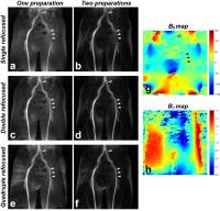
A novel method based on single-voxel-spectroscopy (PRESS) for the analysis and correction of eddy-current induced artefacts in spiral phase-contrast MRI is presented. It is demonstrated, that 0th and 1st order corrections result in residual background offsets of less than 0.5cm/s, inherently correcting for geometrical misalignments between flow acquisitions as well as 2nd order spatial phase offsets. The method does not require special hardware and can be applied as a pre-scan.


2698.   
75 
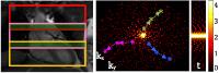
ktv-ARC reconstruction for 4D flow MRI using correlations between velocity encodings
Fatih Suleyman Hafalir1,2, Ana Beatriz Solana2, Peng Lai3, Malek Makki4, Anja C.S. Brau5, Axel Haase1, and Martin A. Janich2
1Technischen Universität München, Munich, Germany, 2GE Global Research, Munich, Germany, 3GE Healthcare, Menlo Park, CA, United States, 4MRI Research Center, University Children Hospital, Zurich, Switzerland, 5GE Healthcare, Munich, Germany
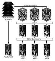
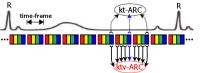
4D flow MRI is a powerful tool for visualization and quantification of blood flow. Repeated acquisition of 4 echoes with different velocity encoding is needed to measure flow in 3D. In this study, we propose a new ktv-ARC reconstruction by incorporating correlations between velocity encoded echoes (v) to the spatiotemporal correlations (kt). The error behavior of the method was analyzed on retrospectively undersampled in vivo cardiac data and resulted in more accurate velocity images with ktv-ARC compared to kt-ARC. 


2699.   
76 Accuracy of relative pressure measurements from 3D PC-MR data using realistic aortic coarctation phantoms
Jesús Urbina1,2, Julio Sotelo1,3, Cristian Montalba1, Felipe Valenzuela1,3, Cristián Tejos1,3, Pablo Irarrázaval1,3, Marcelo Andia1,4, Israel Valverde5,6, and Sergio Uribe1,4
1Biomedical Imaging Center, Pontificia Universidad Católica de Chile, Santiago, Chile, 2School of Medicine, Pontificia Universidad Católica de Chile, Santiago, Chile, 3Electrical Engineering Department, Pontificia Universidad Católica de Chile, Santiago, Chile, 4Radiology Department, Pontificia Universidad Católica de Chile, Santiago, Chile, 5Pediatric Cardiology Unit, Hospital Virgen del Rocio, Seville, Spain, 6Institute of Biomedicine of Seville, Universidad de Sevilla, Seville, Spain
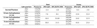
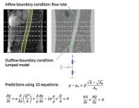
The aim of this work was to evaluate the accuracy of relative pressures obtained from 3D PC-MRI in a realistic aortic phantom with different grades of aortic coarctations at rest and stress conditions. We also evaluated the accuracy of the relative pressures when subjected to different aortic segmentation and spatial resolutions. The accuracy of the 3D PC-MRI is excellent compared with catheterization values with mild to moderate AoCo at rest and stress conditions. Also, relative pressures were in excellent accuracy with catheterization values when the aortic segmentation only included laminar flow and with higher spatial resolution at rest and stress conditions. However, its accuracy decreases for severe AoCo cases.


2700.   
77 4D flow MRI-derived Wall Shear Stress Correlates with Vessel Wall Thickness: Atlases of the Carotid Bifurcation - Permission Withheld
Pim van Ooij1, Merih Cibis2, Wouter V. Potters1, Oscar H Franco3, Meike Vernooij4, Aad van der Lugt4, Frank J Gijsen2, Jolanda J Wentzel2, and Aart J Nederveen1
1Radiology, Academic Medical Center, Amsterdam, Netherlands, 2Biomedical Engineering, Erasmus MC, Rotterdam, Netherlands, 3Epidemiology, Erasmus MC, Rotterdam, Netherlands, 4Radiology, Erasmus MC, Rotterdam, Netherlands
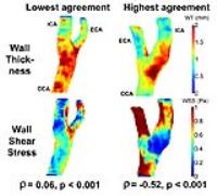
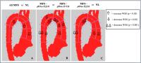
The purpose of this study is to investigate if, already in early disease, a correlation exists between wall thickness (WT) and wall shear stress (WSS) in the carotid bifurcation. Eleven subjects with plaques in the left carotid arteries underwent 3D-flow-MRI and proton-density-weighted-EPI for WSS and WT quantification, respectively. Relationships between WT and WSS were investigated on an individual basis and using cohort-averaged maps (atlases). Spearman’s ρ averaged over all subjects was -0.28, which was significantly different from 0 (p<0.001). For the atlases, ρ was -0.66 (p<0.001). The atlas approach facilitates more statistical power to show that wall thickening occurs in low WSS regions.


2701.   
78 Flow and Structure with Simultaneous Visualization of Registered 4D Flow and Black Blood MRI
Dahan Kim1,2, Carson Hoffman1, Oliver Wieben1,3, and Kevin M. Johnson1
1Department of Medical Physics, University of Wisconsin, Madison, WI, United States, 2Department of Physics, University of Wisconsin, Madison, WI, United States, 3Department of Radiology, University of Wisconsin, Madison, WI, United States
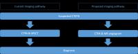
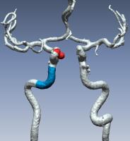
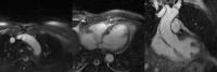
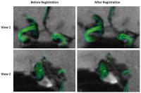
In this work, we examined the feasibility of registering 4D-flow MRI scans with different scan sequences, and demonstrate how the incorporation of complementary, registered data can enhance characterization of hemodynamic information. Black blood (BB) and 4D-flow magnitude images demonstrated excellent registration between the pre- and post-rotation data sets, with high values of correlation and good overlap of the vessels between head rotation. Joint visualization of aneurysm 4D flow and BB shows accurate lesion depiction only after registration and is a promising technique for the comprehensive evaluation of vascular pathology.


2702.   
79 
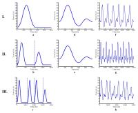
A Validation Study of Real-time Phase Contrast MRI with Low-Rank Modeling
Aiqi Sun1, Bo Zhao2, Yunduo Li1, Qiong He1, Zechen Zhou1, Shuo Chen1, Rui Li1, and Chun Yuan1,3
1Center for Biomedical Imaging Research, Department of Biomedical Engineering, School of Medicine, Tsinghua Universiy, Beijing, China, People's Republic of, 2Martinos Center for Biomedical Imaging, Harvard Medical School, Chalestown, MA, United States, 3Department of Radiology, University of Washington, Seattle, WA, United States
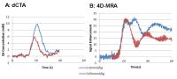
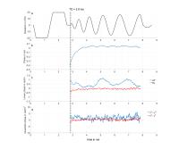
Conventional phase-contrast (PC) MRI method relies on ECG-synchronized cine acquisition to acquire data over multiple cardiac cycles. The underlying spatiotemporal averaging limits this method to studying pathological irregularities. Real-time PC-MRI is a promising approach to overcome these limitations. Although several techniques have been developed to real-time PC-MRI, few have been fully validated due to the difficulty of acquiring a reference data set as gold standard. This study aims at validating the accuracy of a novel real-time PC-MRI technique through both flow phantom experiments and in vivo experiments. 


2703.   
80 
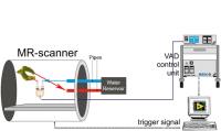
Validation of compressed sensing accelerated 2D flow MRI in the common carotid arteries
Eva S. Peper1, Wouter V. Potters1, Bram F. Coolen1, Henk A. Marquering1, Gustav J. Strijkers1, Pim van Ooij1, and Aart J. Nederveen1
1Radiology, Academic Medical Center (AMC), Amsterdam, Netherlands
Flow MRI of the carotid arteries has emerged as an important imaging field during the last decade and has been shown valuable for the assessment of hemodynamics in the context of atherosclerosis. In this study a 2D flow acquisition was accelerated using random undersampling and compressed sensing reconstruction. For validation of the reconstructed flow a phantom experiment was performed at different acceleration factors followed by an in vivo scan of the carotid arteries.


2704.   
81 
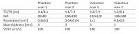
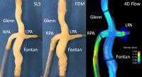
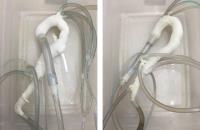
Effects of 3D-printing technology on flow measurements in patient-specific models of total cavo-pulmonary connection
Christopher J Francois1, Zachary Borden1, Sylvana Garcia-Rodriguez1, Jon Wrobel1, and Alejandro Roldan-Alzate1
1Radiology, University of Wisconsin - Madison, Madison, WI, United States
This study investigated the effects of 3D printing technology on flow rates in patient-specific total cavo-pulmonary connection models. 4D flow MRI was used to quantify flow through the Fontan, Glenn, left pulmonary artery and right pulmonary artery in three models at four different flow rates. No statistically significant differences in flow in any of the regions of interest were observed.


2705.   
82 
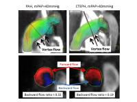
4D flow MR imaging for differentiation of pulmonary arterial hemodynamics in pre-capillary pulmonary hypertension
Hideki Ota1, Koichiro Sugimura2, Haruka Sato2, Yuta Urushibata3, Yoshiaki Komori3, Hiroaki Shimokawa2, and Kei Takase1
1Diagnostic Radiology, Tohoku University Hosipital, Sendai, Japan, 2Cardiovascular Medicine, Tohoku University Hosipital, Sendai, Japan, 3Research&Collaborations, Siemens Japan KK, Tokyo, Japan
Etiologies of pre-capillary pulmonary hypertension may be associated with pulmonary arterial hemodynamics. This study included 64 patients (pulmonary arterial hypertension [PAH], 25, chronic thromboembolic pulmonary hypertension [CTEPH], 39) who underwent 4D flow and cardiac MR imaging. Backward flow ratio and forward flow eccentricity in the pulmonary trunk as assessed by 4D flow MR and several cardiac MR parameters were different between two diseases. After controlling for age and mean pulmonary arterial pressure, backward flow ratio was the strongest differentiator of PAH from CTEPH.  4D flow has a potential to visualize different pulmonary arterial hemodynamics according to etiologies in pulmonary hypertension.


2706.   
83 4D flow MRI Improves Computational Fluid Dynamics Analysis of Aortic Dissection
Sylvana García-Rodríguez1, Jon Wrobel1, Alejandro Roldán-Alzate1,2, and Christopher J. François1
1Radiology, University of Wisconsin-Madison, Madison, WI, United States, 2Mechanical Engineering, University of Wisconsin-Madison, Madison, WI, United States
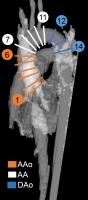
The effects of MRI-derived three-directional velocity profiles implemented at the inlet of aortic dissection (AD) computational fluid dynamics (CFD) simulations were investigated. Two AD models were generated from in vivo MRA data using 3D printing. In vitro 4D Flow MRI was performed on the AD phantoms at two flow rates. Normal and multidirectional blood flow vectors at the AD inlet was measured from 4D Flow MRI data and used in CFD simulations. Significant differences were found in pressure distribution in response to inlet boundary condition definitions. Peak velocity and wall shear stress were also affected by inlet condition definition.


2707.   
84 
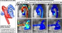
4D flow MRI derived energetic biomarkers are abnormal in repaired tetralogy of Fallot patients and may predict deteriorating hemodynamics
Joshua Daniel Robinson1,2, Cynthia K Rigsby3,4, Michael Rose3, Susanne Schnell4, Alex J Barker4, and Michael Markl4,5
1Pediatric Cardiology, Ann & Robert H Lurie Children's Hospital, Chicago, IL, United States, 2Pediatrics, Northwestern University Feinberg School of Medicine, Chicago, IL, United States, 3Medical Imaging, Ann & Robert H Lurie Children's Hospital, Chicago, IL, United States, 4Radiology, Northwestern University Feinberg School of Medicine, Chicago, IL, United States, 5McCormick School of Engineering, Northwestern University, Evanston, IL, United States
Tetralogy of Fallot (TOF) is the most common form of cyanotic heart disease. As life expectancy continues to increase, MRI plays a central role in evaluation for post-operative complications and reintervention. Current assessment is based on simplified parameters that measure late expression of underlying physiologic changes, with poor outcome prediction. In this study, we explore quantitative 4D flow metrics which may be important measures of hemodynamic efficiency. We found that energetic metrics are abnormal in TOF compared to healthy controls. While these metrics correlated only modestly with routine measurements of ventricular efficiency, they may represent earlier biomarkers of disease progression.


2708.   
85 Highly Accelerated 4D Flow MRI with CIRcular Cartesian UnderSampling (CIRCUS) in Patients with Intracranial Aneurysms - Permission Withheld
Jing Liu1, Yan Wang1, Farshid Faraji1, Sarah Kefayati1, Henrik Haraldsson1, and David Saloner1,2
1University of California San Francisco, San Francisco, CA, United States, 2VA Medical Center, San Francisco, CA, United States
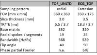
A highly accelerated 4D flow MRI method with a high tempospatial resolution has been validated in healthy volunteers by comparing to the conventional method. The proposed method been demonstrated to be very promising for imaging the patients with intracranial aneurysms, by achieving an isotropic resolution of 1.3mm and a temporal resolution of 26ms within a 5-minute scan time (R=12). 


2709.   
86 
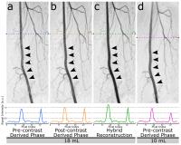
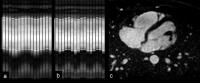
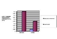
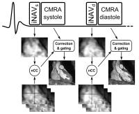
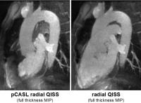
Non-Contrast Cardiac 4D Flow with Bright Blood and Improved Robustness Using Multiple Thin Slab Acquisition and Variable Density Radial Sampling
Peng Lai1, Ann Shimakawa1, Joseph Yitan Cheng2, Marcus T Alley2, Shreyas S Vasanawala2, and Anja C.S Brau3
1Global MR Applications and Workflow, GE Healthcare, Menlo Park, CA, United States, 2Radiology, Stanford University, Stanford, CA, United States, 3Global MR Applications and Workflow, GE Healthcare, Munich, Germany
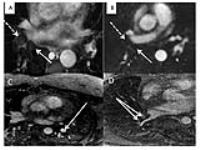
Cardiac 4D Flow suffers from blood signal saturation due to whole-volume imaging and limited accuracy if acquired without contrast agents. This work developed and investigated a new multiple thin slab scheme for non-contrast whole-chest 4D Flow. With in-flow enhancement and bright blood, the new sequence provides higher SNR and motion robustness than conventional 4D Flow. The proposed radial golden angle view order ensures smooth slab merging that is insensitive to cardiac and respiratory variations during the scan.


2710.   
87 
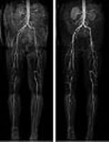
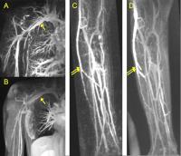
Using MRI to Observe Increased Venous Flow Collateralization in Subjects with Anomalous Jugular Veins
SEAN KUMAR SETHI1, Giacomo Gadda2, Ana M. Daugherty3, David T. Utriainen1, Jing Jiang1, Naftali Raz3, and Ewart Mark Haacke4
1The MRI Institute of Biomedical Research, Detroit, MI, United States, 2Physics, University of Ferrara, Ferrara, Italy, 3Department of Gerontology, Wayne State University, Detroit, MI, United States,4Biomedical Engineering, Wayne State University, Detroit, MI, United States
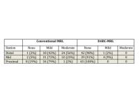
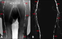
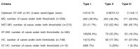
We have established in previous works that a subset of multiple sclerosis (MS) patients show abnormal structure and flow in the internal jugular veins (IJV) when measured with MRI. In this retrospective analysis, we classified and compared extracranial venous collateral flow in MS and normal control samples using MR venography and Phase-contrast flow quantification with a large, standardized dataset. Over 50% of the MS cohort shows a jugular anomaly. The stenotic-MS group shows reduced Type I venous flow compared to healthy controls and non-stenotic MS, while having elevated Type II and Type III flows.


2711.   
88 Design and Validation of a Minimum Time Verse Pulse for 4D Flow MRI
Patrick Magrath1,2, Eric Aliotta1,3, Shams Rashid1, Yutaka Natsuaki4, Xiaoming Bi4, Zhe Wang1,2, Kyung Sung1,2,3, Peng Hu1,2,3, Holden Wu1,2,3, and Daniel B Ennis1,2,3
1Department of Radiological Sciences, University of California, Los Angeles, CA, United States, 2Department of Bioengineering, University of California, Los Angeles, CA, United States, 3Physics and Biology in Medicine IDP, University of California, Los Angeles, CA, United States, 4Siemens Healthcare, Los Angeles, CA, United States
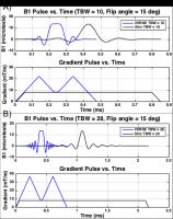
4D-flow MRI is used to quantify blood flow in a variety of neurovascular pathologies including intracranial aneurysms [1], but is limited by long scan times as well as moderate spatial and temporal resolution. Conventional RF pulses have poor slab profiles that contribute to low sequence efficiency by increasing the field-of-view needed to avoid aliasing in the slab direction. Our objectives were to design a minimum time, high Time Bandwith Product (TBW) VERSE pulse for 4D flow and to validate the improvement in sequence efficiency, confirm flow accuracy, and evaluate total SAR deposition for VERSE+4D-flow compared to our clinical 4D flow imaging protocol.


2712.   
89 Contrast-Enhanced 4D Flow Imaging with Reduced Fat Signal
Joseph Y. Cheng1, Tao Zhang1, Adam B. Kerr2, Michael Lustig3, John M. Pauly2, and Shreyas S. Vasanawala1
1Radiology, Stanford University, Stanford, CA, United States, 2Electrical Engineering, Stanford University, Stanford, CA, United States, 3Electrical Engineering & Computer Sciences, University of California, Berkeley, CA, United States
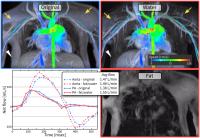
Volumetric time-resolved velocity imaging (4D flow) can be used as a single comprehensive sequence to quantify blood flow, evaluate cardiac function, and assess anatomy. However, fat signal can reduce tissue contrast, introduce high-signal-intensity artifacts from motion, and cause errors in velocity quantification. Two approaches are presented for reducing fat signal in contrast-enhanced 4D flow imaging with minimal time penalty. The first approach is a short spectral-spatial RF pulse that reduces fat signal below the level of contrast-enhanced blood pool. The second approach is the introduction of one additional echo with a different TE to separate fat/water for all flow encoding echoes. The performance of these approaches are evaluated in a static phantom study and in patient volunteer studies. 


2713.   
90 The Communicating Arteries Redistribute Blood Flow in the Circle of Willis with Hypoplastic Segments: Intracranial 4D flow MRI at 7 Tesla
Pim van Ooij1, Matthan Caan1, Bart M. W. Cornelissen2, Henk A Marquering2, Pieter Buur3, Gustav J Strijkers2, Jeroen Hendrikse4, and Aart J Nederveen1
1Radiology, Academic Medical Center, Amsterdam, Netherlands, 2Biomedical Engineering & Physics, Academic Medical Center, Amsterdam, Netherlands, 3Spinoza Center for Neuroimaging, Amsterdam, Netherlands, 4Radiology, University Medical Center Utrecht, Utrecht, Netherlands
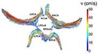
In this study it was investigated if the communicating arteries redistribute blood flow in the circle of Willis (coW) with hypoplastic arteries. For this purpose, 4D flow MRI at 7 Tesla was used in ten healthy volunteers. 50% of the participants had a full coW, whereas 50% had hypoplastic or missing segments. Significant correlations were found for time-averaged blood flow (mL/s) between the left /right posterior communicating artery and the left/right posterior cerebral artery and between the anterior communicating artery and the right anterior cerebral artery. This finding illustrates that flow is redistributed through the communicating arteries in the coW.


2714.   
91 Aortic hemodynamics in pediatric Marfan patients compared to healthy pediatric subjects: heterogeneity in the Marfan population
Roel LF van der Palen1,2, Alex J Barker2, Emilie Bollache2, Michael J Rose3, Pim van Ooij4, Julio Garcia2, Luciana Young5, Arno AW Roest1, Michael Markl2,6, Cynthia K Rigsby3, and Joshua D Robinson5,7
1Department of Pediatric Cardiology, Willem-Alexander Children and Youth Center, Leiden University Medical Center, Leiden, Netherlands, 2Department of Radiology, Feinberg School of Medicine, Northwestern University, Chicago, IL, United States, 3Department of Medical Imaging, Ann & Robert Lurie Children’s Hospital of Chicago, Chicago, IL, United States, 4Department of Radiology, Academic Medical Center, Amsterdam, Netherlands, 5Division of Pediatric Cardiology, Ann & Robert Lurie Children’s Hospital of Chicago, Chicago, IL, United States, 6Department of Biomedical Engineering, McCormick School of Engineering, Northwestern University, Chicago, IL, United States, 7Department of Pediatrics, Ann & Robert Lurie Children’s Hospital of Chicago, Chicago, IL, United States
Marfan syndrome (MFS) is a connective tissue disease with high risk of aortic dissection/rupture. Two-thirds of dissections occur in the ascending aorta, one-third in the descending aorta. Diameter plays an important role in risk stratification. However, recent literature has shown diameter only accounts for 50% of the dissections in the descending aortic region. It is not well known how aortic hemodynamics interact with the altered vascular structure of these aortas and how it may impact dilatation. A cohort of MFS children and an age appropriate control group were evaluated with 4D flow MRI: already distinct abnormalities are present in childhood.


2715.   
92 Model-based estimation of arterial pulse wave velocity from MRI velocity data
Prem Venugopal1, Ek Tsoon Tan1, Peter Lamb1, Christopher J Hardy1, and Thomas K Foo1
1GE Global Research, Niskayuna, NY, United States
Pulse wave velocity (PWV) is a commonly used surrogate for arterial stiffness. This abstract describes a new method to estimate arterial PWV by using MRI phase-contrast data to tune a 1D blood flow model describing the hemodynamics and propagation of the arterial pulse wave. Results obtained in a single volunteer indicate that the proposed approach could be used with low time resolution methods such as 4D Flow MRI to obtain PWV in the aorta with much lower variability than the foot-to-foot method.


2716.   
93 10 fold accelerated 4D flow in the carotid arteries at high spatiotemporal resolution in 7 minutes using a novel 15 channel coil
Eva S. Peper1, Qinwei Zhang1, Bram F. Coolen2, Wouter V. Potters1, Pim van Ooij1, Dennis W.J. Klomp3, and Aart J. Nederveen1
1Radiology, AMC, Amsterdam, Netherlands, 2Biomedical Engineering and Physics, AMC, Amsterdam, Netherlands, 3Radiology, UMCU, Utrecht, Netherlands
Using a novel 15 channel coil for carotid artery imaging we could prove that the g-factor loss at higher acceleration factors is limited. This permits the use of higher parallel imaging factors than commonly exploited in carotid MRI. A 4D flow scan at high spatiotemporal resolution was accelerated 10 fold (SENSE 4 x 2.5), resulting in high quality images and consistent flow values acquired in a scantime as short as 7 min.


2717.   
94 Lower Wall Shear Stress and Abnormal Hemodynamics within the Saccular Aneurysm in Contrast to Fusiform Aneurysm in the Abdominal Aorta.
Masataka Sugiyama1, Yasuo Takehara2, Hatsuko Nasu1, Shuhei Yamashita1, Mika Kamiya1, Nobuko Yoshizawa1, Yuki Hirai1, Takasuke Ushio1, Naoko Hyodo1, Yohei Ito1, Naoki Oishi2, Marcus Alley3, Tetsuya Wakayama4, and Harumi Sakahara1
1Radiology, Hamamatsu University School of Medicine, Hamamatsu, Japan, 2Radiology, Hamamatsu University Hospital, Hamamatsu, Japan, 3Department of Radiology, Stanford University School of Medicine, Stanford, CA, United States, 4Applied Science Laboratory Asia Pacific, GE Healthcare Japan, Hino, Japan
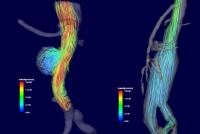
3D cine PC MRI (4D-flow) study of abdominal aorta was conducted to measure wall shear stress (WSS) and to characterize aortic blood flow dynamics within saccular and fusiform abdominal aortic aneurysm and non-dilated aorta.  Peak systolic WSS was significantly lower within saccular aneurysm, and stream line analysis depicted  separated vortex flow within the saccular aneurysm.  The abnormal vortex flow and consequent low WSS of saccular aneurysmal wall may be reflecting the continuing risk of atherogenic changes of the saccular aneurysm in contrast to fusiform aneurysm.


2718.   
95 3D Printed Patient-Specific Model for In Vitro Hemodynamic Studies and Comparison with In Vivo Findings Using 4D Flow MRI
Rouzbeh R Ahmadian1, Austin P Boyd2, Jeremy D Collins1, James C Carr1, Alex J Barker1, and Michael Markl3
1Radiology, Northwestern University, Chicago, IL, United States, 2Northwestern University, Chicago, IL, United States, 3Radiology & Biomedical Engineering, Northwestern University, Chicago, IL, United States
The advent of 3D printing has opened exciting possibilities for biomedical applications. One of the most intriguing of these possibilities is the ability to use images obtained from radiology scanners (CT or MR) to create 3D models of patient anatomy followed by 3D printing. These models will have all the same geometries of patient anatomy to very high detail. In order to have practical applications, however, these 3D models need to behave similarly to human tissue under standard conditions. Our research utilizes patient-specific 3D printed models in an in vitro fluid dynamic circuit to compare 4D Flow MRI data to that of the patient (in vivo). This direct comparison will allow for validation of the 3D printed model for further biomedical application. 


2719.   
96 
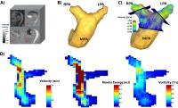
Volumetric assessment of kinetic energy and vorticity in the pulmonary artery: alteration of flow hemodynamics in patients with repaired tetralogy of Fallot using 4D flow MRI
Julio Garcia1, Silvia Hidalgo Tobon2,3, Guadalupe Sagaon Rojas4, Benito de Celis Alonso5, Manuel Obregon2, Porfirio Ibanez2, Julio Erdmenger6, and Pilar Dies-Suarez2
1Radiology, Northwestern University, Chicago, IL, United States, 2Investigacion en Imagen y Resonancia Magnetica Nuclear, Hospital Infantil de Mexico Federico Gomez, Mexico City, Mexico, 3Physics, Universidad Autonoma Metropolitana, Mexico, Mexico, 4Physics, Universidad Autonoma Metropolitana, Mexico City, Mexico, 5Faculty of Physics and Mathematics, Benemérita Universidad Autónoma de Puebla, Puebla, Mexico, 6Pediatric Cardiology, Hospital Infantil de Mexico Federico Gomez, Mexico City, Mexico
Flow alterations in the pulmonary artery (PA) of patients with repaired tetralogy of Fallot (rTOF) may be link with elevated kinetic energy (KE). 4D flow MRI allows for the non-invasive volumetric assessment of flow hemodynamics, vorticity, and KE in patients with rTOF in the pulmonary (PA). Thus, the aim was to investigate the impact of flow alterations in the PA and its association with KE and vorticity.
 

The International Society for Magnetic Resonance in Medicine is accredited by the Accreditation Council for
Continuing Medical Education to provide continuing medical education for physicians.