ISMRM 24th Annual Meeting & Exhibition • 07-13 May 2016 • Singapore

Electronic Poster Session: Contrast Mechanisms

2816 -2839 Relaxation Applications
2840 -2863 Magnetic Susceptibility
2864 -2887 Perfusion & Permeability
2888 -2911 CEST & MT
2982 -3005 Contrast Mechanisms: Electromagnetic Based & Others

Exhibition Hall 

14:15 - 15:15

    Computer #

2816.   
1 trueFLASH: Model-Based Iterative T1 Mapping using Variable-Flip-Angle Fast Low-Angle Shot
Tom Hilbert1,2,3, Damien Nguyen4,5, Jean-Philippe Thiran2,3, Gunnar Krueger2,3,6, Tobias Kober1,2,3, and Oliver Bieri4,5
1Advanced Clinical Imaging Technology (HC CMEA SUI DI BM PI), Siemens Healthcare AG, Lausanne, Switzerland, 2Department of Radiology, University Hospital (CHUV), Lausanne, Switzerland, 3LTS5, École Polytechnique Fédérale de Lausanne, Lausanne, Switzerland, 4Radiological Physics, Department of Radiology, University of Basel Hospital, Basel, Switzerland, 5Department of Biomedical Engineering, University of Basel, Basel, Switzerland, 6Siemens Medical Solutions USA, Inc., Boston, MA, United States
Various methods have been published to quantify the longitudinal relaxation T1; amongst others, a variable-flip-angle fast low-angle shot acquisition can be used. Here, we suggest applying an 8-fold undersampling to such an acquisition, subsequently using a model-based iterative optimization to estimate T1 maps. This approach allows the acquisition of whole-brain 1.3mm isotropic T1 maps within 3:20min using 16 different flip angles. Aliasing artifacts due to the undersampling were successfully removed by the iterative reconstruction. However, the T1 maps show a slight overestimation of T1 values and require a B1-field correction as it is typical for variable-flip-angle approaches.


2817.   
2 Fast 3D Acquisition for Quantitative Mapping and Synthetic Contrasts Using MIRACLE and trueCISS
Tom Hilbert1,2,3, Damien Nguyen4,5, Jean-Philippe Thiran2,3, Gunnar Krueger2,3,6, Oliver Bieri4,5, and Tobias Kober1,2,3
1Advanced Clinical Imaging Technology (HC CMEA SUI DI BM PI), Siemens Healthcare AG, Lausanne, Switzerland, 2Department of Radiology, University Hospital (CHUV), Lausanne, Switzerland, 3LTS5, École Polytechnique Fédérale de Lausanne, Lausanne, Switzerland, 4Radiological Physics, Department of Radiology, University of Basel Hospital, Basel, Switzerland, 5Department of Biomedical Engineering, University of Basel, Basel, Switzerland, 6Siemens Medical Solutions USA, Inc., Boston, MA, United States
Quantitative imaging promises to be a good biomarker of disease but requires long acquisition times, especially when 3D acquisition techniques are used. Here we propose to use a highly undersampled 3D phase-cycled balanced steady-state free-precession sequence in combination with the reconstruction methods MIRACLE and trueCISS, providing quantitative maps (T2,T1,M0). Additionally, synthetic contrasts can be generated using the previously calculated quantitative maps and signal models, exemplarily shown for bSSFP, T2- and T1-weighted contrast. In summary, the proposed method provides a set of quantitative maps and various conventional contrasts using a single 3:31min acquisition.


2818.   
3 Rapid Multislice T1 Mapping of Contrast-Enhanced Mouse Tumor Using Saturation Recovery Look-Locker Method with Spiral Readout
Yuchi Liu1, Zheng Han1, Kai Jiang2, Yun Jiang1, Yajuan Li1, Zheng-Rong Lu1, and Xin Yu1,3,4,5
1Biomedical Engineering, Case Western Reserve University, Cleveland, OH, United States, 2Division of Nephrology and Hypertension, Mayo Clinic, Rochester, MN, United States, 3Radiology, Case Western Reserve University, Cleveland, OH, United States, 4Physiology and Biophysics, Case Western Reserve University, Cleveland, OH, United States, 5Case Center for Imaging Research, Case Western Reserve University, Cleveland, OH, United States
In this study, a rapid multislice saturation-recovery Look-Locker (MSRLL) T1 mapping method with spiral readout was developed for the application on tissues with long T1 value, such as tumor. This method was validated in vitro and evaluated in a dynamic contrast-enhanced MRI (DCE-MRI) study on mouse tumor. The spiral MSRLL method showed a strong agreement with Cartesian method, and achieved temporal resolution of 2 min 40s with a voxel size of 0.23×0.23×1 mm3 in vivo.


2819.   
4 Comparison of Signal intensity measures with absolute T1 value quantification in patients undergoing serial follow up imaging in patients with neurocysticercosis
Pradeep Kumar Gupta1, Prativa Sahoo2, Alok Kumar Singh3, Ravindra Kumar Garg3, Rupsa Bhattacharjee4, and Rakesh Kumar Gupta1
1Radiology and Imaging, Fortis Memorial Research Institute, Gurgaon, India, 2Healthcare, Philips India ltd, Bangalore, India, 3Department of Neurology, KG Medical University, Lucknow, India,4Healthcare, Philips India ltd, Gurgaon, India
Gadolinium based contrast agents are necessary for MRI for disease detection and therapy. Studies reported gadolinium remains in brain for long time after injecting contrast particularly in follow up patients undergoing serial contrast applications. T1 signal intensity increases slowly due to contrast accumulation in brain. Our study quantifies T1 relaxation time as a measure for follow up contrast patients. Results show absolute T1 significantly decreases in Gray Matter regions, putamen, thalamus and globus pallidus. We conclude that absolute T1 quantification can efficiently pick up deposition of gadolinium much earlier than T1 signal intensity changes as observed in Gray matter regions.


2820.   
5 NOVIFAST: A fast non-linear least squares method for accurate and precise estimation of T1 from SPGR signals
Gabriel Ramos-Llordén1, Arnold J. den Dekker1,2, Marcus Björk3, Marleen Verhoye1, and Jan Sijbers1
1University of Antwerp, Antwerp, Belgium, 2Delft University of Technology, Delft, Netherlands, 3Uppsala University, Uppsala, Sweden
The longitudinal relaxation time T1 can be estimated from a set of spoiled gradient recalled echo (SPGR) magnetic resonance images acquired over a range of flip angles with constant repetition time (TR). While linear estimators are widely used because of their low computation time, they are less accurate than their slower nonlinear counterpart1. In this work, we introduce a novel  NOn-linear VarIable Flip Angle data baSed T1 (NOVIFAST) estimator  that combines the best of both worlds: high accuracy and precision in a low computation time.


2821.   
6 The effect of through-plane gradients on 2D gradient-echo acquisitions: are sinc-term corrections appropriate for R2* mapping?
Mukund Balasubramanian1,2 and Robert V. Mulkern1,2
1Department of Radiology, Boston Children's Hospital, Boston, MA, United States, 2Harvard Medical School, Boston, MA, United States
We made slice profile measurements and acquired 2D multi-gradient-echo data on a phantom in the presence of various through-plane gradients. The sinc-term correction for R2* mapping that is commonly used to compensate for the dephasing effects of these gradients was substantially outperformed by a correction based on the measured slice profiles. Our results highlight the importance of incorporating realistic slice profiles into the estimation of “intrinsic” R2* values, which may contain information about tissue structure at microscopic-to-mesoscopic spatial scales.


2822.   
7 Molecular Mechanism of Transverse Relaxation in Whole Blood with Plasma Contrast Reagent: Simulations of Bulk Magnetic Susceptibility and Water Exchange
Gregory J Wilson1, Charles S Springer2, Sarah Bastawrous1,3, and Jeffrey H Maki1
1University of Washington, Seattle, WA, United States, 2Oregon Health and Science University, Portland, OR, United States, 3Puget Sound VA HCS, Seattle, WA, United States
Previously reported T2* values in oxygenated whole blood with gadolinium based contrast reagents are very small. This has important implications for optimization of contrast-enhanced MR angiography and quantification of arterial input functions. To investigate the molecular mechanism of these short T2* values, we have performed Monte Carlo simulations of signal dephasing that predict the reported values remarkably well. Intracellular water signal experiences a frequency shift that is dependent on the orientation of the RBC in the magnetic field. The resulting frequency distribution in combination with trans-membrane water exchange results in rapid dephasing of water signal.


2823.   
8 Relaxation Properties of Human Umbilical Cord Blood at 1.5 Tesla
Sharon Portnoy1, Mark Osmond2, Meng Yuan Zhu3, Mike Seed4, John G. Sled1,2,5, and Christopher K. Macgowan1,4
1Department of Medical Biophysics, University of Toronto, Toronto, ON, Canada, 2Department of Obstetrics and Gynaecology, University of Toronto, Toronto, ON, Canada, 3Institute of Medical Science, University of Toronto, Toronto, ON, Canada, 4Diagnostic Imaging, The Hospital for Sick Children, Toronto, ON, Canada, 5Mouse Imaging Centre, The Hospital for Sick Children, Toronto, ON, Canada
Relaxometry measurements were performed on umbilical cord blood samples (N=88) spanning a broad range of hematocrits (0.19<Hct<0.76) and oxygen saturations (4%<sO2<100%).  Simple biophysical models were used to describe variations in T1 and T2 as a function of these blood properties.  The data and fitted model parameters presented here can be used for calibration of future MRI investigations of fetal and neonatal blood physiology.  


2824.   
9 Quantitative T2 mapping using a modified BIR-4 T2 preparation in the presence of large B0 offsets
Dominik Weidlich1, Hendrik Kooijman2, Peter Börnert3, Jan S. Kirschke4, Ernst J. Rummeny1, Axel Haase5, and Dimitrios C. Karampinos1
1Department of Diagnostic and Interventional Radiology, Technische Universität München, Munich, Germany, 2Philips Healthcare, Hamburg, Germany, 3Philips Research Laboratory, Hamburg, Germany,4Section of Neuroradiology, Technische Universität München, Munich, Germany, 5Zentralinstitut für Medizintechnik, Technische Universität München, Garching, Germany
T2 mapping has been emerging as a tool to quantitatively assess tissue edema and inflammation. Sequences employing T2 preparation have been suggested for accurate and precise T2 quantification. The use of the modified BIR-4 pulse has been proposed to minimize the sensitivity of T2 mapping to B1 inhomogeneities. However, in the presence of large B0 offsets, part of the magnetization can experience both T1 and T2 relaxation during the BIR-4 pulse leading to T2 quantification errors. The present work proposes a novel methodology to make T2 quantification based on the modified BIR-4 robust to the presence of large B0 offsets.


2825.   
10 Contrast optimization using Fast Field Cycling techniques and fast low angle sequences
Nicolas Chanet1 and Ludovic De Rochefort1
1IR4M (Imagerie par Résonance Magnétique Médicale et Multi-modalités), Univ. Paris-Sud, CNRS, UMR8081, Université Paris-Saclay, Orsay, France
Fast Field Cycling MRI offers the possibility to explore new contrasts generated from NMR dispersion (NMRD) profiles of tissue or contrast agents. Here, it is shown that dreMR contrast can be generated and optimized in fast low flip angle sequences extending the range of sequences that can be used with this new degree of freedom. The expression of a generalized Ernst angle maximizing signal with dreMR pulses is derived as well as the angle maximizing the dispersive contrast.


2826.   
11 Whole brain adiabatic T1rho and Relaxation Along a Fictitious Field imaging in healthy volunteers: feasibility and initial findings
Aida Kiviniemi1, Harri Merisaari1, Marko Pesola1, Timo Liimatainen2, Hannu Juhani Aronen1, and Ivan Jambor1
1University of Turku, Turku, Finland, 2A.I. Virtanen Institute for Molecular Sciences, Kuopio, Finland
The adiabatic T and RAFF measurements, RAFF2 and RAFF4, were performed in 28 healthy volunteers (24-69 years) and four of them had repeated MR scan within 4 weeks to evaluate short term repeatability. The relative differences on the voxel level were below 5% in gray and white matter for all of the relaxation parameters except of TRAFF4 in gray matter which was 7.3%. No statistically significant age related changes in white and gray matter were present as evaluated by adiabatic T, RAFF2 and RAFF4 imaging.


2827.   
12 T1? dispersion in human calf muscle
Ping Wang1,2, Henry Zhu1,2, Hakmook Kang3, Jake Block2, and John C. Gore1,2
1Institute of Imaging Science, Vanderbilt University Medical Center, Nashville, TN, United States, 2Department of Radiology and Radiological Sciences, Vanderbilt University Medical Center, Nashville, TN, United States, 3Department of Biostatistics, Vanderbilt University Medical Center, Nashville, TN, United States
 
T imaging is sensitive to slow macromolecular interactions which may be generally characterized by a correlation time (τc) but also varies with the strength of the locking field used (ω1).  At higher fields (3T and beyond) T is also strongly influenced by chemical exchange processes and the dispersion of the relaxation rate R (=1/T) with locking field may be used to quantify exchange processes.  We imaged normal muscles from individuals of different ages and found that R value is negatively correlated with age in normal muscle, and there is a small dispersion of R that appears to increase with age.


2828.   
13 Evaluation of Renal Oxygenation using a Simultaneous Dynamic R2, R2' and R2* Quantification Approach – Under the Influence of Hyperoxic Challenge
CY Wang1, R Zhang2, L Jiang3, R Wang4, XD Zhang4, H Wang3, K Zhao4, LX Jin3, J Zhang1,2, XY Wang1,4, and J Fang1,2
1Academy for Advanced Interdisciplinary Studies, Peking University, Beijing, China, People's Republic of, 2College of Engineering, Peking University, Beijing, China, People's Republic of, 3Philips Healthcare, Suzhou, China, People's Republic of, 4Department of Radiology, Peking University First Hospital, Beijing, China, People's Republic of
This study demonstrates the feasibility of dynamic renal R2, R2’ and R2* measurement with the proposed psMASE-ME method. High temporal resolution was achieved by combining psMASE sequence and moving estimation strategy. The superiorities of this method could potentially be used in the renal oxygenation evaluation.


2829.   
14 Highly Accelerated, Intravascular T1, T2, and Proton Density Mapping with Linear Algebraic Modeling and Sensitivity Profile Correction at 3T
Guan Wang1,2, Yi Zhang2, Shashank Sathyanarayana Hegde2,3, and Paul A. Bottomley2
1Dept. of Electrical & Computer Engineering, Johns Hopkins University, Baltimore, MD, United States, 2Russell H. Morgan Dept. of Radiology & Radiological Sciences, Johns Hopkins University, Baltimore, MD, United States, 3(currently) Philips Innovation Campus, Bangalore, India
Vessel wall MRI with intravascular (IV) detectors can produce superior local signal-to-noise ratios (SNR) and generate high-resolution T1, T2, and proton density (PD) maps that could be used to automatically classify atherosclerotic lesion stage. However, long acquisition times potentially limit multi-parametric mapping. Here, for the first time, spectroscopy with linear algebraic modeling (SLAM) is applied to yield accurate compartment-average T1, T2 and PD measures at least 10 times faster compared to a standard full k-space reconstructed MIX-TSE sequence at 3T. Simple phase and magnitude sensitivity corrections are incorporated into the SLAM reconstruction to compensate for IV detector non-uniformity. 


2830.   
15 A Pulse Sequence for T1rho Imaging of Liver in Human Subjects
Weitian Chen1, Queenie Chan2, Min Deng1, and Yixiang Wang1
1Imaging and Interventional Radiology, the Chinese University of Hong Kong, New Territory, Hong Kong, 2Philips Healthcare, New Territory, Hong Kong
T1rho is a potential biomarker for  evaluation of liver fibrosis. However, the application of T1rho imaging for liver disease in human subjects remains challenging. We propose a pulse sequence based on single shot Turbo (Fast) Spin Echo (SSTSE or SSFSE) acquisitions to address the problems.


2831.   
16 Fast Fluid Suppressed T1? Imaging of the Human Brain
Eberhard Daniel Pracht1 and Tony Stöcker1,2
1German Center for Neurodegenerative Diseases (DZNE), Bonn, Germany, 2University of Bonn, Department of Physics and Astronomy, Bonn, Germany
The longitudinal relaxation time in the rotating frame (T) is a non-invasive biomarker, which is sensitive to slow macromolecular interactions. It might potentially be useful to detect pathological changes in neurodegenerative diseases such as Alzheimer’sDisease. However, long scan times and high SAR limit the application of this technique in a routine setting. Furthermore, high CSF signal modulates the image intensity at CSF/tissue boundaries leading to an overestimation of T1ρ in these regions. To overcome these issues we developed a fast and robust approach to assess T in the human brain at high image resolution.


2832.   
17 Colloidal Nanoparticles for MRI Contrast Agents
Akbar Alipour1,2,3, Vijay Kumar Shamra3,4, Zeliha Soran Erdem3, Zaliha Gamze Aykut5, and Hilmi Volkan Demir2,3,4
1UMRAM-National Magnetic Resonance Research Center, Bilkent University, Ankara, Turkey, 2Department of Electrical and Electronics Engineering, Bilkent University, Ankara, Turkey, 3UNAM – Institute of Materials Science and Nanotechnology, Bilkent University, Ankara, Turkey, 4School of Mathematical and Physical Sciences, Nanyang Technological University, Singapore, Singapore, 5Department of Molecular Biology and Genetics, Bilkent University, Ankara, Turkey
Magnetic resonance imaging (MRI) contrast agents, employed as image contrast enhancement mechanism, are able to change either negative or positive signal intensity. T2 contrast normally offers magnetic susceptibility artifacts and decreases the resolution. On the other hand, T1 contrast provides high spatial resolution, but it suffers from the deficiency of high contrast-to-noise ratio, which limits their clinical applications. To overcome the aforementioned limitations, we offer cubic shape super-paramagnetic iron oxide nanoparticles (SPIONs) as dual-mode colloidal MRI contrast agents. The in vitro and in vivo Tand TMR images promise the great potential clinical application of SPIO nanocubes as dual-mode MR contrast agent.


2833.   
18 Quantifying Chemical Exchange Contributions in Mixtures Using Spin-Lock MRI
John Thomas Spear1,2 and John Gore1,2,3,4
1Physics and Astronomy, Vanderbilt University, Nashville, TN, United States, 2Institute of Imaging Science, Vanderbilt University, Nashville, TN, United States, 3Radiology, Vanderbilt University, Nashville, TN, United States, 4Biomedical Engineering, Vanderbilt University, Nashville, TN, United States
R dispersion provides insight into the rates of molecular processes that give rise to relaxation, and a technique called Exchange Rate Contrast (ERC) can differentiate proton pools based on chemical exchange rates. Double dispersion phenomena may occur when three exchanging proton pools are present, and parametric images may be calculated in which the image intensity scales with the concentration of the exchanging pools. A theoretical equation was derived for this contrast and shown to align well with Bloch-McConnell simulations. Various applications with exogenous contrast agents present a great deal of potential for utilizing this technique in practice.


2834.   
19 Paradoxical Narrowing of the Spectroscopy Water Peak in the Presence of Iron Overload
Diego Hernando1, Samir D Sharma1, Debra E Horng2, and Scott B Reeder1,2,3,4,5
1Radiology, University of Wisconsin-Madison, Madison, WI, United States, 2Medical Physics, University of Wisconsin-Madison, Madison, WI, United States, 3Biomedical Engineering, University of Wisconsin-Madison, Madison, WI, United States, 4Medicine, University of Wisconsin-Madison, Madison, WI, United States, 5Emergency Medicine, University of Wisconsin-Madison, Madison, WI, United States
In phantoms with large iron particles, as well as in patients with liver iron overload, a paradoxical relationship was observed where the linewidth measured from MRS was smaller than the linewidth obtained from multi-echo spoiled gradient echo MRI. Assuming a model of “apparent” R2 decay in the presence of iron where single-echo acquisitions are essentially diffusion-weighted by the iron-induced B0 heterogeneities, we speculate that different isochromats within the observed spectra undergo different diffusion weighting related to their location relative to nearby large iron particles. These observations may have implications for the characterization of iron deposition in tissue. 


2835.   
20 Towards Optimized Experiment Design for Magnetic Resonance Fingerprinting
Bo Zhao1, Justin P. Haldar2, Kawin Setsompop1, and Lawrence L. Wald1
1Martinos Center for Biomedical Imaging, Chalestown, MA, United States, 2Signal and Image Processing Institute, University of Southern California, Los Angeles, CA, United States
A principled framework is proposed to optimize the experiment design for magnetic resonance fingerprinting (MRF) based on the Cramer-Rao bound. Within this framework, we optimize the acquisition parameters (flip angle, TR, etc.)  to maximize the SNR efficiency of quantitative parameter estimation. Preliminary results indicate that the optimized experiments allow for substantially reducing the length of an MRF acquisition and substantially improving estimation performance for the T2 map, while maintaining similar accuracy level for the T1 map. The proposed framework should prove useful for fast quantitative MR imaging with MRF. 


2836.   
21 Myelin Water Imaging using 3D Radial multi-echo GRE acquisition
Hongpyo Lee1, Yoonho Nam2, Min-Oh Kim1, Dongyeob Han1, Sung-Min Gho1, and Dong-Hyun Kim1
1School of Electrical and Electronic Engineering, Yonsei University, Soeul, Korea, Republic of, 2Department of Radiology, Seoul St. Mary Hospital, College of Medicine, The Catholic University of Korea, Seoul, Korea, Republic of
Recently, myelin water fraction was investigated using multi-echo GRE data. In case of complex model fitting for MWF, only a small fraction of the total signal is used, small artifacts caused by physiological motion can induce severe noise in MWF. To overcome this problem, radial trajectory which is known to have robustness against object motion can be used. Also, this trajectory has the additional advantage of acquiring the more center of k-space than Cartesian trajectory which is helpful for detecting accurate T2* decay curve, and it may provide higher SNR and improved quantification. In this abstract, we demonstrated that the radial acquisition can reduce artifacts and improve image quality in MWF.


2837.   
22 Myelin Water Imaging in Multiple Sclerosis Post-Mortem Spinal Cord
Amy McDowell1, Natalia Petrova1, Irene Vavasour2, David Thomas3, Daniele Carassiti1, Marc Miquel4, Shannon Kolind2, and Klaus Schmierer1,5
1Neuroscience and Trauma, Queen Mary University of London, London, United Kingdom, 2University of British Columbia, Division of Neurology, Department of Medicine, Vancouver, BC, Canada,3Neuroimaging Analysis Centre, Department of Brain Repair and Rehabilitation, London, United Kingdom, 4Clinical Physics, Barts Health Trust, London, United Kingdom, 5Department of Neurology, Barts Health Trust, London, United Kingdom
Evidence obtained from post mortem multiple sclerosis (MS) brain suggest that ‘myelin water fraction’ (MWF) acquired using the CPMG sequence is strongly associated with myelin.  However, relaxation times differ significantly between brain and spinal cord (SC), a key area of the clinical manifestations of MS. We investigated the histological correlates of MWF in post mortem MSSC. Good separation of the short T2 components, and correlation between MWF and myelin content detected using immunohistochemistry were observed.


2838.   
23 Linear models for estimating myelin and iron content in the brain
Riccardo Metere1 and Harald E. Möller1
1Max Planck Institute for Human Cognitive and Brain Sciences, Leipzig, Germany
Quantitative MRI maps are believed to strongly correlate with myelin and and ferritin content. Recently, a model combining prior information from non-MRI quantitative techniques was proposed for the quantification of myelin and ferritin based on relaxometry information $$$R_1$$$ and $$$R_2^*$$$ in post mortem human brain samples. Here, we propose four different simple linear models for tentatively obtaining semi-quantitative information using simultaneous quantitative acquisitions under in vivo conditions. Three of the four model are relatively consistent, but do not agree with previously published measurements. A fourth model, although not validated, is compatible with previous works.  


2839.   
24 Inter-scanner reproducibility of 4 minute whole brain myelin mapping using FAST-T2
Thanh D. Nguyen1, Kofi Deh1, Sneha Pandya1, Yi Wang1, and Susan A. Gauthier1
1Weill Cornell Medical College, New York, NY, United States
The purpose of this study was to measure the inter-scanner reproducibility of fast myelin water fraction (MWF) mapping using a 4 min FAST-T2 sequence on three scanners from two vendors at 1.5T and 3T. Regional MWF measurements obtained in 7 healthy volunteers were highly reproducible with excellent inter-scanner correlation (R>0.95) and small MWF bias of less than 1%.
Exhibition Hall 

14:15 - 15:15

    Computer #

2840.   
25 Absolute quantification of white matter lesion iron and myelin using QSM and FAST-T2 at 3 Tesla
Thanh D. Nguyen1, Pascal Spincemaille1, Sneha Pandya1, Susan A. Gauthier1, and Yi Wang1
1Weill Cornell Medical College, New York, NY, United States
The purpose of this study was to develop a clinically feasible method for quantifying myelin and iron distribution in white matter lesions by integrating absolute myelin water mapping using Fast Acquisition using Spiral Trajectory and T2prep (FAST-T2) sequence with Quantitative Susceptibility Mapping (QSM). Preliminary results from 8 healthy volunteers and 3 MS patients demonstrated the feasibility of the developed method.


2841.   
26 Susceptibility Changes in Dentate Nucleus on Quantitative Susceptibility Mapping  due to serial GBCAs administration. - Permission Withheld
Takuya Hinoda1, Yasutaka Fushimi1, Tomohisa Okada1,2, Tsutomu Okada1, Akira Yamamoto1, and Togashi Kaori1
1Radiology, Graduate school of Medicine, Kyoto University, Kyoto, Japan, 2Human Brain Research Center, Graduate school of Mediine, Kyoto University, Kyoto, Japan
Gadolinium-based contrast agents (GBCAs) have been widely used for contrast material-enhanced magnetic resonance (MR) imaging. However, gadolinium accumulation in the dentate nucleus (DN) has gained attention due to recent studies. In this study, we retrospectively evaluated the susceptibility values of DN, using quantitative susceptibility mapping. The susceptibility values of the patients with GBCA administration were significantly higher than those of normal controls.


2842.   
27 The Absence of Phase Information in the Signal-Deprived Image Background is an Important Source of Error in Susceptibility Mapping
Russell Dibb1,2 and Chunlei Liu3,4
1Center for In Vivo Microscopy, Duke University, Durham, NC, United States, 2Biomedical Engineering, Duke University, Durham, NC, United States, 3Brain Imaging & Analysis Center, Duke University, Durham, NC, United States, 4Radiology, Duke University, Durham, NC, United States
Susceptibility mapping is limited in part due to reconstruction artifacts spawning from errors in tissue frequency data. Through simulations of susceptibility tensor imaging (STI) data, we show that a lack of phase information in the image background (where there is typically no signal) engenders artifacts in reconstructed susceptibility property maps. These errors are most egregious in the boundary regions of the object and affect mean susceptibility, susceptibility anisotropy, and tissue structure orientation measurements. Greater understanding of susceptibility mapping artifacts will aid in developing the tools necessary for accurate and reliable susceptibility imaging techniques.


2843.   
28 Improved magnetic dipole kernel for reconstruction methods in quantitative susceptibility mapping
Carlos Milovic1,2, Jose Miguel Pinto1,2, Julio Acosta-Cabronero3, Petr Dusek4,5, Vince Istvan Madai6, Till Huelnhagen7, Thoralf Niendorf7, Jens Wuerfel8, and Cristian Tejos1,2
1Department of Electrical Engineering, Pontificia Universidad Catolica de Chile, Santiago, Chile, 2Biomedical Imaging Center, Pontificia Universidad Catolica de Chile, Santiago, Chile, 3German Center for Neurodegenerative Deceases (DZNE), Magdeburg, Germany, 4Institute of Neuroradiology, University Medicine Goettingen, Goettingen, Germany, 5Department of Neurology and Center of Clinical Neuroscience, Charles University in Prague, 1st Faculty of Medicine and General University Hospital in Prague, Praha, Czech Republic, 6Department of Neurology and Center for Stroke Research Berlin (CSB), Charité-Universitaetsmedizin, Berlin, Germany, 7Berlin Ultrahigh Field Facility (B.U.F.F.), Max-Delbrueck Center for Molecular Medicine, Berlin, Germany, 8Medical Image Analysis Center, Basel, Switzerland
We propose a new magnetic dipole kernel for QSM reconstructions based on the discrete cosine transform and discrete derivative operators. The method minimises aliasing artefacts, reduces noise and improves detection of small objects and tissue interfaces. This is demonstrated numerically with a synthetic phantom and qualitatively with an ultra-high resolution QSM of post-mortem brain tissue.


2844.   
29 Simultaneous Quantification of Blood Vessel Caliber and Oxygenation via Multi-Voxel Joint Utilization of Magnitude and Phase (MV-JUMP)
Patrick McDaniel1, Berkin Bilgic2, Audrey Fan3, Jeffrey Stout4, and Elfar Adalsteinsson1,4
1Electrical Engineering and Computer Science, MIT, Cambridge, MA, United States, 2Athinoula A Martinos Center for Biomedical Imaging, Massachusetts General Hospital, Charlestown, MA, United States, 3Richard M Lucas Service Center for Imaging, Stanford University, Stanford, CA, United States, 4Health Sciences and Technology, Harvard/MIT, Cambridge, MA, United States
Accurate, quantitative determination of blood vessel oxygenation and caliber from GRE data would be useful for assessing local perfusion and oxygen consumption. However, partial-volume effects confound both measurements. Vessel edges are blurred and discretized, hampering assessment of vessel size.  Oxygenation can be measured from GRE phase, but partial-volume effects with other tissues often contaminate these measurements. In this work, we present a novel approach that simultaneously obtains accurate measurements of vessel caliber and oxygenation state for vessels nearly parallel to B0. This approach is demonstrated in numerical and in vivo experiments.


2845.   
30 Estimating Shared Relaxation and Susceptibility Tensor Eigenvectors Enhances STI Tractography in the Heart, Kidney, and Brain
Russell Dibb1,2, Luke Xie1,3, and Chunlei Liu4,5
1Center for In Vivo Microscopy, Duke University, Durham, NC, United States, 2Biomedical Engineering, Duke University, Durham, NC, United States, 3Utah Center for Advanced Imaging Research, University of Utah, Salt Lake City, UT, United States, 4Brain Imaging & Analysis Center, Duke University, Durham, NC, United States, 5Radiology, Duke University, Durham, NC, United States
Conjoint relaxation and susceptibility tensor imaging (CRSTI) uses both magnitude- and phase-derived tensor data to compute susceptibility-based tractography in magnetically anisotropic tissues. CRSTI reduces image artifacts that appear in traditional susceptibility tensor imaging by taking advantage of mutual eigenvector data in relaxation and susceptibility tensors. We present an efficient conjoint tensor estimation algorithm and demonstrate improved susceptibility-based tractography in myofibers, renal tubules, and axon fiber bundles. As susceptibility imaging is sensitive to both microstructure and cellular content, CRSTI is a potential tool for studying disease in tissues throughout the body.


2846.   
31 Assessing Response to Hyperoxic Respiratory Challenges with Quantitative Susceptibility Mapping in Primary Malignant Brain Tumors
Pinar Senay Özbay1,2, Cristina Rossi1, Sonja Stieb3, Oliver Riesterer3, Andreas Boss1, Klaas Paul Pruessmann2, and Daniel Nanz1
1Department of Radiology, University Hospital Zurich, Zurich, Switzerland, 2Institute of Biomedical Engineering, ETH Zurich, Zurich, Switzerland, 3Department of Radio-Oncology, University Hospital Zürich, Zurich, Switzerland
Glioblastoma multiforme and anaplastic astrocytoma are aggressive brain tumors which can form large heterogeneous lesions, parts of which respond with varying sensitivity to radiotherapy. The long-term goal of the current study is to characterize the oxygenation state of tumors or parts of heterogeneous large tumors by quantitative-susceptibility-mapping, under hyperoxic respiratory challenge, and yield valuable information, e.g., for an improved dose shaping, which may lead to an improved therapy outcome. The preliminary results suggest that QSM may indeed be capable of differentiating the response of well vascularized tumor-tissue volumes to respiratory-induced hypoxia from the response of likely necrotic and edematous volumes.


2847.   
32 High-Resolution Quantitative Susceptibility Mapping of Free-Breathing Human Lung
Hongjiang Wei1, Luke Xie2, Yuyao Zhang1, and Chunlei Liu1,3
1Brain Imaging and Analysis Center, Duke University, Durham, NC, United States, 2Utah Center for Advanced Imaging Research, Department of Radiology, University of Utah, Salt Lake City, UT, United States, 3Department of Radiology, School of Medicine, Duke University, Durham, NC, United States
In the pulmonary tissue, strong-susceptibility-induced field perturbation is mostly a source of artifact resulting in severe distortion. Breathing artifacts pose further challenges, especially in pediatric populations. These artifacts coupled with the intrinsic low proton SNR make it extremely difficult to perform quantitative susceptibility mapping in the lung.  State-of-the-art STAR-QSM method combined with the recently developed locally low rank parallel imaging method could further reduce motion artifacts and allow free-breathing high resolution QSM of the lung.


2848.   
33 How to compare SHARP parameters? New definitions in physical rather than numerical space
Pinar Senay Özbay1,2, Andreas Deistung3, Xiang Feng3,4, Daniel Nanz2, Jürgen Reichenbach3,5, and Ferdinand Schweser4,6
1Institute of Biomedical Engineering, ETH Zurich, Zurich, Switzerland, 2Department of Radiology, University Hospital Zurich, Zurich, Switzerland, 3Medical Physics Group, Institute of Diagnostic and Interventional Radiology, Jena University Hospital - Friedrich Schiller University Jena, Jena, Germany, 4Buffalo Neuroimaging Analysis Center, Department of Neurology, Jacobs School of Medicine and Biomedical Sciences, The State University of New York at Buffalo, Buffalo, NY, United States, 5Friedrich Schiller University Jena, Michael Stifel Center for Data-driven and Simulation Science Jena, Jena, Germany, 6MRI Clinical and Translational Research Center, Jacobs School of Medicine and Biomedical Sciences, The State University of New York at Buffalo, Buffalo, NY, United States
Sophisticated Harmonic Artifact Reduction for Phase data (SHARP) is a method widely used for removal of background fields, which is one of the steps of Quantitative susceptibility mapping (QSM). In this work we analyzed SHARP using different radii between 1 and 15mm, with varying regularization parameters in mm-1, determined optimum values and showed two cases that can arise due to wrong interpretation of the original parameters. A direct conversion of the old-parameters-scheme to the new one is presented. Best and extreme cases for parameters are demonstrated for simulated models and an in-vivo case, and the effects on images are discussed.


2849.   
34 Automated tissue phase and QSM estimation from multichannel data
Berkin Bilgic1, Jonathan R Polimeni1, Lawrence L Wald1, and Kawin Setsompop1
1Martinos Center for Biomedical Imaging, Charlestown, MA, United States
An automated method for tissue phase and susceptibility estimation from multichannel data is proposed. Using ESPIRiT with virtual body coil calibration on multichannel data, phase-sensitive coil combination is achieved without an additional reference acquisition. This is shown to perform similarly to SNR-optimal Roemer combination that requires additional reference body and head coil acquisitions. Estimation of the tissue phase from the combined data is posed as a regularized inverse problem using the spherical mean value property. The extension of the technique to single-step Quantitative Susceptibility Mapping is also demonstrated on 2D, 3D and Simultaneous MultiSlice data. 


2850.   
35 Susceptibility-driven oscillations in Multi-Gradient-Echo MRI reflect axonal microstructures
Daniel Nunes1, Tomás Cruz1, Sune Nørhøj Jespersen2, and Noam Shemesh1
1Champalimaud Neuroscience Program, Champalimaud Center for the Unknown, Lisbon, Portugal, 2CFIN/MindLab, Aarhus Universitet, Århus C, Denmark
Characterizing white matter (WM) microstructure in terms of axon size and volume fraction remains elusive and typically requires extremely strong gradients to resolve water diffusion in micron-sized axons. Here, we develop and apply a new approach for quantifying WM microstructure using a simple multi-gradient-echo (MGE) sequence. We use rat spinal cord as our experimental system, and show that, at long echo times, oscillations in the signal decay emerge, which can be used to robustly quantify the WM microstructure. The ensuing parameteric maps segment the SC into its underlying microstructures. The simplicity of the approach bodes well for many future applications.


2851.   
36 Magnetic Saturation and Field Dependency on Magnetic Susceptibility of Ferumoxytol
Jean-Christophe Brisset1, Pippa Storey1, Saifeng Liu2, E. Mark Haacke3, and Yulin Ge1
1Radiology, New York University School of Medicine, New York, NY, United States, 2The MRI Institute for Biomedical Research, Waterloo, ON, Canada, 3Radiology, Wayne State University, Detroit, MI, United States
The magnetization of diamagnetic and paramagnetic materials is proportional to the strength of the applied magnetic field, with a constant of proportionality (χ) known as the magnetic susceptibility. While the magnetization of ferromagnetic and superparamagnetic materials is much higher than that of diamagnetic and paramagnetic materials, it becomes saturated in strong magnetic fields. We show that the magnetization of USPIO agents such as Ferumoxytol saturates at around 1.5T. This introduces a perceived inverse correlation between apparent susceptibility and field strength. By contrast, the magnetization of GdDTPA is proportional to field strength up to 7T, as expected for a paramagnetic material.


2852.   
37 Evaluation of an Iterative Phase Replacement Method for Susceptibility Mapping in Regions with no MRI Signal - Permission Withheld
Emma Dixon1, Anna Barnes2, and Karin Shmueli1
1Department of Medical Physics and Biomedical Engineering, University College London, London, United Kingdom, 2Institute of Nuclear Medicine, UCLH-NHS Foundation Trust, London, United Kingdom
Magnetic susceptibility mapping has the potential to facilitate segmentation of air and teeth in the head due to their different magnetic susceptibilities, though there is no phase signal in these regions. An iterative phase replacement method to improve the calculation of susceptibility distributions in regions with no phase signal is validated using a numerical phantom consisting of three classes: air, teeth and tissue with the phase image set to zero in the air and teeth to simulate the real case. Calculated susceptibility distributions in regions with no phase signal were not accurate and standard deviations were seen to increase in some regions, though the iterative technique improved a simple segmentation.


2853.   
38 Quantitative susceptibility mapping with discrete wavelet transform at 7T & 3T MRI - Permission Withheld
Young Joong Yang1,2, Jong-Hyun Yoon1, Jinho Park1, Hyeon-Man Baek2, and Chang-Beom Ahn1
1Kwangwoon University, Seoul, Korea, Republic of, 2Korea Basic Science Institute, Ochang, Korea, Republic of
A tissue characterization with susceptibility e.g., susceptibility weighted imaging (SWI), quantitative susceptibility mapping (QSM), susceptibility tensor imaging (STI), gets more attention in high field MRI. Using a numerical phantom, various processing elements for QSM are analyzed. We propose a method to remove background phase using a discrete wavelet transform (DWT).  The proposed DWT method shows superb performances over exiting techniques including SHARP in the QSM experiments with volunteer subjects at 7.0T and 3.0T MRI, without distortions at the susceptibility maps.


2854.   
39 Preserving vessel conspicuity and contrast in local frequency maps by processing channel phase images prior to combination
Zahra Hosseini1, Junmin Liu2, and Maria Drangova3
1Biomedical Engineering Graduate Program, Western University, London, ON, Canada, 2Imaging Research Laboratories, Robarts Research Institute, London, ON, Canada, 3Department of Medical Biophysics, Western University, London, ON, Canada
Local frequency shift (LFS) mapping from multi-channel acquisition is traditionally performed with channel-combined data set prior to background field removal. In this work we utilize inter-echo variance channel combination to evaluate the effect of pre-channel combination (pre-CC) phase image processing on the quality of LFS maps. We compare our results with post-channel combination (post-CC) LFS maps. The results illustrate superior performance in the contrast of LFS maps resulting from pre-CC processing compared to the post-CC LFS maps. Several examples where vessel conspicuity is lost through post–CC processing but preserved through pre-CC processing are presented.


2855.   
40 WHOLE-BODY QUANTITATIVE SUSCEPTIBILITY MAPPING IN HEALTHY SUBJECTS AND IN PATIENTS WITH IRON OVERLOAD
Samir D. Sharma1, Jens-Peter Kühn2, Marie-Luise Kromrey2, Scott B. Reeder1,3, and Diego Hernando1
1Radiology, University of Wisconsin - Madison, Madison, WI, United States, 2Experimental Radiology, Greifswald University, Greifswald, Germany, 3Medical Physics, University of Wisconsin-Madison, Madison, WI, United States
Magnetic susceptibility is a fundamental property of all tissues. The presence of biomaterials (e.g. iron, gadolinium) changes the susceptibility of the tissue in a direct and well-understood manner, allowing quantification of biomaterial concentration. Quantitative susceptibility mapping (QSM) techniques have been developed, largely to investigate iron and calcium deposits in the brain. The development and application of whole-body QSM may enable improved characterization and quantification of tissue pathophysiology based on a fundamental property of tissue. Thus, the purpose of this work was to develop and demonstrate the feasibility of whole-body QSM in healthy subjects and in patients with suspected iron overload.


2856.   
41 Gauging the stability of susceptibility and R2* mapping across ten sites
Bing Wu1, Dandan Zheng1, and Zhenyu Zhou1
1GE healthcare MR Research China, Beijing, China, People's Republic of
In this work, the stability of simultaneously obtained R2* and susceptibility are investigated and compared across 10 sites with identical experiment setup and post-processing hardware, as may be the case for a multi-center study.


2857.   
42 Investigation of intrascanner reproducibility of quantitative susceptibility mapping (QSM) and R2* at 3T
Xiang Feng1, Andreas Deistung1, Marianne Cleve1, Ferdinand Schweser2,3, and Juergen Reichenbach1
1Medical Physics Group, Institute of Diagnostic and Interventional Radiology, Jena University Hospital - Friedrich Schiller University Jena, Jena, Germany, 2Buffalo Neuroimaging Analysis Center, Department of Neurology, Jacobs School of Medicine and Biomedical Sciences, The State University of New York at Buffalo, Buffalo, NY, United States, 3MRI Molecular and Translational Research Center, Jacobs School of Medicine and Biomedical Sciences, The State University of New York at Buffalo, Buffalo, NY, United States
In the present work, we quatitatively assessed the reproducibility of Quantitative susceptibility mapping (QSM) and effective transverse relaxation rate (R2*) in healthy volunteers at 3T scanner during multiple scanning sessions. We also investigated the QSM normalization based on 2 reference structures, Cerebrospinal fluid (CSF) and frontal White Matter (fWM).Our results indicated that QSM using CSF as reference region reveals increased scan-rescan reliability than referencing with fWM.


2858.   
43 Optimising SWI in neonates
Rui Pedro A. G. Teixeira1,2, Christopher Kelly1, Tomoki Arichi1, Shaihan J. Malik2, Serena Counsell1, and Joseph V. Hajnal1,2
1Perinatal Imaging and Health, King's College London, London, United Kingdom, 2Imaging Sciences and Biomedical Engineering, King's College London, London, United Kingdom
Susceptibility-weighted imaging (SWI) is sensitive to susceptibility changes associated with blood and and shows excellent performance in detecting hemorrhage. Infants born preterm and those with congenital heart disease are at risk of parenchymal, cerebellar or intraventricular/ germinal layer haemorrhage.As such, there is a clinical need for rapid, high resolution SWI to assess brain injury in these infants.In this work a dedicated neonatal SWI protocol is proposed that aims to maximize the obtainable signal while minimizing the expected WM/GM contrast to improve sensibility to local field variations.


2859.   
44 Quantification of Liver Iron Concentration using the Apparent Susceptibility of Vessels
Saifeng Liu1, Chaoyue Wang2, Xiaoqi Zhang3, Hongyan Ni3, Panli Zuo4, Jiani Hu5, and E. Mark Haacke1,2,5
1The MRI Institute for Biomedical Research, Waterloo, ON, Canada, 2School of Biomedical Engineering, McMaster University, Hamilton, ON, Canada, 3Tianjin First Central Hospital, Tianjin, China, People's Republic of, 4Siemens Healthcare, MR Collaborations NE Asia, Beijing, China, People's Republic of, 5Department of radiology, Wayne State University, Detroit, MI, United States
Quantification of liver iron concentration (LIC) is critical for the diagnosis of patients with liver iron overload. LIC is conventionally measured using R2/R2* mapping methods which have limited accuracy and precision, partly due to the non-linear relation between relaxation rate and iron concentration. Quantitative susceptibility mapping (QSM) has been shown to be effective in quantifying cerebral iron deposition. For LIC quantification using QSM, the challenges include dealing with air-tissue interface in the abdomen in background field removal and solving the ill-posed inverse problem. Here we show a method which uses the apparent susceptibility of liver vessels for LIC quantification. 


2860.   
45 Investigating the magnetic susceptibility of skeletal muscle at 7 T
Benjamin Tendler1 and Richard Bowtell1
1Sir Peter Mansfield Imaging Centre, University of Nottingham, Nottingham, United Kingdom
There has been longstanding interest in the anisotropic magnetic properties of muscle tissue, but to date there has been little examination of these properties using MRI. Here we describe an initial examination of muscle via field mapping measurements carried out at 7T on phantoms containing small pieces of muscle embedded in agar.  The results indicate that the susceptibility of this muscle is  diamagnetic (~-100ppb) with respect to agar and only weakly anisotropic (<5 ppb).  There was a significant uniform and orientation-independent positive frequency offset inside the muscle of 30 ppb most likely due to chemical exchange effects.


2861.   
46 Quantitative susceptibility mapping including a white matter Lorenzian correction - Permission Withheld
Diana Khabipova1,2, Rita Gil2, Marcel Zwiers2, and José Pedro Marques2
1CIBM-AIT, EPFL, Lausanne, Switzerland, 2Centre for Cognitive Neuroimaging, Donders Institute, Nijmegen, Netherlands
Quantitative susceptibility mapping (QSM) has been shown to provide quantitative measures of iron concentration in deep gray matter structures. In white matter, QSM is affected not only by local susceptibility but also by the local organized microstructure of axons and its myelin coating where reduced water signal exists. Recently, the anisotropic effect of myelin susceptibility was shown to be minor compared to the effect of its compartmentalization. In this work, the Lorentzian correction was, for the first time, implemented in a COSMOS like QSM reconstruction. The correction does not affect deep gray matter structure values, but creates QSM maps of isotropic susceptibility and maps of the susceptibility of cylindrically organized inclusion spaces.


2862.   
47 Combining phase information from multi-channel coils using a short TE reference scan (COMPOSER): evaluation with a range of coils and comparison with other approaches at 7T - Permission Withheld
Simon Daniel Robinson1,2, Barbara Dymerska1,2, Wolfgang Bogner1,2, Markus Barth3, Zaric Olgica1,2, Sigrun Goluch1,4,5, Günther Grabner1,2, Xeni Deligianni6,7, Oliver Bieri6,7, and Siegfried Trattnig1,2
1High Field Magnetic Resonance Centre, Medical University of Vienna, Vienna, Austria, 2Department of Biomedical Imaging and Image-guided Therapy, Medical University of Vienna, Vienna, Austria,3Centre for Advanced Imaging, The University of Queensland, Brisbane, Australia, 4Center for Medical Physics and Biomedical Engineering, Medical University of Vienna, Vienna, Austria, 5Division of Endocrinology and Metabolism, Department of Internal Medicine III, Medical University of Vienna, Vienna, Austria, 6Department of Radiology, Division of Radiological Physics, University of Basel Hospital, Basel, Switzerland, 7Department of Biomedical Engineering, University of Basel, Basel, Switzerland
A new method for combining phase images from multi-channel radio-frequency coils in the absence of a volume reference coil is presented and tested with calf, breast and head coils at 7 Tesla. This approach, called COMbining Phased array data using Offsets from a Short Echo-time Reference, or COMPOSER, is shown to yield phase matching between channels which is better than a rival, widely adopted reference-free method (MCPC-3D) and comparable with the reference-based Roemer method. COMPOSER can be used with all coil arrays, including the next generation of PTx coils where the transmit array may not be engineered to receive signal.


2863.   
48 Quantitative susceptibility mapping (QSM) as a means to monitor cerebral hematoma treatment
Hongjiang Wei1, Yuyao Zhang1, Yan Zhou2, Yawen Sun2, Jianrong Xu2, Nian Wang1, and Chunlei Liu1,3
1Brain Imaging and Analysis Center, Duke University, Durham, NC, United States, 2Department of Radiology, Ren Ji Hospital, Shanghai Jiaotong University, Shanghai, China, People's Republic of,3Department of Radiology, School of Medicine, Duke University, Durham, NC, United States
Quantitative susceptibility mapping (QSM) based on GRE phase data can provide an accurate measurement of the hemorrhage volumes by removing blooming artifacts inherent in traditional T2* weighted imaging.  It is shown here that new developed STAR-QSM can effctively remove the streaking artifact and provide more reiable measure for hematoma volume and suceptibility quantification, e.g., for evaluating the patients per- and post-treatment. By taking the avantage of high quality susceptibility maps, susceptibility information can help to provide furhter classification of the hemorrhage stage.
Exhibition Hall 

14:15 - 15:15

    Computer #

2864.   
49 Magnetic Resonance Perfusion Quantification using QR-based Deconvolution - Permission Withheld
Phaneendra Kumar Yalavarthy1, Kasireddy Viswanatha Reddy1, and Junki Lee2
1Samsung R & D Institute, Bangalore, India, 2Medical System Lab, Samsung Electronics, Suwon, Korea, Republic of
The standard approaches for performing the deconvolution in post-processing of DSC-MRI data is  singular value decomposition (SVD) and Frequency-Domain Deconvolution (FDD), which are known to be relatively less accurate and requires a careful choice of threshold to obtain physiologically meaningful results. In this work, a method that utilizes QR decomposition to perform the deconvolution is proposed. The QR method is a well-known dimensionality reduction technique for solving ill-conditioned linear system of equations and known to have better numerical properties in obtaining a regularized solution. It is shown that the proposed method provides superior results compared to the standard deconvolution methods.


2865.   
50 Assessment of an automated method for AIF voxel selection for renal filtration rate estimation from DCE-MRI data.
Anita Banerji1, Derek Magee2, Constantina Chrysochou3, Philip Kalra3, David Buckley1, and Steven Sourbron1
1Department of Biomedical Imaging, The University of Leeds, Leeds, United Kingdom, 2School of Computing, The University of Leeds, Leeds, United Kingdom, 3Department of Renal Medicine, Salford Royal hospital NHS foundation trust, Salford, United Kingdom
In this work we present an automated arterial input function voxel selection method for estimation of glomerular filtration rate (GFR) from renal DCE-MRI data. We assessed the agreement of GFR values estimated using the automated method with values estimated using semi-automatic expert selection and nuclear medicine techniques using 16 acquired data sets. The automated method successfully selected voxels within the aorta in all cases. The agreement between the expert and automated method was in some cases poor. However, the agreement of the automated method with the nuclear medicine results was similar to that of the expert method. 


2866.   
51 The quantitative research of the vertebral microvascular permeability and fat fraction in alloxan-induced diabetic rabbits - Video Not Available
lei hu1, fei yun zha1, dong xing1, wei gong1, jiao wang1, yuan lin1, hui lin2, and xiao xu2
1Renmin Hospital of Wuhan University, wuhan, China, People's Republic of, 2GE healthcare, wuhan, China, People's Republic of
To estimate the effect of diabetes on vertebral bone marrow, twelve young New Zealand White rabbits including alloxan-induced diabetic rabbits (n=6) and the controls (n=6) underwent sagittal magnetic resonance imaging (IDEAL-IQ, DCE-MRI) of lumbar at each time point. At the week of 16, all rabbits were killed. L7 sampling, HE staining and immunoperoxidase CD31 labeling were performed. According to statistical analysis, there were statistically significant differences of the vertebral DCE-MRI parameters and fat fraction (FF) between the diabetic groups and controls at each time point. The variety of vertebral microvascular permeability was strongly associated with the increasing vertebral fat deposition.


2867.   
52 Saturation Recovery Fast Spin Echo Method for Rapid T1 Mapping of Mouse Kidney
Kai Jiang1, Hui Tang1, Prassana K. Mishra2, Slobodan I. Macura2, and Lilach O. Lerman1
1Division of Nephrology and Hypertension, Mayo Clinic, Rochester, MN, United States, 2Department of Biochemistry and Molecular Biology, Mayo Clinic, Rochester, MN, United States
In this study, a saturation recovery fast spin echo method was developed for fast T1 mapping of mouse kidney at a temporal resolution of ~3 min. The validity of this method was first demonstrated in vitro on a phantom with different concentrations of MnCl2 by comparing to the conventional spin echo T1 mapping method and a previously validated saturation recovery Look-Locker (SRLL) method and then in vivo on mouse kidneys at both pre- and post-MnCl2 infusion by comparing to SRLL. 


2868.   
53 SPIO-enhanced MRI as a nondestructive in vivo Method to assess Vascularization of a 3D Matricel Collagen Scaffold planted on the Chorioallantoic Membrane of the Chick Embryo in ovo
Conny F. Waschkies1,2, Fatma Kivrak Pfiffner3, Yinghua Tian1, Maurizio Calcagni3, Pietro Giovanoli3, Markus Rudin2, and Johanna Buschmann3
1Division of Visceral and Transplantation Surgery, University Hospital Zurich, Zurich, Switzerland, 2Institute for Biomedical Engineering, ETH and University Zurich, Zurich, Switzerland, 3Plastic Surgery and Hand Surgery, University Hospital Zurich, Zurich, Switzerland
MRI has been presented as a nondestructive in vivo readout to report perfusion capacity in biomaterials planted on the CAM in the living chick embryo in ovo. Perfusion capacity was assessed through changes in T1 relaxation pre and post injection of a paramagnetic contrast agent, Gd-DOTA (Dotarem®, Guerbet S.A.). Hence local contrast agent concentration was dependent on perfusion, vascular permeability and extravascular compartment size. In the present study we explore intravascular SPIO particles of 30-40 nm size (FeraSpin series M, Viscover™, Miltenyi Biotec, Germany) that stay in the vasculature to deliver a more direct measure of vascularization.  


2869.   
54 Influence of Linear and Non-linear Conversions of T1-weighted Signal Intensity to Gadolinium Concentration on Contrast Kinetic Model Parameters: A Simulation Study
Nicole Wake1, Hersh Chandarana1, Koji Fujimoto1, Daniel K Sodickson1, and Sungheon Gene Kim1
1Bernard and Irene Schwartz Center for Biomedical Imaging, Center for Advanced Imaging Innovation and Research, Department of Radiology, New York University School of Medicine, New York, NY, United States
The purpose of this study was to assess the effect of linear and non-linear signal-to-concentration conversion methods on the estimation of contrast kinetic parameters in T1-weighted dynamic contrast-enhanced MRI. A simulation study was conducted, using the Generalized Kinetic Model with a population-based arterial input function, to compare the two conversion methods in terms of the uncertainty in contrast kinetic parameter estimation, influence by the error in the pre-contrast T1 value, and the effect of flip angle, one of the important scan parameters. The results provide useful information on how to interpret the results with the linear conversion method.  


2870.   
55 Measurement of Absolute Cerebral Blood Volume using Velocity-Selective Pulse Trains
Feng Xu1,2 and Qin Qin1,2
1Radiology, Johns Hopkins University, Baltimore, MD, United States, 2F.M. Kirby Research Center for Functional Brain Imaging, Kennedy Krieger Institute, Baltimore, MD, United States
Current MRI techniques for quantification of absolute cerebral blood volume (CBV) are all contrast-based. To reduce associated risks and cost, we proposed a non-contrast-enhanced (NCE) MRI method using a velocity-selective (VS) spin labeling approach for CBV measurement at 3T. Gray matter CBV values across 5 subjects are 2.4±0.2 mL/100g for blood flowing between the encoded cutoff velocities of 0.5cm/s and 3.1cm/s, with a GM/WM ratio of 1.9±0.3. 


2871.   
56 Pulmonary Perfusion using pseudo-Continuous Arterial Spin Labeling
Joshua S. Greer1,2, Xinzeng Wang2, Ivan Pedrosa2,3, and Ananth J. Madhuranthakam2,3
1Bioengineering, University of Texas at Dallas, Richardson, TX, United States, 2Radiology, UT Southwestern Medical Center, Dallas, TX, United States, 3Advanced Imaging Research Center, UT Southwestern Medical Center, Dallas, TX, United States
Cardiac triggering is regularly used in arterial spin labeled (ASL) pulmonary perfusion imaging to minimize pulsatile flow effects. A cardiac-triggered pseudo-continuous ASL (pCASL) sequence was previously implemented to improve the SNR of lung perfusion, but was found to be sensitive to variations in heart rate due to the prolonged TR, causing the data acquisition to occasionally occur during systole. The purpose of this study was to investigate methods to reduce this cardiac cycle sensitivity in pulmonary perfusion images using pCASL, including cardiac triggering the acquisition and alternative readouts such as a projection acquisition.


2872.   
57 Comparison of pseudo-continuous arterial spin labeling with [15O]-water PET at baseline and after Diamox: a simultaneous PET-MRI study
Audrey Fan1, Praveen Gulaka1, Mohammad Mehdi Khalighi2, Bin Shen1, Aileen Hoehne1, Prachi Singh1, Jun H Park1, Dawn Holley1, Frederick T Chin1, and Greg Zaharchuk1
1Radiology, Stanford University, Stanford, CA, United States, 2Applied Science Lab, GE Healthcare, Menlo Park, CA, United States
The ability to noninvasively image cerebral blood flow (CBF) would help with assessment of many cerebrovascular disorders including stroke. We compared simultaneous PET-MRI measurements of CBF by arterial spin labeling MRI and the [15O]-water PET reference standard in healthy volunteers. ASL and PET revealed similar spatial distributions of perfusion in the brain and reliably detected CBF augmentation due to Diamox administration. ASL MRI also demonstrated lower scan-rescan coefficient of variation across the gray matter relative to PET. Going forward, we will perform kinetic modeling of absolute CBF from [15O]-water PET and consider potentially different radiotracer arterial input functions (derived from the PET-MRI images themselves) that occur in different brain perfusion states.


2873.   
58 Improving ASL Perfusion MRI-based Functional Connectivity Analysis with Robust Principal Component Analysis
Ze Wang1
1Hangzhou Normal University, Hangzhou, China, People's Republic of
ASL perfusion fMRI has much less neurovascular effects than BOLD fMRI, but its application in time-series analysis is still depreciated due to the low signal-to-noise-ratio (SNR). Robust principal component analysis (RPCA) decomposing the original data into a smoothly varying low-rank component and a residual component with sparse signal. In this study, we used RPCA to denoise ASL MRI. Our results showed that RPCA can markedly increase the sensitivity of ASL MRI-based functional connectivity analysis. 


2874.   
59 Reduction of motion artefacts in multi-shot 3D GRASE Arterial Spin Labelling using Autofocus
David L Thomas1,2, Fabio Nery3, Isky Gordon3, Chris A Clark3, Sebastien Ourselin2, Xavier Golay1, David Atkinson4, and Enrico De Vita1,5
1Department of Brain Repair and Rehabilitation, UCL Institute of Neurology, London, United Kingdom, 2Translational Imaging Group, UCL, London, United Kingdom, 3Developmental Imaging and Biophysics Section, UCL Institute of Child Health, London, United Kingdom, 4Centre for Medical Imaging, UCL, London, United Kingdom, 5Lysholm Department of Neuroradiology, National Hospital for Neurology and Neurosurgery, London, United Kingdom
Multi-shot 3D acquisition schemes offer an efficient method to obtain ASL data with good SNR and spatial resolution. However, multi-shot techniques are susceptible to motion-induced artefacts that can severely degrade image quality. In this work, we investigate the use of the autofocus algorithm to correct k-space phase inconsistencies caused by inter-shot motion, and demonstrate its effectiveness at improving image sharpness and removing artefacts in motion-corrupted 3-shot 3D GRASE data. As such, autofocus offers a retrospective approach to improve the quality of multi-shot ASL data, with the associated improvements in CBF quantification accuracy and reproducibility.


2875.   
60 The potential of arterial spin labeling with full labeling duty cycle (FDC-ASL) to measure blood flow and transit delay simultaneously with substantially improved sensitivity and efficiency
Jia Guo1, Richard B. Buxton1, and Eric C. Wong1,2
1Radiology, UC San Diego, La Jolla, CA, United States, 2Psychiatry, UC San Diego, La Jolla, CA, United States
Pulsed arterial spin labeling (PASL) has the potential to achieve full labeling duty cycle with almost instant labeling. We explore here a new PASL-based fast labeling and imaging method for measuring blood flow (BF) and transit delay (TD) simultaneously, through a series of simulations. The results show that the BF and the TD can be accurately estimated. This new approach shows promise for substantially improved sensitivity and efficiency compared with conventional methods using PASL or pseudo-continuous ASL.


2876.   
61 CBF correction in vessel selective ASL perfusion using vessel selective MRA as a calibration
Bing Wu1, Jianxun Qu1, Ziyang Meng2, and Zhenyu Zhou1
1GE healthcare MR Research China, Beijing, China, People's Republic of, 2Department of Precision Instrument, Tsinghua University, Beijing, China, People's Republic of
CBF quantification in territorial arterial spin labeling (tASL) is difficult in practice due to unknown labeling efficiency as compared to non-selective ASL perfusion. It is also difficult to derive this labeling efficiency based on perfusion map due to the irregular cerebral region and potential multi-vessel supply. In this work, tASL MRA is used as a reference scan to derive the needed labeling efficiency for correct derivation of the CBF quantification.


2877.   
62 ASL in the MriCloud: a platform-independent, installation-free tool for arterial-spin-labeling analysis
Peiying Liu1, Yue Li2, Angelica Herrera3, Andreia Vasconcellos Faria1, Can Ceritoglu4, Michael Miller4, Susumu Mori5, and Hanzhang Lu1
1Department of Radiology, Johns Hopkins University School of Medicine, Baltimore, MD, United States, 2AnatomyWorks, LLC, Baltimore, MD, United States, 3Department of Biomedical Engineering, Johns Hopkins University, Baltimore, MD, United States, 4Center for Imaging Science, Johns Hopkins University, Baltimore, MD, United States, 5Radiology, Johns Hopkins University School of Medicine, Baltimore, MD, United States
There has been a surging interest in using arterial spin labeling (ASL) MRI to measure cerebral perfusion. Current downloadable ASL analysis toolboxes are still primitive compared those for fMRI and DTI, and involves issues of software compatibility and computational burden. Here we describe a cloud-computing-based tool for comprehensive analysis of ASL-MRI. With this tool, the user can obtain CBF maps and ROI-averaged CBF values without having to install any software on the local computer. The maintenance of software upgrades will be performed by the developer. This tool may prove valuable and timely in accommodating the recent surging interest in ASL-MRI.


2878.   
63 Partial Volume Effects Correction for QUASAR ASL: A Spatially Regularised Approach
Moss Y Zhao1, Egill Rostrup2,3, Otto Mølby Henriksen3, and Michael A Chappell1
1Institute of Biomedical Engineering, University of Oxford, Oxford, United Kingdom, 2Functional Imaging Unit, Department of Clinical Physiology, Nuclear Medicine and PET, Copenhagen University Hospital Rigshospitalet Glostrup, Glostrup, Denmark, 3Department of Clinical Physiology, Nuclear Medicine and PET, Copenhagen University Hospital Rigshospitalet Blegdamsvej, Copenhagen, Denmark
The precise quantification of cerebral blood flow (CBF) using ASL MRI is affected by signal contaminations and partial volume (PV) effects. Among the numerous ASL techniques, QUASAR ASL exhibits unique characteristics of separating tissue and arterial blood components, allowing the bias from the arterial blood component being controlled. However, PV effects remain a critical issue that has not been fully addressed for QUASAR ASL. Here, we investigate a spatially regularised method to correct PV effects in QUASAR ASL using a three-component model. The results indicate that the proposed method preserves more spatial variations than the linear regression method.


2879.   
64 Time Efficient ASL Imaging with Segmented Multiband-acquisition (TEAISM)
Xiufeng Li1, Dingxin Wang1,2, Edward J. Auerbach1, Steen Moeller1, Gregory J. Metzger1, and Kamil Ugurbil1
1Center for Magnetic Resonance Research, University of Minnesota, Minneapolis, MN, United States, 2Siemens Medical Solutions USA Inc., Minneapolis, MN, United States
        Recent studies have demonstrated the benefits of multi-band (MB) EPI for high-resolution whole brain PCASL imaging. However, the intrinsic nature of spatially interleaved simultaneous slice acquisition of MB-EPI requires a post-labeling delay larger than the longest arterial transit time in the brain, resulting in SNR efficiencies in the inferior or middle-inferior slices only comparable to or lower than those in SB-EPI PCASL imaging. To overcome the limitations of traditional MB-EPI and further improve SNR efficiencies of whole-brain high-resolution PCASL imaging, an alternative imaging acquisition strategy has been proposed and demonstrated: Time Efficient ASL Imaging with Segmented Multiband-acquisition (TEAISM).


2880.   
65 SCRUB: A Structural Correlation and Empirical Robust Bayesian Method for ASL Data
Sudipto Dolui1,2, David A. Wolk2, and John A. Detre1,2
1Department of Radiology, University of Pennsylvania, Philadelphia, PA, United States, 2Department of Neurology, University of Pennsylvania, Philadelphia, PA, United States
We propose SCRUB, a data cleaning technique to improve cerebral blood flow (CBF) estimation based on arterial spin labeling (ASL) data. The method consists of (i) an outlier detection and removal stage and (ii) a subsequent voxel-wise empirical robust Bayesian estimation step. Compared to alternative options, SCRUB provided (i) better retest agreement between CBF values obtained from ASL scans of elderly Controls in ADNI database acquired 3 months apart, and (ii) better discrimination between Controls and patients with Alzheimer’s disease (AD) based on CBF values in several regions of interest which are sensitive to AD related changes.


2881.   
66 Time Varying Velocity Labeling Efficiency Correction in Arterial Spin Labeling
Adam Bush1, Yaqoing Chia2, Julie Coloigner2, Thomas Coates3, Natasha Lepore4, and John Wood1
1Biomedical Engineering/ Cardiology, University of Southern California/ Children's Hospital Los Angeles, Los Angeles, CA, United States, 2Cardiology, Children's Hospital Los Angeles, Los Angeles, CA, United States, 3Hematology, Children's Hospital Los Angeles, Los Angeles, CA, United States, 4Children's Hospital Los Angeles, Los Angeles, CA, United States
In this study we propose a time varying correction technique for pseudo continuous arterial spin labeling efficiency.  We compare this correction method to other correction methods and phase contrast MRI. 


2882.   
67 A Theoretical Comparison of Multi-Delay Arterial Spin Labeling Methods
Joseph G Woods1, Michael A Chappell2, and Thomas W Okell1
1FMRIB Centre, NDCN, University of Oxford, Oxford, United Kingdom, 2Institute of Biomedical Engineering, University of Oxford, Oxford, United Kingdom
This study aims to investigate the ideal labeling and delay parameters for single-delay, multi-delay and time-encoded pCASL and compare their theoretical performance in cerebral blood flow (CBF) and bolus arrival time (BAT) estimation under realistic noise levels over two BAT ranges: healthy (500ms-1500ms) and disease (500ms-3000ms).


2883.   
68 Effect of labeling position on MT effects and Bolus Arrival Time
Marta Vidorreta1,2, Yulin V Chang2, and John A Detre1,2
1Neurology, University of Pennsylvania, Philadelphia, PA, United States, 2Radiology, University of Pennsylvania, Philadelphia, PA, United States
The effects of labeling location were assessed in pseudo-continuous ASL by performing kinetic modeling of data acquired at multiple post-labeling delays and by assessing the ratio of mean ASL intensity to M0.  Labeling location differences resulted in significant MT effects on raw ASL data, and produced nonlinear effects on bolus arrival time across vascular distributions.


2884.   
69 Pharmacokinetic modeling driven Rapid Adaptive Spatio-Temporal Resolution for Accelerated (RASTRA) DCE-MRI
Rashmi Reddy1, Imam Shaik1, Nithin N Vajuvalli1, Dharmendra Kumar K C1, and Sairam Geethanath1
1Dayananda Sagar Institutions, Bangalore, India
The proposed method combines Compressed Sensing (CS) framework with view sharing to accelerate Dynamic Contrast Enhanced Magnetic Resonance Imaging (DCEMRI). A novel method to combine k-spaces of different regions (Temporal Region (TR) and Spatial Region (SR)) is carried out in this work. View sharing is adapted to combine the kspace, by mapping samples from SR region frames onto corresponding frames in TR region to improve the edge information, achieving better reconstruction with higher acceleration. It is shown that the proposed method provides better Pharmacokinetic(PK) maps at higher acceleration rates than existing methods with minimum error, as qualified by NRMSE values.


2885.   
70 A simple and low cost Dynamic contrast enhancement MRI perfusion phantom
Nithin N Vajuvalli1, Amaresh Konar1, Shivaprasad Ashok Chikop1, Darshan S Keelara1, Ashwini Kumnoor1, and Sairam Geethanath1
1Medical Imaging Research Centre, Dayananda Sagar Institutions, Bangalore, India
DCE MRI technique is widely used in tissue characterization and tissue perfusion. Current work focuses on the design and development of a low cost in vivo DCE phantom for providing a reference standard for quantitative validation and the ability to generate Signal Intensity curves similar to arterial input functions. Poly vinyl Alcohol material was used in the phantom to mimic tissue perfusion characteristics and obtain signal intensity curves for different flow rates. We obtained similar curves compared to Arterial Input Function and were able to control flow rates through different tube and pore sizes.


2886.   
71 A Correlation Study between DCE-MRI Quantitative Parameters and Immunohistochemical Labeling Indices in Nasopharyngeal Carcinoma
Yunbin Chen 1, Meng Liu2, Dechun Zheng1, Qiuyuan Yue1, Xiaoxiao Zhang1, Hao Lin1, Weibo Chen 3, Queenie Chan4, Wang Ren1, and Xiangyi Liu1
1Fujian Provincial Cancer hospital, Fuzhou, China, People's Republic of, 2Fujian Provincial Cancer Hospital, Fuzhou, China, People's Republic of, 3Philips Healthcare, Shanghai, China, People's Republic of,4Philips Healthcare, Hong Kong, China, People's Republic of
We assessed correlations between DCE-MRI quantitative parameters with immunohistochemical labeling indices which reflect tumor hypoxia (HIF-1α), tumor angiogenesis (VEGF, MVD) and tumor proliferation (Ki67) in nasopharyngeal carcinoma(NPC). We found that HIF-1α positive group was significantly correlated with VEGF positive expression and MVD count respectively. Ktrans and Kepwas positively associated with HIF-1α positive expression, VEGF positive expression and MVD respectively .There were significantly positive correlations between vp and the VEGF expression, MVD count respectively. However, there were no significant relationship between the Ki67 expression and any DCE-MRI quantitative parameters in our study.


2887.   
72 The effect of blood brain barrier disruption on the cerebral blood flow measurement with dynamic susceptibility contrast MRI: Comparison study between SPION and Gd-DOTA
SEOKHA JIN1 and HyungJoon Cho2
1UNIST, Ulsan, Korea, Republic of, 2Ulsan, Korea, Republic of
Dynamic susceptibility contrast MRI (DSC MRI) is widely used for cerebral blood flow (CBF) measurement for diseases, such as stroke and cancer.1-2 Fundamental assumption of DSC-MRI based CBF measurement is that contrast agent is non-leaking with intact blood brain barrier (BBB).3 Here, we investigate the effect of BBB disruption on the CBF measurement, especially for stroke model utilizing serial dual acquisitions using DOTAREM and SPION. As SPION remains as intravascular agent, while DOTAREM becomes extravasating agent with BBB disruption, dual CBF acquisitions with both agents provide quantitative way to characterize the effect of BBB disruption on CBF measurements with DSC-MRI.
Exhibition Hall 

14:15 - 15:15

    Computer #

2888.   
73 
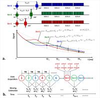
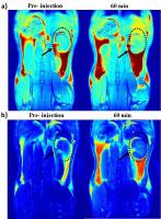
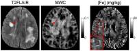
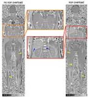
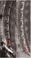
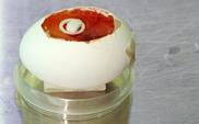
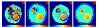
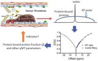
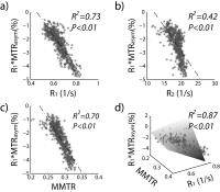
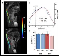
Staging deep vein thrombosis using quantitative Magnetization Transfer
Huanling Liu1,2, Wenbo Li1,3, Yuguo Li1,3, Dexiang Liu1,2, Hanwei Chen2,3, Peter C.M Van Zijl1,3, and Guanshu Liu1,3
1F.M. Kirby Research Center for Functional Brain Imaging, Kennedy Krieger Institute, Baltimore, MD, United States, 2Department of Radiology, Guangzhou Panyu Central Hospital, Guangzhou, China, People's Republic of, 3The Russell H. Morgan Department of Radiology and Radiological Science, Division of MR Research, Johns Hopkins University School of Medicine, Baltimore, MD, United States
There is an urgent need for a quantitative imaging technique that can stage deep vein thrombosis (DVT) and guide thrombolysis treatment.  In the present study, we explored the ability of quantitative Magnetization transfer (qMT) technique as a non-invasive means to stage thrombi based on their macromolecular content. Thrombi in the inferior vena cava were formed using a Mouse Complete Stasis Model. A two-pool MT mathematical model was adapted to fit high-resolution MT data of excised thrombi samples. The results clearly showed that bound proton fraction (BPF) is a useful parameter for distinguishing aged blood clots from freshly formed ones. 


2889.   
74 
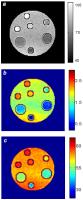
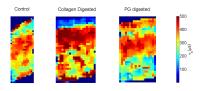
Noninvasive Assessment of Renal Fibrosis Using Magnetization Transfer MRI in Murine Renal Artery Stenosis
Kai Jiang1, Christopher M. Ferguson1, Behzad Ebrahimi1, Hui Tang1, Timothy L. Kline2, Prassana K. Mishra3, Joseph P. Grande4, Slobodan I. Macura3, and Lilach O. Lerman1
1Division of Nephrology and Hypertension, Mayo Clinic, Rochester, MN, United States, 2Department of Radiology, Mayo Clinic, Rochester, MN, United States, 3Department of Biochemistry and Molecular Biology, Mayo Clinic, Rochester, MN, United States, 4Department of Laboratory Medicine and Pathology, Mayo Clinic, Rochester, MN, United States
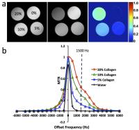
In this study, magnetization transfer was used to measure renal fibrosis in a murine model of renal artery stenosis. A collagen phantom study was performed to optimize the irradiation-offset frequency of MT pulses. Renal fibrosis by in vivo MT and ex vivo histology or hydroxproline assay showed a good correlation, suggesting that MT could be used to assess renal fibrosis. In addition, MT successfully captured the physiological changes at different stages of renal fibrosis, indicating that MT was capable of monitoring the longitudinal development of functionally significant renal fibrosis.


2890.   
75 
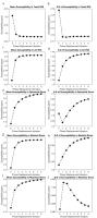
Increasing the inhomogeneous magnetization transfer (ihMT) signal in vivo with high amplitude, low duty cycle irradiation
Gopal Varma1, Olivier M Girard2, Valentin H Prevost2, Guillaume Duhamel2, and David C Alsop1
1Radiology, Division of MR Research, Beth Israel Deaconess Medical Center, Harvard Medical School, Boston, MA, United States, 2CRMBM-CEMEREM UMR 7339, CNRS-AMU, Aix Marseille Université, Marseille, France
High power, off-resonance irradiation as utilized in ihMT/MT can saturate pools of bound magnetization before they exchange in tissues. Observation of such saturation phenomena is limited by power constraints in-vivo and by 
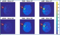
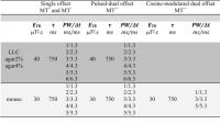
attenuation of the free pool magnetization. A technique for studying and enhancing saturation effects using relatively 
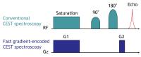
short bursts of higher power irradiation is evaluated. The results provided 2-5 fold increases in ihMTR (depending on 
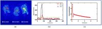
average power and offset frequency). Such short duration, high power pulses offer a new window to probe exchange 
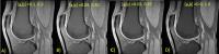
kinetics and dipolar order effects, as well as enhancing the 
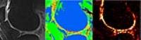
quality and feasibility of ihMT imaging.
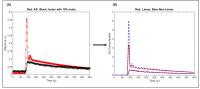

2891.   
76 On-resonance Variable Delay Multi Pulse Scheme for Imaging of Fast-exchanging Protons and semi-solid Macromolecules
Jiadi Xu1,2, kannie W. Y. Chan1,2, Xiang Xu2, Nibhay Yadav1,2, Guanshu Liu1,2, and Peter C. M. van Zijl1,2
1F. M. Kirby Center, Kennedy Kriger Institute, Baltimore, MD, United States, 2Russell H. Morgan Department of Radiology and Radiological Science, Johns Hopkins University School of Medicine, Baltimore, MD, United States
An on-resonance variable delay multi-pulse (onVDMP) scheme was developed for separation and quantification of magnetization transfer contrast (MTC) and total fast-exchanging protons (TFP) contributions. Phantom studies of glucose, bovine serum albumin (BSA) and hair conditioner show the capability of onVDMP to separate out exchangeable protons with different exchange rates by their unique signal buildup curves. Quantitative MTC and TFP maps acquired on healthy mouse brains showed strong gray/white matter contrast for the slowly transferring MTC protons while the TFP map was more uniform across the brain. 


2892.   
77 
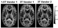
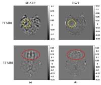
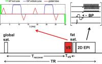
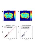
Theoretical and Experimental Optimization of a 3D Steady-State Inhomogeneous Magnetization Transfer (ihMT) Gradient Echo Sequence: Boosting the ihMT Sensitivity with Sparse Energy Deposition
Olivier M. Girard1,2, Gopal Varma3, Samira Mchinda1,2, Valentin Prevost1,2, Arnaud Le Troter1,2, Stanislas Rapacchi1,2, Maxime Guye1,2, Jean-Philippe Ranjeva1,2, David C. Alsop3, and Guillaume Duhamel1,2
1CRMBM, UMR 7339 CNRS, Aix-Marseille University, Marseille, France, 2Pôle d'Imagerie Médicale, CEMEREM, APHM, Marseille, France, 3Radiology, Division of MR Research, Beth Israel Deaconess Medical Center, Harvard Medical School, Boston, MA, United States
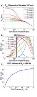
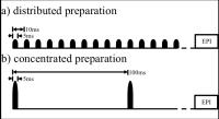
Inhomogeneous Magnetization Transfer (ihMT) has shown improved specificity for myelinated tissue as compared to conventional MT. Recently, fundamental developments have led to theoretical modeling of the ihMT effect. In this study forward modeling of a steady-state ihMT gradient echo (GRE) sequence is used to guide experimental optimization for various TRs, power levels and ihMT pulse-train duration. An efficient RF-energy deposition scheme is demonstrated for relatively long TRs, leading to ihMTRs as high as 15-17% and 10-12% in WM at 1.5T and 3T, respectively. This opens new perspectives for patient studies at clinical field strength and ihMT implementation at higher field strength.


2893.   
78 
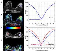
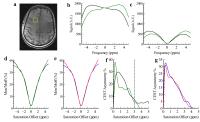
Stratification of graded acute stroke metabolic injury with magnetization transfer and relaxation-normalized amide proton transfer (MRAPT) pH-weighted MRI
Phillip Zhe Sun1, Yingkun Guo1,2, Iris Yuwen Zhou1, Suk-Tak Chan1, Yu Wang3, Emiri Mandeville4, Eng H Lo4, and Xunming Ji3
1Athinoula A. Martinos Center for Biomedical Imaging, Department of Radiology, Massachusetts General Hospital and Harvard Medical School, Charlestown, MA, United States, 2Department of Radiology, Key Laboratory of Obstetric & Gynecologic and Pediatric Diseases and Birth Defects of Ministry of Education, West China Second University Hospital, Sichuan University, Chengdu, China, People's Republic of, 3Cerebrovascular Diseases Research Institute, Xuanwu Hospital of Capital Medical University, Beijing, China, People's Republic of, 4Neuroprotection Research Laboratory, Department of Radiology and Neurology, Massachusetts General Hospital and Harvard Medical School, Charlestown, MA, United States
Amide proton transfer (APT) MRI probes amide protons from endogenous proteins/peptides, which has shown promising results in defining tissue acidosis. pH MRI complements perfusion and diffusion MRI for enhanced stratification of heterogeneous ischemic tissue injury. However, the endogenous APT effect depends not only on pH but also on tissue water content, MRI relaxation rates, and experimental conditions. There are also concomitant RF irradiation effects including direct RF saturation, magnetization transfer and nuclear overhauser effects (NOE). Our study evaluated magnetization transfer and relaxation-normalized APT (MRAPT) MRI in an animal model of acute ischemic stroke that enabled semiautomatic segmentation of graded ischemic tissue injury.


2894.   
79 
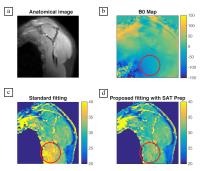
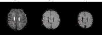
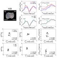
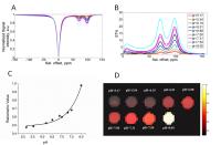
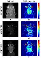
CEST-mDixon for Breast Lesion Characterization at 3T
Shu Zhang1, Stephen Seiler1, Ananth Madhuranthakam1,2, Jochen Keupp3, Ivan E Dimitrov2,4, Robert E Lenkinski1,2, and Elena Vinogradov1,2
1Radiology, UT Southwestern Medical Center, Dallas, TX, United States, 2Advanced Imaging Research Center, UT Southwestern Medical Center, Dallas, TX, United States, 3Philips Research, Hamburg, Germany, 4Philips Medical Systems, Cleveland, OH, United States
In this work, the feasibility of mDixon-based CEST-MRI for breast lesions characterization at 3T was explored. The mDixon technique was used to acquire pure water CEST images without fat contamination. The B0 maps derived from mDixon technique were used for field inhomogeneity correction. Human studies demonstrated marked differences in MTRasym between malignant and healthy tissue in hydroxyl range (0.8-1.8 ppm) and amide range (3.1-4.1 ppm). In addition, the width of the Z-spectrum was reduced in malignant vs healthy breast tissue. The results suggest that the CEST-mDixon has the potential as a robust detection and characterization tool of breast malignancy.


2895.   
80 Identification of the Origin for Amide Proton Transfer (APT) Imaging Signals in Multiple Pathologies
Dong-Hoon Lee1, Hye-Young Heo1, Yi Zhang1, Shanshan Jiang1, and Jinyuan Zhou1
1Department of Radiology, Johns Hopkins University School of Medicine, Baltimore, MD, United States
APT imaging can provide endogenous contrast related to the mobile amide proton concentration, amide proton exchange rate (depending on tissue pH), and other tissue and experimental parameters. Here, based on the correlations between quantified APT signals and combined parameter of water proton concentration and water T1 (T1w/[water proton]), we attempted to reveal the origin for APT imaging signals in multiple disease models. The results showed no significant correlations between APT signals and the T1w/[water proton]. Our findings clearly indicated that the APT signals is primarily related to the mobile amide proton concentration and amide proton exchange rate.


2896.   
81 
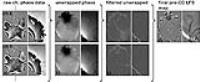
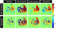
B0-calibrated and motion-registered dynamic CEST MRI of muscles undergoing exercise
Alessandro M Scotti1,2,3, Rong-Wen Tain1,3, Xiaohong Joe Zhou1,2,3, and Kejia Cai1,3
1Radiology, University of Illinois, Chicago, IL, United States, 2Bioengineering, University of Illinois, Chicago, IL, United States, 3Center for MR Research, University of Illinois, Chicago, IL, United States
Artifacts arising from tissue motion and static field inhomogeneities can heavily impact the measurement of metabolites concentration in CEST-MRI experiments in vivo. We present a correction strategy applied to CEST-MRI of creatine concentration during muscle exercise. Corrections consisted in image registration by means of a demons algorithm and signal calibration over static field offsets map at baseline, without the need of additional scanning. After correction, results are in accord with conventionally corrected data and published results. This method is shown to be effective in dynamic studies, where a high temporal resolution and coverage is required.


2897.   
82 Localized, gradient-reversed ultrafast z-spectroscopy in vivo at 7T
Neil Wilson1, Kevin D'Aquilla1, Catherine Debrosse1, Hari Hariharan1, and Ravinder Reddy1
1Department of Radiology, Center for Magnetic Resonance and Optical Imaging (CMROI), University of Pennsylvania, Philadelphia, PA, United States
Ultrafast z-spectroscopy can be collected by saturating the nuclear spins with an RF pulse in the presence of a gradient, effectively encoding the offset frequency spatially across a voxel and allowing full z-spectra to be collected in a single shot. When asymmetry analysis is applied, frequencies on one physical side of the voxel are compared with those on the other physical side. This can be a problem if there is inhomogeneity or partial voluming. By acquiring an additional z-spectrum with the gradient polarity reversed, mixed z-spectra can be created in which the positive and negative offset frequencies come from the same side of the voxel. This method is more robust to inhomogeneity and partial voluming typically found in vivo as demonstrated here with studies on 7T in human brain.


2898.   
83 
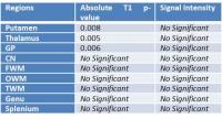
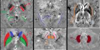
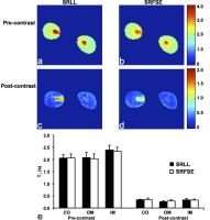
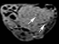
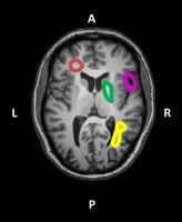
Can CEST be used as biomarker in Huntington’s disease?
Marilena Rega1,2, James Fairney1, Francisco Torrealdea1,3, Blair Leavitt4, Rachel Schahill1, Raymund Roos5, Bernhard Landwehrmeyer6, Beth Borowsky7, Sarah Tabrizi1, and Xavier Golay1
1Institute of Neurology, University College London, London, United Kingdom, 2Medical Physics, University College London Hospital, London, United Kingdom, 3Center of Medical Imaging, University College London, London, United Kingdom, 4Department of Medical Genetics, University of British Columbia, Vancouver, BC, Canada, 5Department of Neurology, University Medical Center, Leiden, United Kingdom, 6Department of Neurology, Ulm University, Ulm, Germany, 7HighQ foundation, CHDI, New York, NY, United States
Huntington’s is a hereditary disease caused by the HTT gene, resulting in the aggregation of mutant huntingtin in the cytoplasm. CEST, known to be affected by protein concentration and structure, was considered a potential biomarker for a clinical trial and comparison with MT, T1 and T2 relaxometry. Data were acquired in n=54 HD patients and n=46 healthy individuals. Comparison of CEST revealed significant differences (p<0.05) in the putamen and globus pallidus regions which did not correlate with any changes in relaxometry or MT, suggesting that CEST might be able to provide additional contrast to the already existing methods. 


2899.   
84 
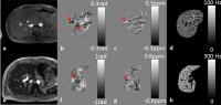
Imaging the pH by means of CEST-responsive iopamidol: normal and AKI model studies
Wei Hu1, Zhuozhi Dai1, Zhiwei Shen1, Yuanyu Shen1, Xiangyong Tang1, Zhiyan Zhang1, Gang Xiao2, Phillip Zhe Sun3, and Renhua Wu1
12nd Affilicated Hospital, Shantou University Medical College, Shantou, China, People's Republic of, 2Hanshan Normal University, Chao zhou, China, People's Republic of, 3Massachusetts General Hospital and Harvard Medical School, Charlestown, MA, United States
The promising imaging technique of chemical exchange saturation transfer (CEST) MRI, which is sensitive to microenvironment properties including pH, metabolites, temperature, metal ions, and enzymatic activities, has been increasingly applied in vivo such as acute ischemia, infection and cancer. The aim of this work was to observe the differentiation between normal and acute kidney injury (AKI) using echo-planar imaging sequence (EPI) as well as the sensitive ratiometric methods under 7T magnetic field.


2900.   
85 
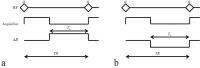
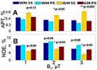
Comparison of 3D CEST acquisition schemes: steady state versus pseudo-steady state
Vitaliy Khlebnikov1, Nicolas Geades2, Dennis WJ Klomp1, Hans Hoogduin1, Penny Gowland2, and Olivier Mougin2
1Radiology, University Medical Center Utrecht, Utrecht, Netherlands, 2Sir Peter Mansfield Imaging Centre, University of Nottingham, Nottingham, Nottinghamshire, United Kingdom, Nottingham, United Kingdom
Chemical Exchange Saturation Transfer (CEST) has gained much popularity due to its unmatched sensitivity to dilute labile protons when compared to other MRI techniques. Of particular interest are two CEST effects: Amide Proton Transfer (APT, CEST of amides) and Nuclear Overhauser Enhancement (NOE). Fast-paced developments for CEST resulted in the design of multiple CEST imaging sequences. This raises the obvious question as to which sequence to use and in what particular applications. Two pulsed volumetric CEST acquisition schemes are currently available in the literature. The first is based on the standard Magnetization Transfer imaging technique: a steady-state (SS) acquisition with alternating brief saturation and image acquisition. The second is based on the preparation of the magnetization before a long readout, where the prolonged saturation reaches a pseudo-steady state (PS) before the image acquisition. In this report, these two CEST acquisition schemes, optimized for maximum sensitivity to APT and NOE effects through Bloch-McConnell simulations, were systematically compared for the same spatial resolution, brain coverage and scan time.


2901.   
86 
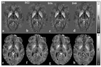
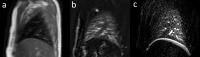
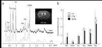
GluCEST imaging of spinal cord in a mouse model of Friedreich ataxia
Jérémy Pépin1,2, Françoise Piguet3,4,5,6, Hélène Puccio3,4,5,6, and Julien Flament1,7
1CEA/DSV/I2BM/MIRCen, Fontenay-aux-Roses, France, 2CNRS Université Paris-Saclay UMR 9199, Fontenay-aux-Roses, France, 3Department of Translational Medecine and Neurogenetics, Institut de Génétique et de Biologie Moléculaire et Cellulaire, Illkirch, France, 4INSERM U596, Illkirch, France, 5CNRS UMR7104, Illkirch, France, 6Université de Strasbourg, Strasbourg, France, 7INSERM UMS 27, Fontenay-aux-Roses, France
Friedreich Ataxia (FA) is the most common form of recessive inherited ataxia which induces severe neurological disabilities and reduced life expectancy. As glutamate has been shown to be a potential biomarker of neurodegenerative diseases, we used Chemical Exchange Saturation Transfer imaging of glutamate (gluCEST) in order to characterize our mouse model of FA and to monitor glutamate alterations in the spinal cord. GluCEST images revealed decrease of glutamate level in FA mouse model compared to control littermates, especially in the lumbar part. These results demonstrate the potential of gluCEST in providing innovative and relevant biomarkers of FA.


2902.   
87 Tissue Characterization with Fast High Resolution Magic Angle Spinning (HRMAS) CEST Spectroscopy
Iris Yuwen Zhou1, Taylor Fuss1,2, Gang Xiao3, Takahiro Igarashi1, Lin Li1, Leo L. Cheng1,2, and Phillip Zhe Sun1
1Athinoula A. Martinos Center for Biomedical Imaging, Department of Radiology, Massachusetts General Hospital and Harvard Medical School, Charlestown, MA, United States, 2Department of Pathology, Massachusetts General Hospital and Harvard Medical School, Boston, MA, United States, 3Department of Mathematics and Statistics, Hanshan Normal University, Chaozhou, China, People's Republic of
Z-spectrum is conventionally acquired through multiple experiments with selective saturation at different frequency offsets of interest, leading to extreme long acquisition time. Here, we employ gradient-encoding to substantially accelerate the acquisition of Z-spectrum. This speedup in combination with higher spectral resolution provided by high resolution magic angle spinning (HRMAS) allows rapid quantification of chemical exchange rates of CEST agents, monitoring dynamic processes and fast tissue characterization. The approach was validated in phantom and used for characterization of brain tissues after ischemic stroke. 


2903.   
88 GluCEST imaging: a relevant biomarker of Huntington’s disease.
Jérémy Pépin1,2, Laetitia Francelle1,2, Maria-Angeles Carillo-de Sauvage1,2, Huu Phuc Nguyen3,4, Nicole El Massioui5,6, Valérie Doyère5,6, Emmanuel Brouillet1,2, and Julien Flament1,7
1CEA/DSV/I2BM/MIRCen, Fontenay-aux-Roses, France, 2CNRS Université Paris-Saclay UMR 9199, Fontenay-aux-Roses, France, 3Institute of Medical Genetics and Applied Genomics, University of Tuebingen, Tuebingen, Germany, 4Centre for Rare Diseases, University of Tuebingen, Tuebingen, Germany, 5Paris-Saclay Institute of Neuroscience, Université Paris-Sud, UMR 9197, Orsay, France, 6Centre National de la Recherche Scientifique, Orsay, France, 7INSERM UMS 27, Fontenay-aux-Roses, France
Huntington’s disease (HD) is an inherited neurodegenerative disease characterized by motor, cognitive and psychiatric symptoms. As glutamate has been shown to be a potential biomarker of neurodegenerative diseases, we used Chemical Exchange Saturation Transfer imaging of glutamate (gluCEST) to map cerebral glutamate distribution in mouse and rat models of HD. A decrease of [Glu] was measured in the striatum by MRS and gluCEST. In addition, good spatial resolution of gluCEST over MRS allowed identification of other afflicted brain regions such as corpus callosum. These results demonstrate the potential of gluCEST in providing relevant biomarkers of HD in the whole brain.


2904.   
89 
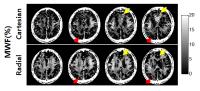
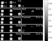
Accelerating CEST Imaging with Spatial-Temporal Sparse Dictionary Learning
Huajun She1, Bian Li1, Robert Lenkinski1, and Elena Vinogradov1
1Radiology, Advanced Imaging Research Center, UT Southwestern Medical Center, Dallas, TX, United States
This work investigates accelerating CEST imaging. The original blind compressive sensing method assumes that a few functions are enough to represent the dynamic behavior, and the coefficient matrix should be sparse. In CEST imaging, z-spectrum shows group sparsity in the same compartment. So not only the coefficients matrix is sparse but also the transformation of the coefficients matrix is sparse, such as total variation and wavelets. The proposed method addresses this prior information and further improves the original BCS method, demonstrating a better estimation of the CEST effect at high reduction factors for both Cartesian and radial sampling patterns.


2905.   
90 Use of Yb-HPDO3A probe for CEST-MRI pH mapping in glioblastoma mouse models
Giuseppe Ferrauto1, Michel Sarraf2, Enza Di Gregorio1, Vincent Auboiroux3, Ulysse Gimenez2, François Berger2, Silvio Aime1, and Hana Lahrech2
1Dept of Molecular Biotechnologies & Health Sciences, University of TORINO (IT), Torino, Italy, 2CLINATEC-CEA-INSERM UA01 – CHU –UJF Grenoble (FR), Grenoble, France, 3CLINATEC-CEA Grenoble (FR), Grenoble, France
  MRI maps of extracellular/extravascular pH distribution in glioblastoma mouse model (U87 cells) have been obtained by administrating YbHPDO3A CEST probe. This molecule has potential in a clinical setting as it displays analogue properties (stability and in vivo pharmacokinetic) of its analogue clinically approved Gd-HPDO3A (ProHance). Furthermore, glioblastoma pH maps have been correlated with histology (H/E, Hif-1a and Ki-67 staining). The assessment of glioblastoma pHe could be used to monitor tumor development and to target acidic tumor regions using innovative responsive pH therapies.


2906.   
91 Comparison of gagCEST and sodium MRI in evaluating knee cartilage in vivo at 7 Tesla - Permission Withheld
Vladimir Mlynarik1,2, Stefan Zbyn1, Markus Schreiner3, Vladimir Juras1, Pavol Szomolanyi1, Didier Laurent4, and Siegfried Trattnig1,2
1Department of Biomedical Imaging and Image-Guided Therapy, High Field MR Center, Medical University of Vienna, Vienna, Austria, 2CD Laboratory for Clinical Molecular MR Imaging, Vienna, Austria,3Department of Orthopedic Surgery, Medical University of Vienna, Vienna, Austria, 4Novartis Institutes for Biomedical Research, Basel, Switzerland
There are several methodological and data processing issues in gagCEST, which complicate the translation of this method into clinical practice. For assessing performance of the gagCEST protocol optimized in our laboratory, corrected signal intensities from sodium images were used as a reference. The results demonstrate a good correlation between both methods, although the small magnitude of the gagCEST effect and the low resolution in sodium images require carefully optimized methodology and long measurement times. It can be concluded that the gagCEST method can be useful in evaluating early degeneration of articular cartilage at 7 Tesla.


2907.   
92 
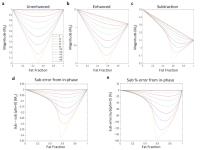
Chemical shift artifact of the third kind: Implications for gradient-echo based contrast enhanced imaging
Jamal J. Derakhshan1, Elizabeth S. McDonald2, Evan S. Siegelman3, Mitchell D. Schnall3, and Felix W. Wehrli4
1Radiology, Hospital of the University of Pennsylvania, Philadelphia, PA, United States, 2Radiology, Breast Imaging Division, Hospital of the University of Pennsylvania, Philadelphia, PA, United States,3Radiology, Abdominal Imaging Division, Hospital of the University of Pennsylvania, Philadelphia, PA, United States, 4Radiology, University of Pennsylvania, Philadelphia, PA, United States
A common subtraction band artifact in breast MRI was not understood, causing reduced confidence in clinical interpretation.  The source of the artifact is shown to be a subtle chemical shift effect between fat and water in the presence of contrast enhancement. The phenomenon is now generalized and characterized at all off-resonance angles. Strong echo-time and fat signal dependence may lead to enhancement errors as a function of scanner hardware, field strength and fat suppression limitations. A time and SNR-equivalent in-phase VIBE sequence eliminates the artifact; gradient-echo based contrast enhanced imaging can be performed in-phase to eliminate these important potential pitfalls.


2908.   
93 
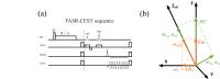
Fixed Angle Single Rotation CEST (FASR-CEST) sequence for reducing saturation time
Yi Wang1, Yang Fan2, Bing Wu2, and Jia-Hong Gao1
1School of Physics, Peking University, Beijing, China, People's Republic of, 2MR Research Group, GE Healthcare China, Beijing, China, People's Republic of
In CEST imaging, when saturation time is not sufficient long (empirically smaller than 0.8s), rotation effect would appear and contaminate with saturation effect, making the signal unavailable for further analysis. Considering the inhomogeneity of $$$B_{0}$$$ and $$$B_{1}$$$ in reality, we propose a novel Fixed Angle Single Rotation CEST(FASR-CEST) sequence to overcome the restriction, successfully reducing the saturation time to about 0.5s while keeping identical effect as CEST sequence with long saturation time, with the help of analytical calibration method in another abstract. Effect of the sequence is verified with vitro and in vivo data. 


2909.   
94 
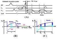
T1D-w ihMT:  Dipolar Relaxation time weighted imaging using inhomogeneous Magnetization Transfer - Permission Withheld
Guillaume Duhamel1, Valentin H Prevost1, Gopal Varma2, David C Alsop2, and Olivier M Girard1
1CRMBM / CNRS 7339, Aix Marseille University, Marseille, France, 2Radiology, Division of MR Research, Beth Israel Deaconess Medical Center, Harvard Medical School, Boston, MA, United States
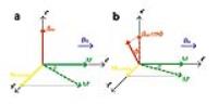
The inclusion of a dipolar reservoir in the existing two pool model for MT allowed interpreting the inhomogeneous MT (ihMT) signal as a dipolar order effect -characterized by a relaxation time T1D - within motion restricted molecules.  In this study, we demonstrate that an ihMT signal can actually be evidenced in any component with non-trivial T1D value. Adjustment of the dual frequency irradiation efficiency by increase of Δt, the repetition rate of consecutive saturation pulses, filters the signal of shorter T1D components. This provides a means to realize T1D-weighted imaging, a new source of MR contrast between tissues.


2910.   
95 Improvement of in vivo glucoCEST imaging in rat brain using inverse z-spectrum analytical scheme at 7.0 T
Ping-Huei Tsai1,2,3, Hua-Shan Liu4,5, Fei-Ting Hsu6, Yu-Chieh Kao3, Chia-Feng Lu3, Li-Chun Hsieh2, Pen-Yuan Liao2, Hsiao-Wen Chung7, and Cheng-Yu Chen1,2,3
1Department of Radiology, School of Medicine, College of Medicine, Taipei Medical University, Taipei, Taiwan, 2Department of Medical Imaging, Taipei Medical University Hospital, Taipei Medical University, Taipei, Taiwan, 3Translational Imaging Research Center, Taipei Medical University, Taipei, Taiwan, 4Graduate Institute of Clinical Medicine, Taipei Medical University, Taipei, Taiwan,5Department of Medical Imaging, Taipei Medical University Hospital, Taipei, Taiwan, 6Taipei Medical University Hospital, Taipei Medical University, Taipei, Taiwan, 7Graduate Institute of Biomedical Electronics and Bioinformatics, National Taiwan University, Taipei, Taiwan
GlucoCEST have been proposed to assess the discrepant concentrations of glucose in vitro. However, it is still a challenge to obtain accurate signals from glucose in vivo.  Our preliminary finding demonstrated that the proposed analytical scheme provides an alternative to extract glucose profile and could be more robust to the field inhomogeneity, which may be helpful in further implementation in disease models.


2911.   
96 A fast chemical exchange saturation transfer imaging scheme based on spatiotemporal encoding
Jianpan Huang1, Miao Zhang1, Shuhui Cai1, Congbo Cai2, Lin Chen1, and Ting Zhang1
1Department of Electronic Science, Xiamen University, Xiamen, China, People's Republic of, 2Department of Communication Engineering, Xiamen University, Xiamen, China, People's Republic of
Chemical exchange saturation transfer (CEST) is widely exploited in magnetic resonance imaging (MRI) because of its special quantitative contrast mechanisms. To overcome the long acquisition time required by fast spin-echo multi-slice imaging and alleviate the sensitivity to field inhomogeneity and chemical shift effects appeared in echo planar imaging, we proposed a CEST imaging scheme based on spatiotemporally encoded magnetic resonance imaging (SPEN MRI). Experimental results validated the feasibility and capability of the new scheme.
Exhibition Hall 

15:15 - 16:15

    Computer #

2982.   
73 Spectral analysis enhances the detectability of rotary saturation contrast
Jingwei Sheng1, Yuhui Chai1, Bing Wu2, Yang Fan2, and Jia-Hong Gao1
1Center for MRI Research, Peking University, Beijing, China, People's Republic of, 2GE Healthcare MR Research China, Beijing, China, People's Republic of
Traditional rotary saturation based methods requires triggered phases to create a robust signal change, which may not be satisfied in practical application. In this work, based on the analysis of magnetization in the double rotating frame, we proposed detecting the signal fluctuations of proposed SLOE method by manipulating TRs, which needs no triggering. Further, a spectral statistical test in the frequency domain was proposed and verified, which featured an enhanced detection sensitivity than that of deviation test. 


2983.   
74 QUANTITATIVE SUSCEPTIBILITY MAPPING OF THE LIVER AND SPLEEN IN SUBJECTS WITH FERUMOXYTOL INJECTION
Samir D. Sharma1, Timothy J. Colgan1, Camilo A. Campo1, Tilman Schubert1, Utaroh Motosugi2, Diego Hernando1, and Scott B. Reeder1,3
1Radiology, University of Wisconsin - Madison, Madison, WI, United States, 2Radiology, University of Yamanashi, Yamanashi, Japan, 3Medical Physics, University of Wisconsin-Madison, Madison, WI, United States
Ferumoxytol is an ultrasmall superparamagnetic iron oxide (USPIO) agent that is taken up by the reticuloendothelial system where it accumulates in organs such as the liver, spleen, and bone marrow. Given the superparamagnetic properties of ferumoxytol, it may be possible for quantitative susceptibility mapping (QSM) techniques to detect and quantify the concentration of ferumoxytol in these organs. Recent technical developments have demonstrated the feasibility of a QSM technique for magnetic susceptibility mapping of the abdomen. Thus, the purpose of this work was to test the feasibility of QSM to assess the longitudinal changes of ferumoxytol in the liver and spleen.


2984.   
75 Contrast-Enhanced Susceptibility Weighted Imaging with Ultrasmall Superparamagnetic Iron Oxide Improves the Detection of Tumour Vascularity in A HCC-LM3 Nude Mouse Model
Shuohui Yang1, Jiang Lin1, Fang Lu2, Yuanyuan Dai1, Zhihong Han3, and Caixia Fu4
1Radiology, Zhongshan Hospital, Shanghai Medical College, Fudan University, Shanghai, China, People's Republic of, 2Radiology, Shuguang Hosipital, Shanghai University of Traditional Chinese Medicine, Shanghai, China, People's Republic of, 3Pathology, Shuguang Hosipital, Shanghai University of Traditional Chinese Medicine, Shanghai, China, People's Republic of, 4Siemens Shenzhen Magnetic Resonance Ltd., Shenzhen, China, People's Republic of
Hepatocellular carcinoma (HCC) is a hyper-vascular tumor and knowledge of the intratumoral vascularity is essential. Susceptibility weighted imaging (SWI) uses magnitude and filtered-phase information to provide high sensitivity to susceptibility changes caused by hemorrhage, calcium, iron, and small veins. It has been used to visualize normal or pathologic vascular structures that are not visible on conventional MRI. Ultrasmall superparamagnetic iron oxide (USPIO) is an intravascular blood pool contrast medium. After intravenous administration, it can cause a greater effect on local magnetic field inhomogeneities and results in higher susceptibility differences between all intratumoral vessels and the tumor on SWI. This study showed that USPIO-enhanced SWI could further enhance the demonstration of tumor vascularity inside HCC when compared to unenhanced SWI in an orthotopic xenograft nude mice HCC model.  


2985.   
76 Using frequency difference mapping to assess white matter microstructure in the human corpus callosum
Benjamin Tendler1 and Richard Bowtell1
1Sir Peter Mansfield Imaging Centre, University of Nottingham, Nottingham, United Kingdom
Frequency difference mapping (FDM) is a recently developed phase-based technique that takes advantage of the non-linear temporal evolution of the phase in GE sequences to produce images that are sensitive to white matter microstructure. Images can be produced simply from raw phase data, with minimal post-processing. In this study 10 subjects underwent six repeats of a single-slice, sagittal multi-echo GE scan on the mid-line.  Frequency difference maps reproducibly depicted white matter tracts oriented perpendicular to the applied field. Fitting the FD and magnitude data to a three-pool model provided insight into the variation of microstructure along the corpus callosum.  


2986.   
77 Can myelin protons be directly imaged with the adiabatic inversion recovery prepared ultrashort echo time (IR-UTE) sequence - a validation study based on D2O exchange in sheep brain specimens
Hongda Shao1, Soorena Azam ZAnganeh1, Rong Luo1, Jun Chen1, Graeme Bydder1, and Jiang Du1
1Radiology, University of California, San Diego, San Diego, CA, United States
Directly assessing the integrity of myelin in white matter is important for diagnosis and assessment of prognosis in multiple sclerosis (MS). However, the protons in myelin have extremely short T2s and cannot be directly imaged with conventional clinical MRI sequences. Adiabatic inversion recovery prepared ultrashort echo time (IR-UTE) sequences can detect signal from myelin protons and efficiently suppress the signal from water. In this study we aimed to further validate the IR-UTE technique in sheep brain using a D2O exchange model.


2987.   
78 Phase Correction of a Bipolar Gradient-Echo Acquisition for Quantitative Susceptibility Mapping - Permission Withheld
Hongfu Sun1, M. Ethan MacDonald1, and G. Bruce Pike1
1University of Calgary, Calgary, AB, Canada
A method to remove phase offsets in bipolar gradient-echo readouts is proposed. Their effects on Quantitative Susceptibility Mapping (QSM) reconstruction are demonstrated by comparing QSM before and after phase offsets removal. 


2988.   
79 High Resolution Blood Vessel Imaging with USPIOs in the Human Brain: T1 vs T2* contrast.
Thomas Christen1, Samantha Holdsworth1, Samuel Cheshier2, Michael Moseley1, Greg Zaharchuk1, and Kristen Yeom1
1Radiology, Stanford University, Palo Alto, CA, United States, 2Neurosurgery, Stanford University, Stanford, CA, United States
In this study, we probed the potential of ultrasmall superparamagnetic iron oxide particles (USPIOs) to provide high-resolution angiographic images of the human brain using T2* and T1 contrasts. 10 paediatric patients suspected of arteriovenous malformations (AVM) were scanned pre and post intravenous USPIOs injection as part of their clinical exam. To test the influence of USPIOs concentration on both T2* and T1 images, a healthy volunteer was scanned before and after injections of 7 incremental doses of the contrast agent. The results suggest that both approaches can provide exquisite details of the neurovascular anatomy while offering complementary information.


2989.   
80 Artifacts Affecting Derivative of $$$B_1^+$$$ maps for EPT Reconstructions
Stefano Mandija1, Alessandro Sbrizzi1, Astrid L.H.M.W. van Lier1, Peter R. Luijten1, and Cornelis A.T. van den Berg1
1Center For Image Sciences, UMC Utrecht, Utrecht, Netherlands
EPT conductivity reconstruction is affected by difficulties to reliably calculate spatial derivatives on voxelized MRI data. Here, we explore the impact of several numerical approximations. In particular: the different size of finite-difference kernels and the k-space truncation (always present in MR images). We also explore a Fourier-domain alternative, which does not require finite-difference approximation kernels.


2990.   
81 Comparison of Multi-transmit Microstrips and Loops for Electrical Properties Tomography (EPT) in the Breast at 3T
Yicun Wang1, Gregor Adriany2, Jiaen Liu1, Xiaotong Zhang1, Pierre-Francois Van de Moortele2, and Bin He1,3
1Department of Biomedical Engineering, University of Minnesota, Minneapolis, MN, United States, 2Center for Magnetic Resonance Research, University of Minnesota, Minneapolis, MN, United States,3Institute for Engineering in Medicine, University of Minnesota, Minneapolis, MN, United States
Electrical Properties Tomography (EPT) is a promising technique to provide high specificity in breast cancer diagnosis. Previous studies have demonstrated that multi-transmit coils array enables reconstruction of the electrical properties free of transmit phase and local homogeneity assumptions. In this study, the strengths and drawbacks of a microstrip array and a loop array for EPT reconstruction in the breast at 3T were investigated based on numerical simulations. It has been discovered that with the same driving power, a loop array provides higher signal to noise ratio on the reconstructed image while a microstrip array performs better at delineating tissue boundaries.


2991.   
82 Generalized Phase based Electrical Conductivity Imaging - Permission Withheld
Necip Gurler1 and Yusuf Ziya Ider1
1Electrical and Electronics Engineering, Bilkent University, Ankara, Turkey
In this study, a new formulation for phase based electrical properties tomography (EPT) has been proposed to eliminate the boundary artifact issue and to provide robustness against noise. The feasibility of the proposed method, which is called "generalized phase based EPT", has been demonstrated using simulation, experimental phantom, and in vivo experiments. 


2992.   
83 Applying „Electric Properties Tomography“ to Low Frequency Conductivity Using Magnetic Particle Imaging
Ulrich Katscher1, Jürgen Rahmer1, Christian Stehning1, Peter Vernickel1, and Bernhard Gleich1
1Philips Research Europe, Hamburg, Germany
MRI-based “Electric Properties Tomography” (MRI-EPT) determines electric conductivity measuring and post-processing the spatial distribution of the TX RF field. Obtained conductivity corresponds to Larmor frequency, reflecting biochemical content of tissue, but not its specific cellular structure. Cellular tissue structure is reflected by conductivity at low frequency (LF, 100-500 kHz), as used for “Magnetic Particle Imaging” (MPI). Similar to MRI, MPI is able to determine the spatial distribution of applied LF field, and thus, a reconstruction of LF conductivity should be possible (“MPI-EPT”) in analogy to MRI-EPT. This study investigated the principle feasibility of MPI-EPT and compared results with MRI-EPT.


2993.   
84 Global Maxwell Tomography: a novel technique for electrical properties mapping without symmetry assumptions or edge artifacts
Jose E.C. Serralles1, Athanasios Polimeridis2, Manushka V. Vaidya3,4, Gillian Haemer3,4, Jacob K. White1, Daniel K. Sodickson3,4, Luca Daniel1, and Riccardo Lattanzi3,4
1Research Laboratory of Electronics, Massachusetts Institute of Technology, Cambridge, MA, United States, 2Center for Computational and Data-Intensive Science and Engineering, Skolkovo Institute of Science and Technology, Moscow, Russian Federation, 3Center for Advanced Imaging Innovation and Research (CAI2R) and Center for Biomedical Imaging, Department of Radiology, New York University School of Medicine, New York, NY, United States, 4The Sackler Institute of Graduate Biomedical Science, New York University School of Medicine, New York, NY, United States
We introduce Global Maxwell Tomography (GMT), a novel volume integral equation-based technique for the extraction of electric properties from MR data. GMT is framed as an unconstrained optimization problem in which the error between measured and simulated B1+ magnitude maps is minimized. Due to its global nature, GMT is not subjected to edge artifacts. By using exclusively B1+ magnitude, GMT does not rely on symmetry assumptions to estimate B1+ phase. In one numerical example, three tumors were inserted into a head model, and starting from a tumorless initial guess, GMT accurately inferred the electrical properties and locations of these tumors.


2994.   
85 Application of Generalized Phase based Electrical Conductivity Imaging in the Subacute Stage of Hemorrhagic and Ischemic Strokes - Permission Withheld
Necip Gurler1, Omer Faruk Oran1, Hava Donmez Keklikoglu2, and Yusuf Ziya Ider1
1Electrical and Electronics Engineering, Bilkent University, Ankara, Turkey, 2Department of Neurology, Yildirim Beyazit University Atatürk Education and Research Hospital, Ankara, Turkey
In this study, clinical applicability of the recently proposed generalized phase based EPT method has been investigated for two patients with neurovascular diseases in the subacute phase, i.e. hemorrhagic and ischemic stroke. In the case of ischemia, conductivity was found to be increased in the lesion area. In the case of hematoma, although the conductivity of the surrounding edema region was found to be increased, the conductivity in the hematoma region itself was found to be similar to that of brain tissue.


2995.   
86 Investigation and Reduction of the Effects of Gibbs Ringing in SSFP Phase Based MR-EPT
Gokhan Ariturk1, Necip Gurler1, and Yusuf Ziya Ider1
1Electrical and Electronics Engineering, Bilkent University, Ankara, Turkey
Phase based MR-EPT methods mainly make use of the balanced SSFP since it is a fast MRI sequence. As SSFP sequence yields high magnitude contrast variations, Gibbs artifacts occur at tissue boundaries. This artifact manifests itself as phase spikes and oscillations at and near tissue boundaries respectively. The commonly used image enhancement methods, for reducing the ringing artifact, mainly focus on the magnitude images, leaving out the phase images. Throughout simulation and phantom experiment results, we investigate and propose a method for the reduction of the effect of ringing artifact on phase based MR-EPT studies.


2996.   
87 Validation study of water-content-assisted electrical properties tomography (wEPT) with an electrolyte protein phantom and B1 inhomogeneity consideration
Eric Michel1 and Soo Yeol Lee1
1Kyung Hee University, Suwon, Korea, Republic of
New MRI-based tissue electrical properties (EPs) mapping techniques had achieved higher accuracy and resolution increasing its chances to be used in several clinical applications. In this work, we describe a validation study for a technique called water-content-assisted electrical properties tomography (wEPT) based on in situ measurements of a brain-tissue-like phantom created by electrolytic protein solutions. The influence of inhomogeneous radiofrequency field distributions and its impact in wEPT reconstructions is also analyzed. Significant consistency between in vivo brain tissue estimations and phantom measurements was found, supporting the formalism and validity of wEPT for EPT studies.


2997.   
88 Peripheral Nerve Imaging using 2-point Dixon 3D Fast Spine Echo (CUBE-FLEX) with Flow-Saturation Preparation (FSP): Initial Feasibility Study
Darryl Sneag1, Mitsuharu Miyoshi2, Maggie Fung3, Daniel Litwiller3, and Hollis Potter1
1Hospital for Special Surgery, New York, NY, United States, 2GE Healthcare, Hino, Japan, 3GE Healthcare, New York, NY, United States
High-resolution MRI currently plays an important role in diagnostic management of peripheral nerve pathology. Peripheral nerves, however, pose particular imaging challenges that conventional sequences frequently cannot address. Their small size and oblique course between muscles and alongside vessels may inhibit reliable identification. We propose a 2-point Dixon fat/water separation 3D fast spin echo technique to achieve uniform fat suppression, combined with a flow-saturation prep pulse to suppress moving vascular spins to improve nerve visualization.  The objective of this study was to assess the technique’s feasibility and potential diagnostic utility in evaluating peripheral nerves throughout the body.


2998.   
89 Enforcing a Physical Tissue Model for Partial Volume MR Fingerprinting
Anagha Deshmane1, Debra F. McGivney2, Yun Jiang1, Dan Ma2, and Mark A. Griswold2
1Biomedical Engineering, Case Western Reserve University, Cleveland, OH, United States, 2Radiology, Case Western Reserve University, Cleveland, OH, United States
We present a method to compute fraction maps of tissue types using a subset of three dictionary entries chosen specifically for each individual, enforcing consistency with a physical tissue model.  This approach removes erroneous partial volumes in the fraction maps, and reduces noise and distortions in T1, T2, and M0 maps.


2999.   
90 Continuously Adjustable Fat Suppression Image Contrast with one MRI Acquisition
Kecheng Liu1, Xiaodong Zhong1, Dan Ma2, Brian Dale1, and Mark Griswold2
1Siemens Healthcare USA Inc., Malvern, PA, United States, 2Case Western Reserve Univisity, Cleveland, OH, United States
Fat suppression or saturation (FatSat) is widely used in MRI applications, which reduces the fat signal and preserves water signal for clinical diagnosis, enhancing potential pathological changes. Different degree of FatSat yields different image contrast. However, when radiologists read FatSat images, they have different contrast preferences. To provide such variable preferences is not trivial because it typically costs more imaging time to re-acquire another contrast of FatSat image by adjusting imaging parameters. This work presents an alternative approach to provide continuously adjustable image contrast of FatSat images with only one acquisition.


3000.   
91 Spin Echo Dynamics as Codes for Quantitative Imaging
Gigi Galiana1
1Diagnostic Radiology, Yale University, New Haven, CT, United States
Recent work has highlighted the complicated and distinctive dynamics that shape the signal observed during an irregularly-spaced train of spin echoes.  Here, we use those signals as codes that allows us to identify mixtures of molecules that are too similar to distinguish by standard spectroscopy.  Extensions to water based systems, where relaxation mechanisms are the primary driver of signal dynamics, will also be presented.


3001.   
92 Dual-pathway sequences for MR thermometry: When and where to use them
Pelin Aksit Ciris1, Cheng-Chieh Cheng2, Chang-Sheng Mei3, Lawrence P. Panych2, and Bruno Madore2
1Department of Biomedical Engineering, Akdeniz University, Antalya, Turkey, 2Department of Radiology, Brigham and Women’s Hospital, Harvard Medical School, Boston, MA, United States,3Department of Physics, Soochow University, Taipei, Taiwan
Dual-pathway sequences have been proposed to improve the temperature-to-noise-ratio (TNR) in MR thermometry. The present work establishes how much improvement these sequences may bring for various tissue types. Simulation results were validated against analytical equations, phantom and in vivo human results. PSIF-FISP thermometry allowed TNR improvements for kidney, pelvis, spleen or gray matter, and up to 2-3 fold reductions in TR with 20% TNR gains were achievable. Further TNR benefits are expected for heated tissues, due to heating-related changes in relaxation rates. In other tissue types such as liver, muscle or pancreas improvements were observed only for short TR settings.  


3002.   
93 T2* Based MRI Temperature Contrast - Video Not Available
Janusz Henryk Hankiewicz1, Jason Nobles1, Zbigniew Celinski1, Karl Stupic2, and Robert Camley1
1UCCS Center for Biofrontiers Institute, University Colorado Colorado Springs, Colorado Springs, CO, United States, 2National Institute of Standards and Technology, Boulder, CO, United States
The aim of this study was to develop a novel temperature-sensitive MRI contrast agent based on temperature changes of the magnetic moment of magnetic particles. Gadolinium was used to test the hypothesis that magnetic particles will create a temperature-dependent local dipole magnetic field. This effect was locally visible as a temperature dependent darkening on gradient-echo MRI images. Shades of gray within the images can then be calibrated to map the local temperatures in specific areas of tissue during medical procedures. The estimated accuracy of temperature determination deep in the phantom using MR image intensity is ±1.8oC, at 37oC.


3003.   
94 Correlation time mapping of degenerated human and bovine articular cartilage reveals tissue structure and degenerative changes
Hassaan Elsayed1,2,3,4, Matti Hanni2,3,4, Jari Rautiainen2,3,5, Mikko Johannes Nissi5,6, and Miika Nieminen2,3,4
1Computer Science and Engineering, University of Oulu, Oulu, Finland, 2Research Unit of Medical Imaging, Physics and Technology, University of Oulu, Oulu, Finland, 3Medical Research Center, University of Oulu and Oulu University Hospital, Oulu, Finland, 4Department of Diagnostic Radiology, Oulu University Hospital, Oulu, Finland, 5Department of Applied Physics, University of Eastern Finland, Kuopio, Finland, 6Diagnostic Imaging Center, Kuopio University Hospital, Kuopio, Finland
Correlation time ($$$\tau_c$$$) maps were obtained by fitting $$$\tau_c$$$ from $$$T_{1\rho}$$$ dispersion measurements of human articular cartilage specimens and enzymatically digested bovine samples to develop a new MRI contrast which characterizes tissue properties at molecular level. $$$\tau_c$$$ revealed the laminar appearance in control and PG depleted bovine specimens, which was not observable in the collagen-digested specimens. The laminar appearance was also visible in the early OA human specimens, in contrast to the advanced OA group. The results demonstrated that $$$\tau_c$$$ mapping is a potential method for providing information about the articular cartilage structure and associated changes with degeneration.  


3004.   
95 Cardiac in vivo T1-Mapping with Novel Reactive Oxygen Species Sensing Agent Specifically Detects Cardiac Oxidative Stress in Doxorubicin-treated Rats
Ronald J Beyers1, Meng Yu2, Dean Schwartz3, Nouha Salibi1,4, Christian Goldsmith5, and Thomas Denney1
1MRI Research Center, Auburn University, Auburn University, AL, United States, 2Chemistry & Biochemistry, Auburn University, Auburn University, AL, United States, 3Anatomy, Physiology and Pharmacology, Auburn University, Auburn University, AL, United States, 4MR R&D, Siemens Healthcare, Malvern, PA, United States, 5Chemistry and Biochemistry, Auburn University, Auburn University, AL, United States
Pathological cardiac oxidative stress causes cardiac dysfunction and possible cardiac failure. We developed a novel reactive oxygen species sensing T1 agent (H4qpt2) and applied it with in vivo cardiac T1 mapping MRI at 7T in a doxorubicin-treated (Dox) rat model.  Cardiac T1 mapping with H4qpt2 specifically detected significantly shortened myocardial T1 in Dox rats while no change in T1 occurred in skeletal muscle or control rats with H4qpt2.  This new H4qpt2 agent combined with cardiac or non-cardiac T1 mapping may advance the early detection of oxidative stress in multiple pathologies and promote their early treatment.


3005.   
96 Heteronuclear Proton MRI - SNR, temperature effects and frequency separation of UTE versus CSI readout
Tim Klasen1, Carsten Höltke1, and Cornelius Faber1
1Department of Clinical Radiology, University of Münster, Münster, Germany
Heteronuclear proton MRI is a recent method for MRI cell tracking and opens up the possibility for Multicolor MRI when using different Ln-DOTMA complexes. Here, we compare UTE and CSI as readout with regard to SNR efficiency, sensitivity towards temperature changes and performance in separating resonance lines from different Ln-DOTMA complexes at 9.4 T. UTE readout provides faster image acquisition and higher SNR efficiency than CSI. CSI on the other hand is insensitive to temperature changes. Multicolor MRI was possible with both readout schemes. Further studies will use these methods for in vivo cell tracking experiments.
 

The International Society for Magnetic Resonance in Medicine is accredited by the Accreditation Council for
Continuing Medical Education to provide continuing medical education for physicians.