|
Exhibition Hall 15:15 - 16:15 |
|
|
|
Computer # |
 |
2912.
 |
1 |
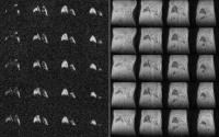
Ventilation imaging with sulfur hexafluoride in free-breathing
mice: initial experience 
Marta Tibiletti1, Martin Tschechne1,
Andrea Bianchi2, Detlef Stiller2, and
Volker Rasche1,3
1Core Facility Small Animal MRI, Ulm University,
Ulm, Germany, 2Target
Discovery Research, In-vivo imaging laboratory, Boehringer
Ingelheim Pharma GmbH & Co. KG, Biberach an der Riss,
Germany,3Department of Internal Medicine II, Ulm
University, Ulm, Germany
Functional information of the lung is of great importance
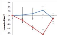
for staging and monitoring lung disease. Imaging of lung
ventilation has been addressed by inhalation of polarized
gases like Helium or Xenon. Major limitation of this
technique rises from the high costs of equipment and gases.
As an efficient alternative to polarized gases, the use of
fluorinated gases has been proposed. In pre-clinical
application these have always been used in combination with
intubation, which does not realistically reflect the
ventilation during free breathing. In this contribution the
imaging of ventilation in mice with fluorinated gases during
free-breathing is addressed.
|
|
2913.
 |
2 |
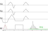
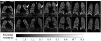
3D Lung Ventilation 1H Imaging Using a Respiratory
Self-Navigated Stack-of-Stars Sequence in Comparison to 2D
Fourier Decomposition
Andreas Voskrebenzev1,2, Marcel Gutberlet1,2,
Frank Wacker1,2, and Jens Vogel-Claussen1,2
1Institute of Diagnostic and Interventional
Radiology, Hanover, Germany, 2German
Centre for Lung Research, Hanover, Germany
Fourier Decomposition (FD) is a lung function imaging
technique with a high clinical potential. Nevertheless the
2D acquisition leads to long acquisition times for complete
lung scans and the 3D breathing motion might lead to errors
in the ventilation measurements. Self-navigated sequences
offer the possibility to reconstruct images in different
respiratory states. Using a stack-of-stars sequence, a
method for 3D fractional ventilation (FV) imaging is
demonstrated for six healthy volunteers and compared with FV
calculated by 2D FD. The two methods show a good agreement.
Additionally, 3D FV depicts 3D lung motion, which is not
adequately detected with 2D FD.
|
|
2914.
 |
3 |
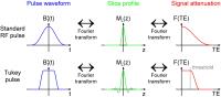
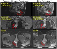
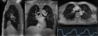
PETRA Lung MRI: Towards Robust Lung Imaging with Patient Comfort
and with Improved Contrast
Yutaka Natsuaki1, Xiaoming Bi1,
Gerhard Laub1, and David Grodzki2
1Siemens Healthcare, Los Angeles, CA, United
States, 2Siemens
Healthcare, Erlangen, Germany
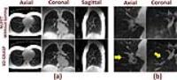
With recent developments in ultra-short TE (UTE) MRI
sequences such as PETRA with the respiratory triggering and
the segmented acquisition, MRI has shown a potential of
being a viable radiation-free alternative to the incumbent
gold standard CT lung imaging. Within a volunteer
validation study (n=14), the current work demonstrates a
recent progress in the PETRA lung MRI towards its robustness
and its applicability to all patient populations. The
proposed solution improves patient comfort and image
contrast by suppressing the unintended high intensity
signals from surrounding tissues.
|
|
2915.
 |
4 |
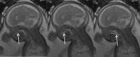
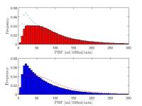
Histogram based Analysis of Lung Perfusion of Children after
Congenital Diaphramatic Hernia Repair
Nora Kassner1, Meike Weis2, Katrin
Zahn3, Thomas Schaible4, Stefan O
Schoenberg2, Lothar R Schad1, K
Wolfgang Neff2, and Frank G Zöllner1
1Computer Assisted Clinical Medicine, Medical
Faculty Mannheim, Heidelberg University, Mannheim, Germany, 2Institute
of Clinical Radiology and Nuclear Medicine, University
Medical Center Mannheim, Heidelberg University, Mannheim,
Germany, 3Department
of Pediatric Surgery, University Medical Center Mannheim,
Heidelberg University, Mannheim, Germany, 4Department
of Neonatology, University Medical Center Mannheim,
Heidelberg University, Mannheim, Germany
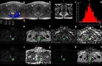
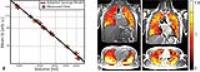
Reported measured lung perfusion data of 2-year old children
after congenital diaphragmatic hernia repair was evaluated
by regions of interest (ROI) within the acquired 3D
volume. In this work a histogram based approach is used to
characterize the distribution of perfusion in the whole left
and right lung, and suitable quantities to characterize the
distribution are extracted.
|
|
2916.
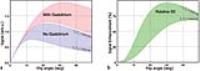 |
5 |
Balanced SSFP pulmonary signal enhancement after contrast agent
injection
Orso Pusterla1,2, Grzegorz Bauman1,2,
and Oliver Bieri1,2
1Radiological Physics, Dep. of Radiology,
University of Basel Hospital, Basel, Switzerland, 2Department
of Biomedical Engineering, University of Basel, Basel,
Switzerland
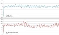
In contrast to the common view that Gd-based contrast agents
have only marginal/limited effect on balanced steady state
free precession (bSSFP) from its $$$T_2/T_1$$$ signal
properties, we will demonstrate that especially for lung
imaging single-dose contrast agent administration increases
the parenchymal signal nearly up two fold.
|
|
2917.
 |
6 |
Fast 3D quantitative 1H ventilation imaging of the human lung at
1.5T with SSFP
Orso Pusterla1,2, Grzegorz Bauman1,2,
Mark Wielpütz3,4, Claus Heussel3,4,
and Oliver Bieri1,2
1Radiological Physics, Dep. of Radiology,
University of Basel Hospital, Basel, Switzerland, 2Department
of Biomedical Engineering, University of Basel, Basel,
Switzerland, 3Department
of Diagnostic and Interventional Radiology with Nuclear
Medicine, Thoraxklinik at University Hospital Heidelberg,
Heidelberg, Germany, 4Department
of Diagnostic and Interventional Radiology, University
Hospital of Heidelberg, Heidelberg, Germany
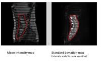
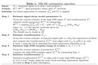
Monitoring lung ventilation is of great interest to assess
pulmonary function and disease progression. Here, a novel,
fast, and simple three-dimensional (3D) quantitative in vivo 1H
imaging method is introduced, reflecting regional
ventilation information. To this end, typically five
ultra-fast balanced steady state free precession (ufSSFP)
scans are repetitively performed in breath-hold from which a
respiratory index map, $$${\gamma}$$$, is derived. The new
measure $$${\gamma}$$$ shows high reproducibility in healthy
volunteers and high sensitivity to respiratory defects, such
as in patients with COPD.
|
|
2918.
 |
7 |
Four-Dimensional Respiratory Motion-Resolved Sparse Lung MRI
Li Feng1, Jean Delacoste2, Hersh
Chandarana1, Davide Piccini2,3,
Francis Girvin1, Matthias Stuber2,4,
Daniel K Sodickson1, and Ricardo Otazo1
1Center for Advanced Imaging Innovation and
Research (CAI2R), New York University School of Medicine,
New York, NY, United States, 2University
Hospital (CHUV) and University of Lausanne (UNIL), Lausanne,
Switzerland, 3Advanced
Clinical Imaging Technology, Siemens Healthcare, Lausanne,
Switzerland, 4Center
for Biomedical Imaging (CIBM), Lausanne, Switzerland
A four-dimensional (4D) respiratory motion-resolved UTE MRI
method is presented for free-breathing lung MRI with
isotropic spatial resolution. Center-out radial
half-projection k-space data are continuously acquired using
a 3D golden-angle UTE sequence. The radial k-space data are
retrospectively sorted into distinct respiratory states,
resulting in an undersampled 4D dataset (kx-ky-kz-respiration)
using a respiratory motion signal extracted from the
acquired data. The undersampled 4D data are reconstructed by
exploiting sparsity along the new respiratory dimension. The
proposed approach enables free-breathing lung MRI with 100%
scan efficiency, allowing for assessment of lung tissue in
arbitrary orientations at different respiratory states.
|
|
2919.
 |
8 |
Fourier Decomposition MRI using the SENCEFUL Approach for
Non-Contrast-Enhanced Ventilation Imaging in Cystic Fibrosis
Patients
Simon Veldhoen1, Andreas Max Weng1,
Clemens Wirth1, Andreas Steven Kunz1,
Janine Nicole Knapp1, Daniel Stäb1,2,
Florian Segerer3, Helge Uwe Hebestreit3,
Thorsten Alexander Bley1, and Herbert Köstler1
1Department of Diagnostic and Interventional
Radiology, University Hospital Würzburg, Würzburg, Germany, 2The
Centre for Advanced Imaging, The University of Queensland,
Brisbane, Australia,3Department of Pediatrics,
University Hospital Würzburg, Würzburg, Germany
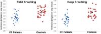
Fourier Decomposition MRI using the SENCEFUL approach is a
recent development in functional lung MRI allowing for
site-resolved assessment of lung function. The purpose of
the present study is to evaluate its feasibility for
ventilation imaging in patients with cystic fibrosis. Seven
cystic fibrosis patients and 7 healthy volunteers were
examined, lung ventilation maps were reconstructed and
quantitative ventilation measurements were performed in
tidal and deep breathing. Mean quantitative ventilation was
significantly lower for patients with cystic fibrosis when
compared to the healthy controls. The ventilation maps
indicated increased ventilation inhomogeneity in cystic
fibrosis patients.
|
|
2920.
 |
9 |
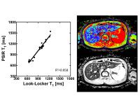
Tobacco smoke shortens T1 in a mouse model of COPD
Daniel Alamidi1, Amir Smailagic2,
Abdel Bidar2, Nicole Parker2, Marita
Olsson2, Sonya Jacksson2, Linda Swedin2,
Paul Hockings3,4, Kerstin Lagerstrand1,
and Lars E Olsson5
1Department of Radiation Physics, Institute of
Clinical Sciences, Sahlgrenska Academy, University of
Gothenburg, Gothenburg, Sweden, 2AstraZeneca
R&D, Mölndal, Sweden, 3Medtech
West, Chalmers University of Technology, Gothenburg, Sweden, 4Antaros
Medical, BioVenture Hub, Mölndal, Sweden, 5Department
of Medical Physics, Lund University, Translational Sciences,
Malmö, Sweden
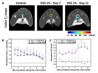
Tobacco smoking is the main cause of COPD. MRI may improve
the characterization of COPD where lung T1 mapping has been
used to study lung disease. We investigated whether tobacco
smoke exposure affects lung T1 in a mouse model with
repeated T1 readouts and biological measurements. Free
breathing 3D-UTE T1 maps of the lungs were weekly performed
over one month in mice exposed to air or tobacco smoke. The
lung T1 was shortened in the tobacco smoke exposed mice,
most likely due to early signs of smoking-induced lung
pathology. Consequently, T1 is a potential biomarker of lung
disease.
|
|
2921.
 |
10 |
Respiratory self-gating in 3D UTE lung acquisition in small
animal imaging
Marta Tibiletti1, Andrea Bianchi2,
Åsmund Kjørstad3, Detlef Stiller2, and
Volker Rasche1,4
1Core Facility Small Animal MRI, Ulm University,
Ulm, Germany, 2Target
Discovery Research, In-vivo imaging laboratory, Boehringer
Ingelheim Pharma GmbH & Co. KG, Biberach an der Riss,
Germany,3Department of Neuroradiology, University
Hospital Hamburg-Eppendorf, Hamburg, Germany, 4Department
of Internal Medicine II, Ulm University, Ulm, Germany
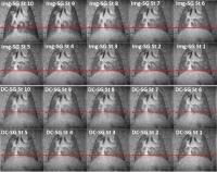
1D (k-space center) and 3D (sliding window 3D
reconstruction) have been evaluated for respiratory
retrospective self-gating for Quasi Random UTE-3D lung
acquisition in freely breathing rats. Low-resolution 3D
GRASP reconstruction allowed the extraction of an effective
gating signal from changes in lung-liver interface position.
The 1D center-of-k-space method did not yield sufficient
gating signal fidelity, most likely caused by the only
limited changes of the anatomy in the investigated volume,
and to a lesser extent intensity modulations introduced by
residual eddy-currents.
|
|
2922.
 |
11 |
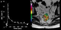
Longitudinal Assessment of Pulmonary Permeability in a Mouse
Model of Lung Fibrosis
Iliyana P Atanasova1, Pauline Desogere1,
Clemens K Probst2, Nicholas Rotile1,
Andrew M Tager2, and Peter Caravan1
1A. A. Martinos Center for Biomedical Imaging,
Massachusetts General Hospital, Charlestown, MA, United
States, 2Center
for Immunology and Inflammatory Diseases, Massachusetts
General Hospital, Boston, MA, United States
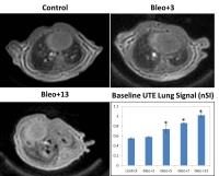
Idiopathic pulmonary fibrosis is a fatal condition without
effective treatment. Given evidence that vascular leak
promotes fibrosis, we assessed whether pulmonary leak could
be quantified using dynamic MRI and an intravascular tracer.
In a mouse model we observed that permeability to albumin
rose sharply on day 3 after insult, returned to baseline by
day 5 and increased moderately between days 5-13. To our
knowledge this is the first report of the time course of
vascular leak in pulmonary fibrosis. The proposed method
could be useful for studying the role of lung permeability
in fibrosis and for monitoring of treatment response.
|
|
2923.
 |
12 |
Imaging of Lung Conductivity Using Ultrashort Echo-Time Imaging
Ulrich Katscher1 and
Peter Börnert1
1Philips Research Europe, Hamburg, Germany
Reliable MR imaging of lung tissue could be an important
element of diagnosing lung-related diseases. The very short
T2 components of lung tissue, one of the main problems of
lung imaging, can be visualized using ultrashort echo times
(UTE). Furthermore, UTE sequences allow the determination of
conductivity of the imaged tissue. This study shows the
principle feasibility of UTE to image lung conductivity,
examining healthy volunteers. Obtained conductivity was
lower for inspiration breath hold than expiration breath
hold, which is the expected behaviour due to corresponding
fraction of air (with nearly zero conductivity) inside lungs
during inspiration and expiration.
|
|
2924.
 |
13 |
Pulmonary Fourier decomposition MRI compared to multiple breath
washout and spirometry: A preliminary study in Primary Ciliary
Dyskinesia
Grzegorz Bauman1,2, Sylvia Nyilas2,3,4,
Orso Pusterla1,2, Christoph M Heyer5,
Cordula Koerner-Rettberg6, Philipp Latzin3,4,
and Oliver Bieri1,2
1Division of Radiological Physics, Deparment of
Radiology, University Hospital of Basel, Basel, Switzerland, 2Department
of Biomedical Engineering, University of Basel, Basel,
Switzerland, 3Department
of Pediatric Pneumology, University Children's Hospital
Basel (UKBB), Basel, Switzerland, 4Division
of Respiratory Medicine, Department of Pediatrics,
University Children's Hospital of Bern, Bern, Switzerland, 5Institute
of Diagnostic Radiology, Interventional Radiology and
Nuclear Medicine, Ruhr-University of Bochum, Bochum,
Germany, 6Department
of Pediatric Pneumology, University Children's Hospital of
Ruhr University Bochum at St Josef-Hospital, Bochum, Germany
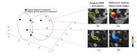
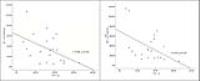
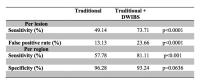
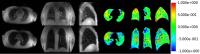
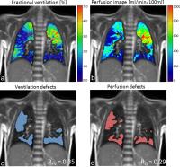
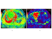
In this work, the feasibility of contrast-media-free
pulmonary Fourier decomposition (FD) MRI is assessed in
patients with primary ciliary dyskinesia (PCD). An automatic
evaluation of regional functional defects on fractional
ventilation and perfusion FD maps has been developed.
Furthermore, the lung function evaluated using FD MRI is
compared to the parameters obtained using multiple breath
washout technique and spirometry. Statistical analysis was
used to find significant correlations between FD MRI and
lung function techniques.
|
|
2925.
 |
14 |
Whole Lung Morphometry with Hyperpolarised 3He Gas Diffusion MRI
- 3D Multiple b-value Acquisition and Compressed Sensing
Ho-Fung Chan1, Neil J. Stewart1, Juan
Parra-Robles1, Guilhem J. Collier1,
and Jim M. Wild1
1Academic Unit of Radiology, University of
Sheffield, Sheffield, United Kingdom
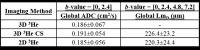
Compressed sensing (CS) was implemented to reduce scan time
and facilitate acquisition of 3D multiple b-value 3He
diffusion-weighted (DW) MRI data for whole lung morphometry.
A fully-sampled 3D DW-MRI dataset was retrospectively
undersampled using CS simulations to determine optimal
k-space undersampling patterns. Whole lung morphometry
measurements derived from prospective 3-fold undersampled 3D
multiple b-value
DW-MRI were compared to 3D and 2D fully-sampled equivalents.
Good agreement was obtained between lung morphometry
measurements indicating 3D multiple b-value 3He
DW-MRI with CS can provide reliable measurements of whole
lung morphometry within a single breath-hold.
|
|
2926.
 |
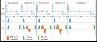
15 |
Single breath washout imaging – regional phase III slope mapping
with rapid hyperpolarized gas MRI - Permission Withheld
Felix C Horn 1 and
Jim M Wild1
1University of Sheffield, Sheffield, United
Kingdom
Single breath washout (SBW) is a whole lung pulmonary
function test that has been shown to be sensitive to early
changes in lung disease. Of particular clinical interest has
been the Phase III slope (S III) as the
concentration decay between 25-75% of the exhaled volume.
In this work rapid lung imaging of exhaled hyperpolarized
gas is used to acquire 2D images of SBW of subjects expiring
to residual lung volume. The ability to calculate regional SIII
from those time resolved images is
demonstrated in healthy volunteers.
|
|
2927.
 |
16 |
Incorporation of prior knowledge of the signal behavior into the
compressed sensing framework for accelerated acquisition in
hyperpolarized gas diffusion MRI
Juan FPJ Abascal1,2, Manuel Desco1,2,3,
and Juan Parra-Robles1,2
1Department of Bioengeering and Aerospace
Engineering, Universidad Carlos III de Madrid, Madrid,
Spain, 2Instituto
de Investigación Sanitaria Gregorio Marañón, Madrid, Spain, 3Centro
de Investigación en Red de Salud Mental (CIBERSAM), Madrid,
Spain
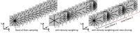
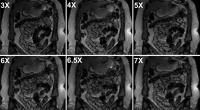
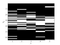
Diffusion MRI
measurements using hyperpolarized gases are generally
acquired during patient breath hold, which yields a
compromise between achievable image resolution, lung
coverage and number of b-values. In this work, we propose a
novel method that incorporates the knowledge of the signal
decay into the reconstruction (SIDER) to accelerate the
acquisition of MR diffusion data by undersampling in both
spatial and b-value dimensions. SIDER is assessed by
restrospectively undersampling diffusion datasets of normal
volunteers and COPD patients. Results suggest that
accelerations of at least x7 are achievable with negligible
effect in the estimates of diffusion parameters.
|
|
2928.
 |
17 |
3D Multi-Parametric Acquisition of 3He Lung Ventilation Images,
Lung Diffusion Morphometry and T2* Maps with Compressed Sensing
Ho-Fung Chan1, Neil J. Stewart1,
Guilhem J. Collier1, and Jim M. Wild1
1Academic Unit of Radiology, University of
Sheffield, Sheffield, United Kingdom
Whole-lung coverage 3He
ventilation images, maps of ADC, alveolar dimension (LmD),
and T2* were acquired in a single breath-hold
using a multiple-interleaved 3D sequence with compressing
sensing (CS). A fully-sampled three-interleaved ADC and T2*
dataset was acquired for CS simulations, to determine the
optimal k-space undersampling patterns. A prospective,
3-fold undersampled 3D five-interleaved dataset was acquired
with CS and parametric maps were compared to those
calculated from fully-sampled datasets. CS-derived ADC and LmD values
showed good agreement with fully-sampled equivalents.
CS-derived T2* values were lower than
fully-sampled ones due to the smoothing process of the CS
reconstruction.
|
|
2929.
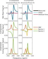 |
18 |
Improved fitting of 129Xe
spectroscopy identifies three dissolved-phase resonances in the
human lung
Scott H. Robertson1,2, Elianna A. Bier1,2,
Rohan S. Virgincar1,3, and Bastiaan Driehuys1,2,3,4
1Center for In Vivo Microscopy, Duke University
Medical Center, Durham, NC, United States, 2Medical
Physics Graduate Program, Duke University, Durham, NC,
United States, 3Department
of Biomedical Engineering, Duke University, Durham, NC,
United States, 4Department
of Radiology, Duke University Medical Center, Durham, NC,
United States
Hyperpolarized 129Xe
experiences chemical shifts between the lung airspaces,
interstitium, and capillary beds, enabling functional
information to be directly probed. However, in order to
realize the potential of these chemical shifts, the spectrum
must be carefully decomposed. Previous methods have assumed
only two dissolved-phase resonances exist in the human lung
and have used inconsistent 0 ppm reference frequencies. Here
we present novel non-linear fitting of complex exponentially
decaying FIDs and demonstrate that the dissolved phase
signal can be robustly decomposed into three dissolved-phase
resonances. We present updated frequencies and widths using
and appropriately adjusted 0 ppm reference value.
|
|
2930.
 |
19 |
Improving quantitative Hyperpolarized 129Xe
gas exchange MRI in idiopathic pulmonary fibrosis
Scott H. Robertson1,2, Jennifer Wang1,
Geoffry Schrank1, Holman P. McAdams3,
and Bastiaan Driehuys1,2,4,5
1Center for In Vivo Microscopy, Duke University
Medical Center, Durham, NC, United States, 2Medical
Physics Graduate Program, Duke University, Durham, NC,
United States, 3Department
of Radiology, Duke University Medical Center, Durham, NC,
United Kingdom, 4Department
of Biomedical Engineering, Duke University, Durham, NC,
United States, 5Department
of Radiology, Duke University Medical Center, Durham, NC,
United States
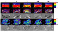
While gas-exchange imaging with 129Xe
holds great potential for enhancing both the diagnosis and
followup of IPF, the quantitative ability of these
techniques is currently limited by the SNR and spatial
resolution afforded by the limited dissolved-phase signal
and highly undersampled acquisition. Here we tune our
reconstruction for these challenging conditions, and
demonstrate improvements in image quality. We then quantify
the loss of gas exchange in the apex and base of the lung
and show that there is significantly reduced gas exchange in
the basal regions of subjects with IPF relative to healthy
controls.
|
|
2932.
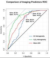 |
20 |
Prediction of Longitudinal FEV1 Decline in Smokers with
Hybrid Hyperpolarized 3He MRI
Hooman Hamedani1,2, Yi Xin1, Stephen J
Kadlecek1, Heather Gatens1, Maurizio
Cereda3, Sarmad M Siddiqui1,2, Mehrdad
Pourfathi1,4, Joseph Naji1, Masaru
Ishii5, and Rahim R Rizi1
1Radiology, University of Pennsylvania,
Philadelphia, PA, United States, 2Bioengineering,
University of Pennsylvania, Philadelphia, PA, United States, 3Anesthesiology
and Critical Care, University of Pennsylvania, Philadelphia,
PA, United States, 4Electrical
and Systems Engineering, University of Pennsylvania,
Philadelphia, PA, United States, 5Johns
Hopkins University, Baltimore, MD, United States
Aside from the superior diagnostic power that HP gas MRI
provides through imaging unique aspects of lung function, it
is evident that the underlying mechanisms that lead to
subsequently evident global changes in the lung function in
future are detectable through regional and functional
imaging using HP gas MRI.
|
|
2933.
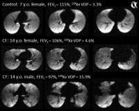 |
21 |
Hyperpolarized 129Xe MRI ventilation in pediatric cystic
fibrosis lung disease: safety and sensitivity
Laura L Walkup1, Robert P Thomen1,2,
Teckla Akinyi1,3, Wolfgang Loew4, Kai
Ruppert1, John P Clancy5, Zackary I
Cleveland1, and Jason C Woods1
1Center for Pulmonary Imaging Research, Division
of Pulmonary Medicine and Department of Radiology,
Cincinnati Children's Hospital Medical Center, Cincinnati,
OH, United States, 2Department
of Physics, Washington University in St. Louis, St. Louis,
MO, United States, 3Department
of Biomedical Engineering, University of Cincinnati,
Cincinnati, OH, United States, 4Imaging
Research Center, Department of Radiology, Cincinnati
Children's Hospital Medical Center, Cincinnati, OH, United
States, 5Division
of Pulmonary Medicine, Cincinnati Children's Hospital
Medical Center, Cincinnati, OH, United States
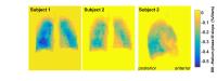
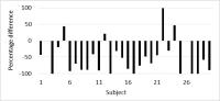
We demonstrate hyperpolarized 129Xe
MRI in healthy pediatric control subjects and cystic
fibrosis patients as young as age 7, for the first time.
Subjects experienced a transient nadir in SpO2 that
quickly returns to baseline with normal breathing. Despite
having similarly high lung function (i.e., normal FEV1),
CF patients had nearly 4-fold increase in 129Xe
ventilation defect volume compared to their healthy peers,
with statistical significance. Importantly, ventilation
defects were present even in CF patients with FEV1 near
or exceeding 100% predicted, suggesting that 129Xe
MRI is more sensitive to early CF lung disease than
traditional clinical spirometry.
|
|
2931.
 |
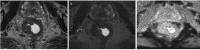
22 |
Magnetic Resonance Elastography of the Anterior Mediastinal Mass
at 3T: a Preliminary Study - Permission Withheld
Wei Tang1, Yao Huang1, Ning Wu1,
Wenwen Lu1, Linlin Qi1, and Bing Wu2
1Diagnostic Radiology, Cancer Hospital, Chinese
Academy of Medical Sciences, Peking Union Medical College,
Beijing, China, People's Republic of, 2GE
heathcare MR research China, Beijing, China, People's
Republic of
Magnetic resonance (MR) elastography depitcs the elastic
properties of tissues of interest has been primarily applied
in the work-up of diagnosis for hepatic fibrosis.
Theoretically, interference fringes could be visualized on
the elastogram due to the miscalculation of the interaction
between the attenuated propagations of shear wave and the
tissue overlying or surrounding to the investigated subject,
which might be one of the main concern that limited the
clinical potentials of MRE. We propose an investigation on
the feasibility of MR elastography in characterizing the
mechanical properties of anterior mediastinal masses with
the consideration of the relatively superficial location of
these entities, therefore few interactions between the shear
wave and subject unexpected were produced during the process
of elasticity mapping. It was demonstrated in our study that
anterior mediastinal mass in various etiology of thymic
carcinoma, thymoma, and lymphoma has distinct elastic
properties. MR elastography was helpful in distinguishing
the thymic carcinoma from lymphoma.
|
|
2934.
 |
23 |
Assessment of Pleural Effusion in Dengue Fever
Therese Sjoholm1, Benjamin A Thomas1,
Yin Mo2, Louisa Sun2, Ashley St. John3,
Paul Anantharajah Tambyah2, and John J Totman1
1A*STAR-NUS Clinical Imaging Research Centre,
Singapore, Singapore, 2Department
of Medicine, National University Hospital Singapore,
Singapore, Singapore, 3Duke-NUS
Graduate Medical School, Singapore, Singapore
In this study we assess the feasibility of using MRI for
measurement of pleural effusion in Dengue Fever. 30 subjects
with confirmed Dengue infection were scanned using
T2-weighted HASTE MRI and chest x-rays (CXRs) at baseline
and 4-8 days follow-up. Fluid accumulation in the pleural
cavity was assessed for both modalities. For MRI,
significantly different fluid accumulations were measured
between baseline and follow-up (p=0.002). The fluid
accumulations were all below the detectability limit of CXR.
As such, MRI provides a sensitive measurement of pleural
effusion in dengue fever and can be used to track fluid
accumulation over time.
|
|