ISMRM 24th Annual Meeting & Exhibition • 07-13 May 2016 • Singapore

Electronic Poster Session: Diffusion 1

3006 -3029 Diffusion: Acquisition
3030 -3053 Diffusion: Analysis
3054 -3077 Diffusion: Analysis & Tractography
3078 -3101 Diffusion: Microstructure

Exhibition Hall 

16:30 - 17:30

    Computer #

3008.   
3 Towards Fast Diffusion-Sensitized MR Imaging of the Eye and Orbit with High Anatomic Fidelity: Combining a Segmented RARE variant with Inner Volume Imaging
Katharina Paul1, Till Huelnhagen1, Oliver Stachs2, and Thoralf Niendorf1,3
1Berlin Ultrahigh Field Facility (B.U.F.F), Max Delbrück Center for Molecular Medicine in the Helmholtz Association (MDC), Berlin, Germany, 2Department of Ophthalmology, University Medicine Rostock, Rostock, Germany, 3Experimental and Clinical Research Center, a joint cooperation between the Charité Medical Faculty and the Max-Delbrueck-Center for Molecular Medicine in the Helmholtz Association, Berlin, Germany
Diffusion-weighted imaging of the eye and orbit is an emerging MRI application to provide guidance during diagnostic assessment and treatment of ophthalmological diseases. It has been shown that RARE based diffusion-sensitized imaging (ms-RARE) provides images free of geometric distortions. Though, artifacts induced by involuntary eye motion remain a concern. Applying inner volume imaging (IVI) offers the possibility to shorten acquisition times by reducing the number of acquired phase encoding lines. This study examines the applicability of IVI in conjunction with ms-RARE with the goal to reduce the propensity to bulk eye motion in diffusion-sensitized ophthalmic imaging.


3026.   
21 Quantitative evaluation of Apparent Diffusion Coefficient (ADC) from two DWI sequences – echo planar spin echo (EPSE) and read-out segmented echo planar (RESOLVE) imaging - Permission Withheld
Eo-Jin Hwang1, Hyun-Seok Choi1, Yoon-Ho Nam1, and Joon-Yong Jung1
1Department of Radiology, St.Mary Hospital, Seoul, Korea, Republic of
The purpose of this study was to quantitatively evaluate ADCs from two different DWI sequences – echo planar spin echo (EPSE) and read-out segmented echo planar (RESOLVE) imaging. The mean ADCs of the whole brain, GM, WM and CSF were compared based on masking templates generated from T2-weighted images. Our results demonstrated that although difference between the two ADCs was statistically significant over the whole brain regions, ADCs from msDWI and ssDWI were highly correlated (R2 = 0.989). The relationship between the two ADCs should be considered to utilize it as an effective quantitative metric.


3027.   
22 The Optimization of Zoomed Field of View DWI (iZOOM) with Echo Planar Imaging (EPI) Sequence Excited by a Two Dimensional Radiofrequency Pulse (2D RF) on Prostate at 3.0 T
Kangjie Xu1, Xiaodong Zhang1, Chengyan Wang2, Hongxia Sun1, Yu Zhang3, Zhigang Wu4, and Xiaoying Wang1
1The Department of Radiology, Peking University First Hospital, Beijing, China, People's Republic of, 2Academy for Advanced Interdisciplinary Studies, Peking University, Beijing, China, People's Republic of, 3Philips Healthcare, Beijing, China, People's Republic of, 4Philips Healthcare (Suzhou) Co., Ltd, Suzhou, China, People's Republic of
This study aims to demonstrate the feasibility of iZOOM using a 2D RF pulse EPI sequence in the application of prostate DWI and the optimization of iZOOM at 3.0 T MRI. 58 patients took prostate MRI examination with 3 iZOOM sequences (1. b=1000, resolution: 1.4×1.4mm; 2. b=1000, resolution: 1.0×1.0mm; 3. b=2000, resolution: 1.4×1.4mm). Images were evaluated with the basic image quality, the display of lesions and overall image quality. The result shows iZOOM is feasible on prostate DWI. High b value and high resolution iZOOM are both favored by radiologists.


3028.   
23 Efficient Macroscopic Motion Correction for Multi-shot DTI
zhongbiao xu1, Yanqiu Feng1, Wufan Chen1, Zhigang Wu2, Ha-kyu Jeong3, Wenxing Fang2, Yingjie Mei1,4, Li Guo1, and Feng Huang2
1School of Biomedical Engineering and Guangdong Provincial Key Laboratory of Medical Image Processing, Southern Medical University, Guangzhou, China, People's Republic of, 2Philips Healthcare (Suzhou), Suzhou, China, People's Republic of, 3Philips Korea, Seoul, Korea, Republic of, 4Philips Healthcare, Guangzhou, China, People's Republic of
Though cMUSE proposed by our group tackles the pixels mismatch of macroscopic motion in multi-shot EPI by clustering and registration, it neglects altered gradient directions. In this work, we treat motion induced variations in gradient direction as addtional diffusion direction(s). The proposed method simply and effectively solves the gradient direction alternation due to macroscopic motion in multi-shot DTI.


3029.   
24 Acceleration motion compensation DWI for measuring intraventricular temperature
Shuhei Shibukawa1,2, Toshiaki Miyati2, Naoki Ohno2, Tetsu NIwa3, Yutaka Imai3, Tetsuo Ogino4, and Isao Muro1
1Depertment of Radiology, Tokai university hospital, Isehara, Japan, 2Division of Health Sciences, Graduate School of Medical Sciences, Kanazawa University, Ishikawa, Japan, 3Depertment of Radiology, Tokai University School of Medicine, Isehara, Japan, 4Healthcare, Philips Electronics Japan Ltd., Tokyo, Japan
Although it has been reported a method for monitoring the intraventricular cerebrospinal fluid (CSF) temperature calculated from the DWI, this method was affected by the CSF pulsation. We proposed the acceleration-motion compensation DWI (aMC-DWI) to the determination of the intraventricular temperature to improve that accuracy and precision.  Compared with conventional DWI, measurement of intraventricular temperature using aMC-DWI was accuracy even in ventricles with high flow, e.g., third ventricle, fourth ventricle. These results suggested that aMC-DWI is a suitable method for analyzing the intraventricular temperature.


3006.   
1 Imaging Restrictive Pore Geometry with Asymmetrical Chirped Pulses - Video Not Available
Qutaibeh Katatbeh1, Alexey Tonyshkin2, and Andrew Kiruluta3
1Jordan University of Science and Technology, Irbid, Jordan, 2MGH, Boston, MA, United States, 3Harvard, Cambridge, MA, United States
Presents a practical way of recovering the phase of diffusing spins to recover the underlying restrictive geometry through a Fourier transform


3007.   
2 Enhanced efficiency and sensitivity in detection of acute ischemic brain injury using fast diffusion kurtosis imaging
Yin Wu1,2, Jinsuh Kim3, Suk-Tak Chan1, Iris Yuwen Zhou1, Yingkun Guo1, Takahiro Igarashi1, Hairong Zheng2, Gang Guo4, and Phillip Zhe Sun1
1Athinoula A. Martinos Center for Biomedical Imaging, Department of Radiology, Massachusetts General Hospital and Harvard Medical School, Charlestown, MA, United States, 2Paul C. Lauterbur Research Centre for Biomedical Imaging, Shenzhen Key Laboratory for MRI, Shenzhen Institutes of Advanced Technology, Chinese Academy of Sciences, Shenzhen, China, People's Republic of, 3Department of Radiology, University of Illusions at Chicago, Chicago, IL, United States, 4Department of Radiology, Xiamen 2nd Hospital, Xiamen, China, People's Republic of
We systematically compare the diagnostic value of conventional tensor-based DKI and recently proposed fast DKI protocols using an acute stroke rodent model. The measures and volumes of diffusion and kurtosis lesions were in good agreement between the two DKI methods. Importantly, contrast-to-noise ratio (CNR) of mean kurtosis using the fast DKI protocol was significantly higher than that of the routine method with its CNR efficiency approximately doubles. Therefore, our results demonstrated excellent performance of the fast DKI protocol in characterizing acute ischemic tissue injury, which may facilitate translation of the fast DKI approach in the acute stroke setting.


3009.   
4 Impact of sequence parameters on the sensitivity of DDE and DODE sequences to microscopic anisotropy
Andrada Ianu?1, Ivana Drobnjak1, Noam Shemesh2, and Daniel C. Alexander1
1CMIC, University College London, London, United Kingdom, 2Champalimaud Neuroscience Programme, Champalimaud Centre for the Unknown, Lisbon, Portugal
In the long mixing time regime, double-diffusion-encoding (DDE) sequences provide contrast capable of reflecting microscopic anisotropy, which may have added value for highly heterogeneous tissues such as the gray matter. Recently, double-oscillating-diffusion-encoding (DODE) sequences, which combine oscillating waveforms and varying gradient orientation, have been proposed to improve sensitivity to microscopic anisotropy. This work investigates the effect of varying different sequence parameters and shows that DODE sequences provide higher sensitivity to pore size for elongated pores, while DDE sequences are more sensitive to pore eccentricity. 


3010.   
5 A novel iterative approach to reap the benefits of multi-tissue CSD from just single-shell (+b=0) diffusion MRI data
Thijs Dhollander1 and Alan Connelly1,2
1The Florey Institute of Neuroscience and Mental Health, Melbourne, Australia, 2The Florey Department of Neuroscience, University of Melbourne, Melbourne, Australia
Constrained spherical deconvolution (CSD) is a robust approach to resolve the fibre orientation distribution (FOD) from diffusion MRI data.  However, the FOD from CSD only aims to represent "pure" white matter (WM) and is inappropriate/distorted in regions of (partial voluming with) grey matter (GM) or cerebrospinal fluid (CSF).  Multi-shell multi-tissue CSD was proposed to solve this issue by estimating WM/GM/CSF components, but requires multi-shell data to do so.  In this work, we provide the first proof that similar results can also be obtained from only simple single-shell (+b=0) data, and propose a novel specialisedoptimiser that achieves this goal.


3011.   
6 Double-Pulsed-Field Gradient MRI in the Long Evans shaker rats' spinal cords - Permission Withheld
Debbie Anaby1, Darya Morozov1, Ian D. Duncan2, and Yoram Cohen1,3
1School of Chemistry, The Raymond and Beverly Sackler Faculty of Exact Sciences, Tel Aviv University, Tel Aviv, Israel, 2Medical Sciences, The University of Wisconsin-Madison, Madison, WI, United States, 3Sagol School of Neurosciences, Tel Aviv University, Tel Aviv, Israel
Double pulsed-field gradient (d-PFG) MRI has recently been suggested as an additional methodology for studying microstructure in the CNS. The Long Evans shaker (les) rats’ CNS has been previously studied by q-space diffusion (QSI) and more recently by angular d-PFG MRI. Here, we characterize the microstructure of the les spinal cords by fitting the angular d-PFG MRI data to the multiple correlation function (MCF) and extracting the unique parameter L/R ratio and its fractions. Clearly, the angular d-PFG methodology is capable of distinguishing between the les and their controls, in white matter and even in some gray matter ROIs.


3012.   
7 Effects of the Number of Diffusion Directions in Diffusion Kurtosis Imaging: a Structural Connectivity Study using in vivo data
Ricardo Loução1, Karolina Elsner1, Rita G. Nunes1, Rafael Neto-Henriques2, Marta Correia2, André Ribeiro3, and Hugo Ferreira1
1Instituto de Biofísica e Engenharia Biomédica, Lisbon, Portugal, 2Cognition and Brain Science Unit, MRC, Cambridge, United Kingdom, 3Centre for Neuropsychopharmacology, London, United Kingdom
Diffusion Kurtosis Imaging Tractography Reconstructions (DKI-TR) are often performed using high quality data. In clinical practice, that is often not possible, as only a lower number of b-values and diffusion gradient directions can be acquired. This study assessed the performance of DKI-TR for the two algorithms currently proposed for DKI-TR using variable amounts of data, and looked at their respective structural connectivity metrics. A 64 gradient direction data set was acquired in six healthy subjects, and down-sampled to 21 and 32 directions. Differences were found between gradient sets and also between algorithms, regarding the reconstructions and the connectivity metrics.


3013.   
8 Assessment of Diffusion Time Dependence of Diffusion Kurtosis in Rat Spinal Cord
Nathaniel D Kelm1,2, Kevin D Harkins2, and Mark D Does1,2,3,4
1Biomedical Engineering, Vanderbilt University, Nashville, TN, United States, 2Vanderbilt University Institute of Imaging Science, Vanderbilt University, Nashville, TN, United States, 3Radiology and Radiological Sciences, Vanderbilt University School of Medicine, Nashville, TN, United States, 4Electrical Engineering, Vanderbilt University, Nashville, TN, United States
Diffusion kurtosis imaging (DKI) is an extension of DTI with the ability to provide additional information about tissue microstructure. To evaluate the diffusion time dependence of diffusion kurtosis, kurtosis is measured perpendicular to white matter tracts in rat spinal cord for diffusion times ranging from 12 to 100 ms. In this study, kurtosis increased as a function of diffusion time in white matter, yet decreased in gray matter. Assessing the change in diffusion kurtosis across diffusion time could potentially inform upon the underlying white matter microstructure.


3014.   
9 Time-dependent diffusion on in vivo human brain data from the Connectom scanner
Uran Ferizi1, Claudia Wheeler-Kingshott2, Daniel Alexander3, and Jose Raya1
1Department of Radiology, New York University School of Medicine, New York, NY, United States, 2Institute of Neurology, University College London, London, United Kingdom, 3Department of Computer Science, University College London, London, United Kingdom
Diffusion MRI models for brain microstructure do not currently incorporate time-dependent diffusivity. Previous studies with ex vivo tissue (muscle and brain) have observed such dependency. The advent of high diffusion gradient scanners have enabled us to probe the tissue to unprecedented. Here we examine and observe the time dependency of the measured diffusion using data from the live human brain.


3015.   
10 Between-Session Variability of Diffusion Kurtosis Metrics in in vivo Brains
Nino Kobalia1, Farida Grinberg1,2, Ezequiel Farrher1, Xiang Gao1, and N. Jon Shah1,2
1Institute of Neuroscience and Medicine - 4, Forschungszentrum Jülich GmbH, Jülich, Germany, 2Department of Neurology, Faculty of Medicine, JARA, RWTH Aachen University, Aachen, Germany
The knowledge of intra- and inter-subject variability of diffusion kurtosis imaging metrics plays an important role in the interpretation of the results in clinical trials. However, it has not been sufficiently studied thus far. The purpose of this work is to investigate between-session variability of a single subject with N-repeated measurements with an identical experimental protocol, and thus to provide the baseline for comparison with phantom measurements and inter-subject in vivo variability. We quantified variability in terms of the coefficient of variation and studied how its value varies between various diffusion tensor and kurtosis metrics estimated in twenty anatomical regions.


3016.   
11 The importance of b-values selection and the precision of diffusion kurtosis estimation by the conventional schemes
Andrey Chuhutin1, Noam Shemesh2, Brian Hansen1, and Sune Nørhøj Jespersen1,3
1CFIN, Aarhus University, Aarhus, Denmark, 2Champalimaud Neuroscience Programme, Champalimaud Centre for the Unknown, Lisbon, Portugal, 3Department of Physics and Astronomy, Aarhus University, Aarhus, Denmark
In DKI imaging studies, a wide range of different gradient strengths is used, which is known to affect the estimated kurtosis. Being driven by a need to assess the validity of the DKI expression and the accuracy of the estimated parameters as a function of b-value we both evaluated the variability of the kurtosis parameter for the in vivo and ex vivo data for different fitting techniques and studied the error in a mean kurtosis parameter with respect to the ground truth. The results and conclusions suggest circumspection while preferring a specific technique.


3017.   
12 Diffusion Kurtosis Breast Imaging model – Which should be the highest b-value?
Filipa Borlinhas1, Luísa Nogueira2, Sofia Brandão3, Rita G. Nunes1, Raquel Conceição1,4, Joana Loureiro3, Isabel Ramos3, and Hugo A. Ferreira1
1Instituto de Biofísica e Engenharia Biomédica, Faculdade de Ciências da Universidade de Lisboa, Lisboa, Portugal, 2Escola Superior de Tecnologia da Saúde fo Porto, ESTSP/IPP, Porto, Portugal, 3Hospital de São João, Porto, Porto, Portugal, 4Institute of Biomedical Engineering, University of Oxford, United Kingdom, Oxford, United Kingdom
For the application of the Diffusion Kurtosis Imaging (DKI) model, the use of higher b values is advised. Here, the diagnostic performance of the DKI model in the differentiation of benign and malignant breast tumors was studied for the first time regarding the most suitable higher b-value (2000, and 3000 s/mm2).   In this study were included 36 benign and 75 malignant lesions, assessed using combinations of b-values 50 to 2000, and to 3000s/mm2. The b-value range 50 to 3000 s/mm2 showed the best results regarding diagnostic performance and so this range is suggested for use in future DKI breast cancer studies.


3018.   
13 A new framework for the optimisation of multi-shell diffusion weighting MRI settings using a parameterised Cramér-Rao lower-bound - Permission Withheld
Ezequiel Farrher1, Ivan I. Maximov2, Vincent Gras1, Farida Grinberg1,3, Rüdiger Stirnberg1,4, and N. Jon Shah1,3
1INM 4 - Medical Imaging Physics, Forschungszentrum Jülich, Jülich, Germany, 2Experimental Physics III, TU Dortmund University, Dortmund, Germany, 3Department of Neurology, Faculty of Medicine, RWTH Aachen University, Aachen, Germany, 4German Center for Neurodegenerative Diseases (DZNE), Bonn, Germany
We propose a parameterisation for multi-shell diffusion gradient settings which is sufficiently flexible to cover a broad range of experimental configurations, on the one hand, but uses only 6 degrees of freedom, on the other. Thus, such parameterisation enables a robust optimisation of the diffusion weighting settings by minimising the Cramér-Rao lower bound of the parameters of interest. Finally, we demonstrate its performance in the context of the biexponential diffusion tensor analysis.


3019.   
14 A Robust Reconstruction Method for High Resolution Multishot DWI: SPIRiT-based SYMPHONY
Zijing Dong1, Fuyixue Wang1, Xiaodong Ma1, Erpeng Dai1, Zhe Zhang1, and Hua Guo1
1Center for Biomedical Imaging Research, Department of Biomedical Engineering, School of Medicine, Tsinghua University, Beijing, China, People's Republic of
SYnergistic iMage reconstruction using PHase variatiOns and seNsitivitY (SYMPHONY) is a reconstruction method for multi-shot DWI which can provide high resolution diffusion weighted images. In this study, we proposed a SPIRiT-based SYMPHONY method in which a self-consistency constraint is applied instead of the conventional GRAPPA kernel to further improve the robustness of SYMPHONY. Simulation and in-vivo experiment validated the benefits of the proposed method which can improve the accuracy of reconstruction with less navigator data.


3020.   
15 Self-calibrated K-space Phase Correction Method for Multi-shot Diffusion Imaging
Zhe Zhang1, Xiaodong Ma1, Erpeng Dai1, Hui Zhang1, and Hua Guo1
1Center for Biomedical Imaging Research, Department of Biomedical Engineering, School of Medicine, Tsinghua University, Beijing, China, People's Republic of
Multi-shot EPI can achieve high resolution diffusion imaging, but the ghost artifacts caused by shot-to-shot phase variations must be corrected. In recent works, k-space phase correction methods have been proposed, which require navigator acquisitions for each excitation for calibrating the k-space interpolation parameters. In this work, a self-calibrated method for multi-shot DWI correction in k-space is proposed, which does not require navigator acquisitions for efficient scanning and does not suffer from the potential mismatch between image and navigator echoes. Experiments on liver DWI demonstrate that the proposed method can correct the motion induced artifacts in diffusion imaging.


3021.   
16 Diffusion Weighted 3D UTE Imaging Using Stimulated Echoes: Technical Considerations
Michael Carl1, Yajun Ma2, Graeme M Bydder2, and Jiang Du2
1Global MR Applications & Workflow, General Electric, San Diego, CA, United States, 2University of California San Diego, San Diego, CA, United States
Due to the fast decay rate of the transverse magnetization, diffusion weighting is challenging in short T2 tissues; only very limited b-values can be achieved before the signal decays to zero. In this work, we used stimulated echoes to allow spin diffusion while the magnetization is stored along the longitudinal axis, therefore achieving useful b-values and allowing examination of short T2 tissues. Several technical challenges have to be addressed however, such as minimizing the oscillatory signal banding due to the low diffusion gradient area and reducing T1 contamination in multi-spoke UTE acquisition. This initial proof-of-principle study shows good quantitative agreement with clinical EPI diffusion sequences.


3022.   
17 EPI-based diffusion-weighted MR imaging of the neck at 3 Tesla: Comparison of sequence technologies for the reduction of susceptibility-related artifacts. - Permission Withheld
Sergios Gatidis1, Mike Notohamiprodjo1, Alto Stemmer2, Konstantin Nikolaou1, and Petros Martirosian1
1University of Tuebingen, Tuebingen, Germany, 2Siemens Healthcare AG, Erlangen, Germany
The aim of this study was to compare different EPI-based MR sequence techniques (conventional EPI, zoomed EPI, readout-segmented EPI and integrated 2D shim EPI (iShim)) for diffusion weighted imaging of the neck at 3 Tesla. To this end, MR measurements of a dedicated phantom and in-vivo measurements in volunteers were performed.

Our results suggest that iShim is the best option for DWI of the neck at 3 Tesla yielding good image quality and efficiently reducing spatial distortions and off-resonance artifacts.


3023.   
18 Optimization of b-value sampling using error propagation methods for intravoxel incoherent motion imaging for various organs
Suguru Yokosawa1, Hisaaki Ochi1, and Yoshitaka Bito2
1Research & Development Group, Hitachi, Ltd., Tokyo, Japan, 2Healthcare Company, Hitachi,Ltd., Chiba, Japan
  In this study, we proposed a method for optimizing b-value sampling using error propagation methods for IVIM imaging. We investigated the difference in an optimal set of b-values depending on the organ and the effect of b-value sampling on the precision of IVIM parameters. The results show that the optimal b-values were divided into four b-values and the combination of b-values varied with the organ. The SNR increased 1.2–1.6 times by optimization. We concluded that the optimization of b-value sampling corresponding to an organ can improve the fitting precision of IVIM parameters.


3024.   
19 Multi-shot Cartesian TSE DWI with Inherent 2D Phase Correction
Zhe Zhang1, Xiaodong Ma1, Bing Zhang2, Ming Li2, Chun Yuan1,3, and Hua Guo1
1Center for Biomedical Imaging Research, Department of Biomedical Engineering, School of Medicine, Tsinghua University, Beijing, China, People's Republic of, 2Department of Radiology, The Affiliated Drum Tower Hospital of Nanjing University Medical School, Nanjing, China, People's Republic of, 3Vascular Imaging Laboratory, Department of Radiology, University of Washington, Seattle, WA, United States
Single-shot EPI DWI suffers from geometric distortion which can mask the pathology. Single-shot TSE DWI can help settle the distortion problems, but the spatial resolution is limited. In this work, a multi-shot Cartesian TSE DWI method with improved in-plane resolution is proposed. The ghost artifacts from shot-to-shot phase variations are corrected using an image domain correction method. The results show that the proposed method provides diffusion images with improved resolution and insensitivity to susceptibility induced distortion artifacts.


3025.   
20 3D Ultrashort Echo Time Cones Sequence with Diffusion Weighted Imaging (3D UTE-Cones-DWI): Evaluation of the Angular Dependence of Diffusion in the Achilles Tendon
Yajun Ma1, Michael Carl2, Graeme Bydder1, and Jiang Du1
1Department of Radiology, UCSD, San Diego, CA, United States, 2Global MR Application & Workflow, General Electric, San Diego, CA, United States
Diffusion weighted imaging (DWI) has been widely used to evaluate the rate of water diffusion in long T2 tissues such as the white matter of the brain. Tissues such as cortical bone, tendons, ligaments and menisci have short T2 and have little or no signal with conventional clinical MR sequences. Water diffusion in these tissues is inaccessible. Ultrashort echo time (UTE) sequences with TEs down to 8 µs have been employed to image the short T2 tissues. The combination of UTE and stimulated echo diffusion preparation (UTE-DWI) allows water diffusion to be evaluated in short T2 tissues. In this study we report the use of a novel stimulated echo (STE) diffusion prepared 3D UTE Cones sequence (3D UTE-Cones-DWI) for assessment of water diffusion in the Achilles tendon using a clinical 3T scanner. The magic angle dependence of apparent diffusion coefficients (ADC) determined through this sequence was also investigated.
Exhibition Hall 

16:30 - 17:30

    Computer #

 
3030.   
25 Graph theoretical analysis of abnormal structural networks in obese patients using DTI tractography
I Ieng Chao1, Vincent Chin-Hung Chen2, Hse-Huang Chao3, Ming-Chou Ho4, and Jun-Cheng Weng1,5
1Department of Medical Imaging and Radiological Sciences, Chung Shan Medical University, Taichung, Taiwan, 2Department of Psychiatry, Chang Gung Memorial Hospital, Chiayi, Taiwan, 3Tiawan Center for Metabolic and Bariatric Surgery, Jen-Ai Hospital, Taichung, Taiwan, 4Department of Psychology, Chung Shan Medical University, Taichung, Taiwan, 5Department of Medical Imaging, Chung Shan Medical University Hospital, Taichung, Taiwan
Obesity is one of the most serious public health concerns among adults and children in the 21st century, which increases risk of many other diseases, including cardiovascular risks, hypertension, dyslipidemia, endothelial dysfunction, etc., and it is commonly measured with body mass index (BMI). Previously studies about obesity mainly used diffusion tensor imaging (DTI) to examine the relationship between BMI and DTI parameters, and found that white matter integrity was reduced in obesity. However, the research about the particular structural brain network change of obese patients was tended to be less. Hence, our study aimed to map the structural connectomic changes over obese patients based on DTI tractography using graph theoretical and network-based statistic (NBS) analyses. In the result of graph theoretical analysis, poor ability of local segregation, global integration, and transitivity in the obese patients was found. In the result of NBS, decreased connections in structural connectivity network, and alterations in the corpus callosum region was observed. 


3031.   
26 Automated rejection of motion-corrupted slices and optimised retrospective ghost correction for multi-shot DTI
Malte Hoffmann1 and Stephen J Sawiak1,2
1Wolfson Brain Imaging Centre, University of Cambridge, Cambridge, United Kingdom, 2Behavioural and Clinical Neuroscience Institute, University of Cambridge, Cambridge, United Kingdom
DTI sequences based on EPI allow rapid acquisitions of image slices by traversing k-space lines in opposite directions following a single RF excitation. During long acquisition trains, phase errors caused by field inhomogeneity can lead to distortion. Acquiring slices in multiple shots can mitigate this effect. Motion between shots, however, results in ghosting that cannot be corrected. We show that slice-wise phase correction by entropy minimisation reduces ghosting compared to the manufacturer software (ParaVision 4.0, Bruker). Second, we propose an algorithm to automatically detect and reject slices with residual motion-induced ghosting, and validate it in a large cohort of mice.


3032.   
27 Using A Hyperspherical Harmonic Basis for Sparse Multi-Voxel Modeling of Diffusion MRI
Evan Schwab1,2, Hasan Ertan Cetingul2, Rene Vidal3, and Mariappan Nadar2
1Electrical and Computer Engineering, Johns Hopkins University, Baltimore, MD, United States, 2Medical Imaging Technologies, Siemens Healthcare, Princeton, NJ, United States, 3Biomedical Engineering, Johns Hopkins University, Baltimore, MD, United States
For diffusion magnetic resonance imaging (dMRI), 3D signals are acquired at each voxel to estimate neuronal fiber orientation in the brain. Traditionally, dMRI signals are reconstructed using the same basis for each voxel with added spatial regularization and sparsity constraints. By repeating the same basis for each voxel, there exist millions of redundant parameters to represent an entire brain volume. In this work, we reconstruct dMRI signals jointly across multiple voxels to reduce the number of parameters needed to represent a brain volume by 65%. Sparse dMRI representation is important for storage, information extraction, and reduction of clinical scan times.


3033.   
28 Caveats of probabilistic tractography for assessing fiber connectivity strength
Hamed Y. Mesri1, Anneriet M. Heemskerk1, Max A. Viergever1, and Alexander Leemans1
1Image Sciences Institute, University Medical Center Utrecht, Utrecht, Netherlands
Despite previous reviews on the limitations of using probabilistic fiber tractography techniques for assessing the “strength” of brain connectivity, these tractography techniques are still being used for this purpose. The objective of this work is to characterize the reliability of probabilistic tractography for investigating connectivity “strength”. To this end, we demonstrate that the sensitivity of reconstructing fiber tracts with respect to the noise level and/or the choice of number of gradient directions is high, suggesting that the interpretation of such results for assessing the “strength” of connectivity should be used with extreme caution.


3034.   
29 Joint reconstruction of high-SNR multiple b-values diffusion-weighted images using projection onto convex sets based multiplexed sensitivity-encoding (POCSMUSE)
Hing-Chiu Chang1,2, Mei-Lan Chu2, and Nan-Kuei Chen2
1Department of Diagnostic Radiology, The University of Hong Kong, Hong Kong, Hong Kong, 2Brain Imaging and Analysis Center, Duke University Medical Center, Durham, NC, United States
Intra-voxel incoherent motion (IVIM) MRI utilizes diffusion-weighted echo-planar imaging (DW-EPI) technique to acquire a set of image data with multiple b-values. The major limitation of IVIM is due to long acquisition time for multiple b-values that can lower the feasibility of clinical use, especially it requires multiple averaging of high-b value to compensate SNR loss. In this study, we propose a joint reconstruction framework of POCSMUSE algorithm, to simultaneously reconstruct under-sampled multi-b interleaved DW-EPI data with reduced noise amplification.


3035.   
30 Sparse re-parametrization of continuous Fibre Orientation Distributions using spherical harmonic delta functions
Robert Elton Smith1 and Alan Connelly1,2
1The Florey Institute of Neuroscience and Mental Health, Melbourne, Australia, 2The University of Melbourne, Melbourne, Australia
Constrained Spherical Deconvolution (CSD) estimates the Fibre Orientation Distribution (FOD) in each voxel in the Spherical Harmonic (SH) basis. Although features of the underlying fibre configuration are captured within the FOD, the SH basis is not wholly appropriate for their analysis. We propose a re-parametrization of the FOD, fitting a sparse set of SH delta functions to each FOD, that additionally captures the anisotropic dispersion of each discrete fibre bundle.


3036.   
31 Reliable estimation of Diffusional kurtosis in the skeletal muscle needs perfusion correction.
Alberto De Luca1,2,3, Alessandra Bertoldo1, and Martijn Froeling3
1Department of Information Engineering, University of Padova, Padova, Italy, 2Neuroimaging Lab, Scientific Institute IRCCS Eugenio Medea, Bosisio Parini (LC), Italy, 3Department of Radiology, University Medical Center, Utrecht, Netherlands
Perfusion is known to affect the estimation of Diffusion Tensor Imaging (DTI) measures on the skeletal muscle. However, no previous works evaluated its effects on kurtosis estimation. Therefore, in this study we investigated the influence of perfusion on kurtosis. Synthetic signals for different perfusion levels with and without isotropic kurtosis were generated and then fitted with and without taking IVIM into consideration. Results showed that IVIM correction was essential to estimate non-biased values of kurtosis in case of increased perfusion. Real data showed that the simultaneous estimation of kurtosis and IVIM is robust and feasible.


3037.   
32 High field DTI and NODDI imaging to assess the effects of systemic inflammation on the pup rat brain.
Yohan van de Looij1,2, Justin M Dean3, Alistair J Gunn3, Petra S Hüppi1, and Stéphane V Sizonenko1
1Division of Child Growth & Development, Department of Pediatrics, University of Geneva, Geneva, Switzerland, 2Laboratory for Functional and Metabolic Imaging, Ecole Polytechnique Fédérale de Lausanne, Lausanne, Switzerland, 3Department of Physiology, University of Auckland, Auckland, New Zealand
Infection-induced inflammation is a major cause of injury to the white matter and grey matter structures of the brain in the early preterm infant. In the present study, we assessed the long-term effects of early-life inflammation on white and grey matter microstructure using diffusion tensor imaging (DTI) and neurite orientation dispersion and density imaging (NODDI) at 9.4T. In this study, we characterized the microstructural consequences of LPS exposure in newborn pup rats recovered to P21. Mild changes in white matter and cortical development were observed without ventriculomegaly. DTI and NODDI can be used to assess subtle changes following LPS exposure. 


3038.   
33 Unsupervised multi-tissue decomposition of single-shell diffusion-weighted imaging by generalization to multi-modal data
Daan Christiaens1,2, Frederik Maes1,2, Stefan Sunaert2,3, and Paul Suetens1,2,4
1ESAT/PSI, Department of Electrical Engineering, KU Leuven, Leuven, Belgium, 2Medical Imaging Research Center, UZ Leuven, Leuven, Belgium, 3Translational MRI, Department of Imaging & Pathology, KU Leuven, Leuven, Belgium, 4Medical IT Department, iMinds, Leuven, Belgium
Recent method developments have improved the reconstruction of fibre orientation distributions in white matter by incorporating partial voluming with isotropic grey matter and CSF. Yet, their use is limited to multi-shell data. Here, we present a generalization of convexity-constrained non-negative spherical factorization to multi-modal data, and illustrate its use for decomposing single-shell diffusion-weighted data and T1 anatomical data in three tissue components. Results show that we can effectively reconstruct fibre orientation distributions and separate isotropic volume fractions of grey matter and CSF in single-shell data, even at low b-values.


3039.   
34 A Trimodal non-Gaussian Diffusion Model for the Full Spectrum of Multi-b Diffusion MRI in Brain Tissue
Nicholas W. Damen1,2, Kejia Cai2,3,4, Yi Sui2,4, Muge Karaman2, and Frederick C. Damen2,3
1Illinois Math and Science Academy, Aurora, IL, United States, 2Center for Magnetic Resonance Research, University of Illinois at Chicago, Chicago, IL, United States, 3Radiology, University of Illinois Medical Center, Chicago, IL, United States, 4Bioengineering, University of Illinois at Chicago, Chicago, IL, United States
When examining brain diffusion weighted MRI (DW-MRI) images acquired at multiple diffusion weightings, i.e., b-values, it has become apparent that the DW-MRI measurements may not be completely explained with a Gaussian mono-exponential model. A trimodal model is proposed to fit the full spectrum of multi-b DW-MRI (from 0 to 5000 s/mm2) by incorporating a perfusion component, a stretch exponential component, and a 3rd distinct diffusion component.  The trimodal model reduced the χ2 fitting residuals, provided an additional 3rd diffusion component with good white matter contrast without sacrificing contrast in the perfusion and stretch exponential components.


3040.   
35 Voxel Level Comparison of Two Anomalous Diffusion Models: Continuous-Time Random-Walk Model versus Fractional Motion Model
Muge Karaman1, Ying Xiong1,2, Kejia Cai1,3, and X. Joe Zhou1,4
1Center for MR Research, University of Illinois at Chicago, Chicago, IL, United States, 2Department of Radiology, Tongji Hospital, Tongji Medical College, Huazhong University of Science and Technology, Wuhan, China, People's Republic of, 3Department of Radiology, University of Illinois at Chicago, Chicago, IL, United States, 4Departments of Radiology, Neurosurgery, and Bioengineering, University of Illinois at Chicago, Chicago, IL, United States
Over the past decades, several non-Gaussian diffusion models have been developed to reveal the underlying structures of the complex and heterogeneous tissue by measuring the anomalous diffusion behavior. Within these, continuous-time random-walk (CTRW) and fractional motion (FM) models have actively been studied by several groups to compare them at cellular or molecular level. In this study, we provide a comparison between the CTRW and FM models on human brain in vivo at the voxel level using high b-value diffusion MRI. It was observed that all CTRW and FM parameters exhibit characteristic contrasts, reflecting different aspects of the complex diffusion process.


3041.   
36 Novel Diffusion-Kurtosis-Informed Template to Reduce Partial Volume Effects in the Atlas-Based Analysis
Farida Grinberg1,2, Xiang Gao1, Ezequiel Farrher1, and N. Jon Shah1,2
1Institute of Neuroscience and Medicine - 4, Forschungszentrum Juelich GmbH, Juelich, Germany, 2Department of Neurology, Faculty of Medicine, RWTH Aachen University, Aachen, Germany
Between-group comparisons of diffusion tensor and diffusion kurtosis imaging metrics are important for elucidation of differences between pathology and healthy state. However, thus far, the methodology of such comparisons has not been well established. Frequently used methods, such as region-of-interest analysis or atlas-based analysis are subject to errors due to partial volume effects, whereas the track-based spatial statistics reduces consideration to a small amount of voxels along the skeleton, thus diminishing useful information. In this work we represent a simple, robust diffusion-kurtosis-informed template effectively reducing partial volume effects in the atlas-based analysis using less restrictive approach. 


3042.   
37 Differentiating Contributions To Diffusional Kurtosis with Symmetrized Double-PFG MRI
Jeffrey Louis Paulsen1, Iris Yuwen Zhou2, Yi-Qiao Song1, and Phillip Zhe Sun2
1Schlummerger-Doll Research, Boston, MA, United States, 2Department of Radiology, Athinoula A. Martinos Center for Biomedical Imaging, Charlestown, MA, United States
Kurtosis imaging enables valuable diagnostics of stroke and other tissue pathologies.  It can arise both directly from restricted diffusion, and indirectly from averaging multiple microscopic environments.  We develop an EPI imaging sequence utilizing double diffusion contrast that isolates the direct contribution. We demonstrate consistency with traditional kurtosis imaging in phantoms yet unique contrast in-vivo in a live rat brain.    


3043.   
38 Whole-body diffusion weighted imaging in multiple myeloma: Temporal changes of Gaussian and non-Gaussian diffusion parameters following treatment. Initial experience at 3.0T
Arash Latifoltojar1, Margaret Hall-Craggs2, Nikolaos Dikaios1, Kwee Yong1, Neil Rabin2, Alan Bainbridge2, Magdalena Sokolska2, and Shonit Punwani1
1University College London, London, United Kingdom, 2University College London Hospital, London, United Kingdom
Diffusion weighted imaging's apparent diffusion coeffiecient (ADC) has been shown to be a potential imaging biomarker for monitoring treatment response in multiple myeloma. However, in most instances, a mono- exponential fitting model is used to assess temporal changes. In this work, we investigated the Gaussian and non-Gaussian fitting algorithms and their respective quantitative biomarkers for assessing response in multiple myeloma using whole body diffusion weighted imaging. 


3044.   
39 Relative enhanced diffusivity in terms of intravoxel incoherent motion
Peter T. While1, Jose R. Teruel2,3, Igor Vidic4, Tone F. Bathen2, and Pål E. Goa4
1Department of Radiology and Nuclear Medicine, St. Olav's University Hospital, Trondheim, Norway, 2Department of Circulation and Medical Imaging, Norwegian University of Science and Technology (NTNU), Trondheim, Norway, 3St. Olav's University Hospital, Trondheim, Norway, 4Department of Physics, Norwegian University of Science and Technology (NTNU), Trondheim, Norway
Relative enhanced diffusivity (RED) is a recently proposed parameter for DWI that is strongly weighted by pseudo-diffusion and provides good tissue discrimination using only three b-values. In this work we perform a theoretical study on the link between the RED parameter and the intravoxel incoherent motion (IVIM) model, and we derive a simple approximate expression for describing this relationship.


3045.   
40 Angular Complexity and Fractional Displacement Probability: New metrics for diffusion propagator imaging
Luis Miguel Lacerda1, Gareth Barker1, and Flavio Dell'Acqua1
1Department of Neuroimaging, The Institute of Psychiatry, Psychology & Neuroscience, King's College London, London, United Kingdom
Several methods have been used to represent the diffusion signal, and recently novel multi-shell approaches like SHORE, have allowed analytical and continuous reconstruction of the diffusion propagator. In this study, we propose two new metrics to better characterize the propagator at different displacement radii, the Angular Complexity Index (ACI), as a measure of propagator anisotropy, and the Fractional Displacement Probability (FDP), as index of relative mean displacement probability within fixed displacement bands. Furthermore, we display preliminary results that suggest sensitivity to microstructural organisation at different displacement scales, as opposed to traditional propagator metrics that reduce it to single scalar maps.


3046.   
41 Emerging applications of intravoxel incoherent motion MRI in primary nasopharyngeal carcinoma - Permission Withheld
Shuixing Zhang1, Long Liang1, Bin Zhang1, Barbara Dong1, Kannie W.Y. Chan2, Guanshu Liu2, and Changhong Liang1
1Department of Radiology, Guangdong Academy of Medical Sciences/Guangdong General Hospital, Guangzhou, Guangdong Province, China, Guangzhou, China, People's Republic of, 2Russell H. Morgan Department of Radiology and Radiological Sciences, Division of MR Research, The Johns Hopkins University School of Medicine, Baltimore 21287, USA, Baltimore, AL, United States
Worldwide, nasopharyngeal carcinoma (NPC) is a rare malignancy, but it shows marked geographic and racial variation in incidence and is particularly endemic in southern China. The aim of our study is to compare pure molecular diffusion (D), perfusion-related diffusion (D*), perfusion fraction (f) and apparent diffusion coefficient (ADC) based on intravoxel incoherent motion (IVIM) theory in patients with NPC. Our results revealed that IVIM DWI is a feasible technique for investigating primary NPC. D was significantly decreased in primary NPC, and increased D* reflected increased blood vessel generation and parenchymal perfusion in primary NPC. 


3047.   
42 Computed FLAIR-DWI Technique combined with DWI, PDW, T2W and T1W Imagings
Yuki Takai1 and Tokunori Kimura2
1MRI development department, Toshiba Medical Systems, Otawara, Japan, 2Clinical Application Research and Development Dept., Toshiba Medical Systems, Otawara, Japan
We proposed a new computed DWI technique allowing to provide quantitative maps of ADC, T2, T1 and DWI images with arbitrary contrast of PDW, T2W, and T1W. We clarified that our techniques enabled to provide FLAIR-DWI images with higher SNR than actually measured FLAIR-DWI, and furthermore enhanced by using SE-based ADC map and SE- or IR-based T1 map with optimal TI for brain tissue. Although further optimization is required, it is expected to be especially useful for clinical brain diagnosis.


3048.   
43 Correlation Time Diffusion Brain Mapping at 1.5T vs. 3.0T
Hernan Jara1, Arnaud Guidon2, Lloyd Estkowski3, Jorge A Soto1, and Osamu Sakai1
1Radiology, Boston University, Boston, MA, United States, 2Global MR Applications and Workflow, GE Healthcare, Boston, MA, United States, 3Global MR Applications and Workflow, GE Healthcare, Menlo Park, CA, United States
Purpose: To test the hypothesis that correlation time diffusion coefficient (DCT) MRI is not be dependent of the main magnetic field strength B0. Methods: A heathy volunteer was scanned (head) at 1.5T and 3.0T with the same diffusion mapping capable scans: quadra-FSE for DCT mapping and DW-SE-sshEPI for DPFG mapping. Results: Despite the B0 dependent shifts of the relaxation time (T1, T2) histograms, the DCT, DPFG, and PD histograms do not exhibited significant B0 shifts. Conclusion: The correlation time diffusion coefficient is theoretically and experimentally a genuine physical property inherent to biological tissue and independent of experimental variables including B0.


3049.   
44 Non-invasive characterization of BI-RADS 4/5 X-ray mammography findings using unenhanced breast MRI with fusion of diffusion weighted and morphological T2-weighted images - Permission Withheld
Sebastian Bickelhaupt1, Jana Tesdorff1, Frederik Bernd Laun2, Wolfgang Lederer3, Heidi Daniel4, Tristan Amseln Kuder2, Susanne Teiner3, Stefan Delorme2, and Heinz-Peter Schlemmer2
1Department of Radiology, German Cancer Research Center, Heidelberg, Germany, 2Department of Medical Physics in Radiology, German Cancer Research Center, Heidelberg, Germany, 3Radiological Practice at the ATOS Clinic Heidelberg, Heidelberg, Germany, 4Radiology Center Mannheim (RZM), Mannheim, Germany
Additive unenhanced breast MRI prior to biopsy might reduce false positive x-ray mammograms. Therefore we evaluated the adjunctive diagnostic value of unenhanced breast MRI with image fusion of DWI and T2-weighted images and compared it to a side-by-side analysis of the sequences. 30 patients were included undergoing a 1.5 T breast MRI examination prior biopsy. We found that reading DWI/T2w-fused images alone decreased the accuracy (81.6% ±2.8%) compared to the side-by-side analysis (91.6% ± 2.3%) and significantly reduced the reader confidence significantly (p<0.001). Therefore fused image series should currently be considered always in addition to morphologic images since they could miss important morphologic information.


3050.   
45 Inherent correction of both geometric distortion and motion-induced aliasing artifacts in multi-shot diffusion-weighted EPI
Mei-Lan Chu1, Arnaud Guidon2, Hing-Chiu Chang3, and Nan-kuei Chen1
1Brain Imaging and Analysis Center, Duke University, Durham, NC, United States, 2Global MR Applications and Workflow, GE Healthcare, Boston, MA, United States, 3Department of Diagnostic Radiology, The University of Hong Kong, Hong Kong, Hong Kong
A new reconstruction framework is developed to simultaneously correct geometric distortion and motion-induced aliasing artifacts in multi-shot DW-EPI data without relying on additional calibration scan or field mapping. Through reversing the polarity of phase-encoding gradient between interleaves in multi-shot EPI, the field inhomogeneities and segment-specific phase information can be inherently estimated from DW-EPI data. High-quality DWI images free from distortions and aliasing artifacts can then be reconstructed with our new POCSMUSE method that uses coil sensitivity profiles, shot-to-shot phase variations, and field inhomogeneities as the input.  


3051.   
46 The effect of subject motion on fractional anisotropy estimates: A simulation study of angular bias
Szabolcs David1, Anneriet Heemskerk1, Max Viergever1, and Alexander Leemans1
1Image Sciences Institute, UMC Utrecht, Utrecht, Netherlands
Subject motion during diffusion weighted MRI acquisition is a well-known confounding factor which can affect diffusion tensor metrics. In this simulation study, we investigated whether this confound is related to the change in distribution of diffusion gradient orientations due to the required “B-matrix rotation” during subject motion. According to our findings, subject motion can indeed generate a significant angular bias in addition to the noise bias, suggesting that subject motion itself may cause group differences in diffusion metrics.


3052.   
47 High-Resolution DWI using reduced FOV multi-shot EPI with IRIS reconstruction - Permission Withheld
Wenxing Fang1, Zhigang Wu1, Feng Huang1, and Jeong Ha-Kyu2
1Philips Healthcare (Suzhou), Suzhou, China, Suzhou, China, People's Republic of, 2Philips Healthcare Korea, Seoul, Republic of Korea, Seoul, Korea, Republic of
Multi-shot EPI using IRIS technique works fantastically in reduced FOV, which can  enhance high resolution diffusion images. It can be a potential application for the research and clinical diagnosis in diffusion imaging.


3053.   
48 Highly accelerated graph theory implementations show benefits of finer cortical parcellations for group connectomic analyses
Greg D Parker1, Mark Drakesmith1,2, and Derek K Jones1,2
1CUBRIC, School of Psychology, Cardiff University, Cardiff, United Kingdom, 2Neuroscience and Mental Health Research Institute (NMHRI), School of Medicine, Cardiff University, Cardiff, United Kingdom
Graph theoretical connectome analysis1 is an increasingly important research area. There is, however, high computational overhead required to: (a) produce whole or partial brain tractographies; (b) convert tractographies into binary or weighted graphs; and (c) analyse those graphs according to multiple, often complex graph metrics. We have developed GP-GPU accelerated implementations of each step. Exploiting the resultant increase in computational power, we examined the effects of increasing streamline sampling densities and number of cortical parcellations on separability of connectomes between first episode psychosis patients and controls. We show finer cortical parcellation increases separability (while increasing streamline density reduces it).
Exhibition Hall 

16:30 - 17:30

    Computer #

 
3054.   
49 Novel diffusion markers for tract-based spatial statistics and bundle-specific analysis – feasibility study in patients with schizophrenia
Stefan Sommer1, Matthias Kirschner2, Oliver Hager2,3, Stefan Kaiser2, Sebastian Kozerke1, Erich Seifritz2, and Philipp Stämpfli2
1Institute for Biomedical Engineering, ETH and University of Zurich, Zurich, Switzerland, 2Department of Psychiatry, Psychotherapy and Psychosomatics Psychiatric Hospital, University of Zurich, Zurich, Switzerland, 3Laboratory for Social and Neural Systems Research, Department of Economics, University of Zurich, Zurich, Switzerland
In the last few years, tract base spatial statistics (TBSS) become prominent tools for analyzing diffusion data in group studies. In this study, we introduce optimized high-resolution tract density (optTD) images based on tractograms. In order to avoid tractography bias, the tractograms are optimized using the convex-optimization modeling for microstructure informed tractography (COMMIT). The optTD were analyzed in a group of patients with schizophrenia and healthy controls using TBSS. Additionally, we have shown significant group differences comparing fiber weights derived from the COMMIT optimization. The introduced measures provide promising tools for investigating white-matter abnormalities in mental disorders.


3055.   
50 Mapping fibre dispersion and tract specific metrics in multiple fibre orientation using multi Bingham distributions
Rafael Neto Henriques1, Marta Morgado Correia1, and Flavio Dell’Acqua2
1Cognition and Brain Sciences Unit, MRC, Cambridge, United Kingdom, 2Netbrainlab, Centre for Neuroimaging Sciences, Institute of Psychiatry, Psychology & Neuroscience, King's College London, London, United Kingdom
In this study, a new approach to obtain improved fiber dispersion estimates by simultaneously fitting multi-Bingham distributions to the fODF is presented.


3056.   
51 Color this! An automatic algorithm to assign visually distinct colors to white matter bundles
Jean-Christophe Houde1 and Maxime Descoteaux1
1Sherbrooke Connectivity Imaging Lab (SCIL), Université de Sherbrooke, Sherbrooke, QC, Canada
We propose a new technique to automatically assign visually distinct colors to streamlines bundles. Our technique takes into account spatial positioning of bundles to avoid assigning similar colors to neighboring bundles. This will help researchers visually disambiguate close bundles. We show that basic techniques simply trying to spread the colors through the available spectrum often fail to take neighbors into consideration, and assign visually indistinguishable colors to close bundles. Our technique is fully automatic and stable, giving the same color assignments for any subject with the same bundles and similar structure.


3057.   
52 Evaluation of tractography using Fiberfox for whole brain connectome analysis - Permission Withheld
Hu Cheng1, Ahmad Abdulrahman M Alhulail2, and Sharlene Newman1
1Department of Psychological and Brain Sciences, Indiana University, Bloomington, IN, United States, 2Department of Physics, Indiana University, Bloomington, IN, United States
Due to the complexity of brain microstructure and organization, the validation of tractography results remains a challenging problem. In this work, effect of SNR and b value of diffusion imaging on the variability and errors of tractography was evaluated through a software phantom developed by Tractometer. Three fiber tracking algorithms based on DTI, CSD, and Q-sampling were tested. Large false positive and false negative rate was seen for all methods, regardless of SNR or b value. A moderate thresholding can reduce false positive rate and total error rate. 


3058.   
53 Comparison of Automated and Hand-Drawn Tractography of the Optic Radiations in Children with NF1-Associated Optic Pathway Gliomas
Peter de Blank1,2, Greg Russell3, Michael J Fisher4, and Jeffrey I Berman5
1Pediatrics, University Hospitals, Cleveland, OH, United States, 2Case Western Reserve University, Cleveland, OH, United States, 3Boston, MA, United States, 4Pediatrics, Children's Hospital of Philadelphia, Philadelphia, PA, United States, 5Radiology, Children's Hospital of Philadelphia, Philadelphia, PA, United States
Previous studies demonstrate that hand-drawn tractography of the optic radiations correlates with visual acuity in children with optic pathway glioma (OPG).  However, automated tractography using a structural atlas would require less time and user expertise. We evaluated 50 children with OPG using both tractography methods.  Both methods demonstrate significant differences in MD and RD between children with and without visual acuity loss. On multivariable analysis, both methods demonstrate a similar association between DTI measures and visual acuity. Automated tractography is a valid method to assess the optic radiations in children with OPG that requires less time and expertise.


3059.   
54 Improvement of probabilistic tractography of the corticospinal tract in human brain
David Neil Manners1, Claudia Testa1, Stefania Evangelisti1, Stefano Zanigni1, Mariagrazia Popeo2,3, Caterina Tonon1, and Raffaele Lodi1
1Department of Biomedical and Neuromotor Sciences, University of Bologna, Bologna, Italy, 2Istituto Italiano di Tecnologia, Rovereto, Italy, 3Center for Neurosciences and Cognitive Systems, University of Trento, Rovereto, Italy
We performed an along tract analysis of the cortico-spinal tract in a group of healthy subjects using a probabilistic tractography algorithm. We were able to evaluate CST reconstruction along the tract using appropriate performance metrics, based on the congruence of fibre paths in the subject population, and the presence or absence of fibre tracks identified as originating from the precentral gyrus. The method was compatible with clinical protocols given spatial definition and tract localization obtained. 


3060.   
55 Diffusion Tensor Imaging (DTI) of the myopathic and dystrophic skeletal muscle
Sarah Keller1, Jerry Zhiyue Wang2, Amir Golsari3, Adam Gerhard1, Hendrick Kooijman4, Mathias Gelderblom3, and Jin Yamamura1
1Diagnostic and Interventional Radiology, University Medical Center Hamburg Eppendorf (UKE), Hamburg, Germany, 2Radiology, University of Texas Soutwestern Medical Center, Dallas, TX, United States, 3Neurology, University Medical Center Hamburg Eppendorf (UKE), Hamburg, Germany, 4MRI, Philips GmbH, Hamburg, Germany
MRI-Diffusion tensor imaging (DTI) based fiber tracking is an emerging tool for the evaluation of alterations in the skeletal muscle architecture caused by trauma and various inflammatory or hereditary diseases. It remains still a matter of debate, whether dystrophic conditions of the skeletal muscle, which are frequently associated with fatty infiltration, can be reliable assessed by DTI, as previous studies showed the potential biasing effect of the fat fraction (%FF) and the concomitant decrease of the signal to noise ratio (SNR) [1, 2]. The goal of this study was to analyze the DTI based apparent diffusion coefficient (ADC), fractional anisotropy (FA) and tractography data in various conditions of muscular disease, either with or without increase of the FF in comparison to healthy controls on a 3T system.  


3061.   
56 A novel diffusion tensor imaging phantom that simulates complex neuro-architecture for potential validation of DTI Processing
Greg Whitton1, Timotheus Gmeiner1, Chad Tyler Harris1, and Fergal Kerins1
1Synaptive Medical, Toronto, ON, Canada
In this work a DTI phantom containing complex geometries and anisotropic microstructure to mimic fiber bundles is presented. The phantom consists of ten individual fiber bundles distributed in different orientations to best test and confirm proper DTI processing. Each fiber bundle contains 4.4 million fibers within a flexible casing. The phantom was imaged using a 2D EPI DTI sequence, and tractography was generated using BrightMatter™ Plan. Agreement was found between fiber location and tractography: FA values averaging 0.51, were within +/- 10% of the overall FA average within the fibers demonstrating consistent diffusion throughout the fiber bundles. 


3062.   
57 Uncover of fiber reorientation in the medial human cortex in vivo
Oleg Posnansky1, Myung-Ho In1, and Oliver Speck1
1Department of Biomedical Magnetic Resonance, Institute of Experimental Physics, Magdeburg, Germany
Higher spatial resolution at ultra-high field (≥7T) magnetic resonance imaging (UHF MRI), with consequent reduction of partial volume effects, enables diffusion tensor imaging (DTI) to visualize sharp turns of the white matter (WM) fibers into grey matter cortex (CTX), which are challenging to see in the lower main magnetic field data. More, UHF DTI data also recover rather high fractional anisotropy (FA) in several CTX regions, which may help to minimize the strong gyral bias in tractography that are found even in the relatively high resolution 3T DTI. In this work we analyse statistical properties of DTI major eigenvectors at UHF as in WM-CTX interface as in medial CTX to understand fiber orientation characteristics in the whole human brain in vivo and allow connectivity analysis from white matter into the cortex.  


3063.   
58 Construction of a diffusion MRI brain template using Human Connectome Project database
Yung-Chin Hsu1 and Wen-Yih Isaac Tseng1
1Institute of Medical Device and Imaging, National Taiwan University College of Medicine, Taipei, Taiwan
The present study uses the diffusion data of the human connectome project (HCP) as well as a state-of-the-art registration strategy to develop a diffusion template in the ICBM-152 space. A total of 489 diffusion datasets were included in the template construction. The HCP diffusion template matches to the ICBM-152 space well, in both the deep and superficial white matter regions. In addition, this template is capable of revealing detailed diffusion-dependent structures, such as the cortical-depth dependence of general fractional anisotropy (GFA). The HCP diffusion template is potentially useful in segmenting fine fiber tracts and parceling thalamic nuclei or hippocampal subfields.


3064.   
59 Examining Global White Matter Development via the Sparse Coding Properties of Diffusion-Weighted MRI
Vishal Patel1, Mariko Fitzgibbons1, Paul M Thompson2, Arthur W Toga2, and Noriko Salamon1
1University of California, Los Angeles, Los Angeles, CA, United States, 2University of Southern California, Los Angeles, CA, United States
To avoid the assumptions inherent in diffusion modeling and tractography, we develop a new approach for studying global white matter development that operates on diffusion-weighted MR images directly.  We apply a sparse coding method, K-SVD, to decompose a diffusion-weighted series.  We quantify the efficiency of the resulting encoding by computing the Gini coefficient.  We then show that this measure increases in a predictable manner throughout normal pediatric development.  Our results support the hypotheses that more organized white matter can be more sparsely encoded and that the sparsity of the encoding may thus be used to infer the state of development.


3065.   
60 DTI with embedded PLACE: geometric distortion correction with no extra scan time and reduced motion sensitivity
Jordan Chad1, Andrew Curtis1, Qing-San Xiang2,3, and Sofia Chavez1,4
1Centre for Addiction and Mental Health, Toronto, ON, Canada, 2Department of Radiology, University of British Columbia, Vancouver, BC, Canada, 3Department of Physics and Astronomy, University of British Columbia, Vancouver, BC, Canada, 4Department of Psychiatry, University of Toronto, Toronto, ON, Canada
A phase-based method, PLACE, is embedded into a stock DTI sequence to enable geometric distortion correction without the need for extra scans. We show that geometric distortion correction via this embedded PLACE DTI sequence performs like the more traditional B0-map based correction schemes when there is no motion between scans. Furthermore, when motion occurs, embedded PLACE yields superior results. This embedded PLACE DTI is thus more efficient and less sensitive to motion than traditional B0 mapping methods.


3066.   
61 A comparison of multiple acquisition strategies to overcome B1 inhomogeneities in diffusion imaging of post-mortem human brain at 7T
Sean Foxley1, Saad Jbabdi1, Moises Hernandez Fernandez1, Connor Scott2, Olaf Ansorge2, and Karla Miller1
1FMRIB Centre, University of Oxford, Oxford, United Kingdom, 2Nuffield Department of Clinical Neurosciences, University of Oxford, Oxford, United Kingdom
In this work we compare a single flip angle and dual flip angle approach to overcome B1 inhomogeneities in DW-SSFP data of post mortem human brain at 7T. Both approaches were designed and optimized to achieve optimal CNR with whole brain coverage. The single flip angle approach displayed slight improvements in orientation estimates; however, the dual flip angle approach is being further investigated as a potential source of microstructural estimates with little to no cost in orientation.


3067.   
62 Deformation Based Morphometry using Diffusion Tensor MRI (DTI) Data
Neda Sadeghi1, Cibu P. Thomas1,2,3, M. Okan Irfanoglu1,3, Amritha Nayaka1,3, Maria Grazia D'Angelo4, Filippo Arrigoni5, and Carlo Pierpaoli1
1Section on Quantitative Imaging and Tissue Sciences, National Institute of Child Health and Human Development, National Institutes of Health, Bethesda, MD, United States, 2Center for Neuroscience and Regenerative Medicine, Uniformed Services University of the Health Sciences, Bethesda, MD, United States, 3Henry Jackson Foundation, Bethesda, MD, United States, 4Functional Rehabilitation Unit - Neuromuscular Disorders, IRCCS E.Medea Institute, Bosisio Parini, Italy, 5Neuroimaging Lab, IRCCS E.Medea Institute, Bosisio Parini, Italy
Tensor based morphometry (TBM) is a class of deformation based morphometry (DBM) methods that is traditionally performed on T1-weighted images (T1W). Here, we investigate the sensitivity of TBM by comparing the results of TBM, based on T1W and diffusion tensor imaging (DTI) data in patients diagnosed with SPG11, a neurological condition with a known genetic basis. TBM based on T1W and diffusion data captured the volumetric changes along the corpus callosum,which is a known characteristic of SPG11 patients, but does not fully explain the disorder. In contrast, only DTI-TBM identified volumetric changes in several association and projection pathways suggesting greater sensitivity of DTI-TBM.


3068.   
63 Motion correction for abdominal diffusion weighted images by using fitting accuracy guided free-form deformation.
Yihao Guo1, Zhentai Lu1, Yingjie Mei2, Jing Zhang3, and Yanqiu Feng1,4,5
1School of Biomedical Engineering and Guangdong Provincial Key Laboratory of Medical Image Processing, Southern Medical University, GuangZhou, China, People's Republic of, 2Philips Healthcare, GuangZhou, China, People's Republic of, 3Department of Medical Imaging Center, Nanfang Hospital, Southern Medical University, GuangZhou, China, People's Republic of, 4Laboratory of Biomedical Imaging and Signal Processing, The University of Hong Kong, Hong Kong SAR, China, People's Republic of, 5Department of Electrical and Electronic Engineering, The University of Hong Kong, Hong Kong SAR, China, People's Republic of
Free-form deformation  registration has been widely used but would lead to unwanted bias in the registration of different b-value images to b0 image, especially high-b-value images, due to signal attenuation dependent on b values. We use fitting accuracy to guid free-form deformation  registration. The results of our proposed method can well realign the different b-value images in liver edges and improve  the fitting accuracy.


3069.   
64 A New Method to Construct and Validate an Optic Radiation Template using Probabilistic Tractography
Chenyu Wang1,2, Alexander Klistorner1,3,4, Linda Ly1,2, Ruth A Oliver1,2, and Michael H Barnett1,2
1Sydney Neuroimaging Analysis Centre, Camperdown, Sydney, Australia, 2Brain and Mind Centre, University of Sydney, Camperdown, Sydney, Australia, 3Ophthalmology, Save Sight Institute, University of Sydney, Sydney, Australia, 4Australian School of Advanced Medicine, Macquarie University, Sydney, Australia
In-vivo delineation the white matter (WM) fibre bundles with diffusion-weighted imaging (DWI) and tractography facilitates the study of tract-specific damage due to neurological disease.  However, this approach is limited by  suboptimal or absent DWI datasets.  Accurate atlas based white matter segmentation may provide an alternative method that can be broadly applied to existing neuroimaging datasets .  Using the optic radiation as an example, we propose a new framework for the mapping and validation of white matter tracts in healthy subjects using probabilistic tractography.


3070.   
65 Recovery of Lost Connectivities in the Human Brain Connectome as Enabled by Ultra High Spatial Resolution Diffusion MRI
Iain P Bruce1, Christopher Petty1, Nan-Kuei Chen1, and Allen W Song1
1Brain Imaging and Analysis Center, Duke University Medical Center, Durham, NC, United States
Typical diffusion tensor imaging datasets are acquired at relatively low spatial resolutions (~2 mm) because of limitations in commonly used single-shot echo-planar imaging (EPI) protocols. However, within a relatively coarse voxel, it is often difficult for fiber tracking algorithms to accurately resolve short association fibers (e.g. cortical u-fibers) with very high curvature. As these fibers play an important role in linking adjacent gyri and constructing the human connectome, this report aims to quantify the recovery of lost connections and the improved accuracy of mutual connectivities between ROIs through high spatial resolution (enabled by multi-shot EPI based on multiplexed sensitivity encoding).


3071.   
66 Assessment of Rotationally-Invariant Clustering Using Streamlet Tractography
Matthew George Liptrot1,2 and Francois Lauze1
1Department of Computer Science, University of Copenhagen, Copenhagen, Denmark, 2DTU Compute, Technical University of Denmark, Lyngby, Denmark
We present a novel visualisation-based strategy for the assessment of a recently proposed clustering technique for raw DWI volumes which derives rotationally-invariant metrics to classify voxels. The validity of the division of all brain tissue voxels into such classes was assessed using the recently developed streamlets visualisation technique, which aims to represent brain fibres by collections of many short streamlines. Under the assumption that streamlines seeded in a cluster should stay within it, we were able to assess how well perceptual tracing could occur across the boundaries of the clusters.


3072.   
67 Comparing the performances of three diffusion kurtosis tensor estimation algorithms via a ground truth diffusion template derived from HCP data
Daniel V. Olson1, Volkan Arpinar2, and L. Tugan Muftuler2
1Biophysics, Medical College of Wisconsin, Milwaukee, WI, United States, 2Neurosurgery, Medical College of Wisconsin, Milwaukee, WI, United States
A diffusion weighted imaging template is proposed, which is derived from the in vivo data from the HCP database. Diffusion weighted signals are generated from this template using the DKI tensor model, and diffusion tensor and kurtosis tensor metric maps are produced. These maps established the ground truth, against which the outputs of different DKI tensor estimation algorithms were compared. Rician noise is added to simulate typical diffusion MRI acquisitions with different SNR levels. The performances of the algorithms are then compared via voxel-wise Mean Square Error and bias-plus-variance decomposition to determine the optimal algorithm for the desired application.


3073.   
68 Automatic DWI denoising using TGV with local dependent noise estimate
Gernot Reishofer1, Kristian Bredies2, Karl Koschutnig3, Christian Langkammer4, Margit Jehna1, and Hannes Deutschmann1
1Neuroradiology, Medical University of Graz, Graz, Austria, 2Mathematics and Scientific Computing, University of Graz, Graz, Austria, 3Psychology, University of Graz, Graz, Austria, 4Neurology, Medical University of Graz, Graz, Austria
High-resolution diffusion weighted imaging (DWI) with reduced susceptibility artifacts can be acquired using readout-segmented echo planar imaging (rs-EPI). The poor SNR that limits the applicability of this technique increases the need for denoising strategies. We introduce a novel user independent algorithm for denoising DWI data utilizing total generalized variation (TGV)regularization under consideration of the spatial dependent noise distribution. The feasibility of the proposed method was tested on synthetic DWI data at different noise levels and compared with non-local-means (NLM) filtering.


3074.   
69 Cortico-Subcortical motor network integrity relates to functional recovery after stroke
Silvia Obertino1, Lorenza Brusini1, Ilaria Boscolo Galazzo2,3, Mauro Zucchelli1, Alessandro Daducci4, Gloria Menegaz1, and Cristina Granziera5,6
1Computer Science, University of Verona, Verona, Italy, 2Institute of Nuclear Medicine, UCL, United Kingdom, 3Neuroradiology, University Hospital Verona, Italy, 4École polytechnique fédérale de Lausanne, Switzerland, 5Martinos Center for Biomedical Imaging, Massachusetts General Hospital and Harvard Medical School, Chalestown, MA, United States, 6Advanced Clinical Imaging Technology (HC CMEA SUI DI BM PI), Siemens Healthcare AG, Lausanne, Switzerland
In this work, we investigated whether the structural properties of cortico-subcortical (CS) motor circuits are related to motor outcome after stroke. To do this, we acquired Diffusion Spectrum Imaging data in 10 stroke patients at 3 time points after stroke. We then performed tractographic reconstruction and estimated a number of microstructural indices, derived from the 3D Simple Harmonic Oscillator based Reconstruction and Estimation (SHORE) model, in the cortico-subcortical motor fiber tracts. Linear regression analysis showed that that SHORE metrics of thalamo-cortical and intrastriatal connections in the first week after stroke are strongly related to stroke recovery at 6 months follow-up.


3075.   
70 Comparisons of cortical depth dependence of diffusion properties over the whole human brain in-vivo
Oleg Posnansky1, Myung-Ho In1, and Oliver Speck1
1Department of Biomedical Magnetic Resonance, Institute of Experimental Physics, Magdeburg, Germany
Ultra-high field (≥7 T) magnetic resonance imaging possesses substantial sensitivity to depict patterns of tissue cytoarchitecture. Recent studies have shown a cortex depth dependence of diffusion tensor invariants such as fractional anisotropy and mean diffusivity. In this study we further probe the potential of diffusion tensor imaging for the whole human brain in vivo by mapping it on the segments of Desikan-Killiany atlas. Such analysis complied via very precise distortion correction procedure and building a series of lamellae, allows one to understand a spatial specificity of marginal distribution of diffusion tensor invariants and reveal unusual effects in fiber topology.


3076.   
71 Asymmetrical cerebral response to appetite in mice
Ania Benítez1,2, Blanca Lizarbe3, Pilar López-Larrubia1, Luis Lago-Fernández2, Manuel Sánchez-Montañés2, and Sebastián Cerdán1
1Instituto de Investigaciones Biomédicas "Alberto Sols", Madrid, Spain, 2Universidad Autónoma de Madrid, Madrid, Spain, 3École Polytechnique Fédérale de Lausanne EPFL, Lausanne, Switzerland
We investigate the global cerebral response to appetite in mice by functional Diffusion Weighted Imaging, implementing two independent, but complementary, methods of data analysis; a) Model-free classification algorithm and b) Biexponential diffusion parameter fittings. The model-free approach allowed the pixel by pixel calculation of appetite index maps, used to classify the brain between fed and fasted conditions. Biexponential model fittings allowed the calculation of diffusion parameter maps, revealing significant increases in diffusion parameters through the whole fasted brain. Both methods detected an asymmetric cerebral response to appetite with the right cerebral hemisphere becoming more responsive.


3077.   
72 3D Curvature of Medial Gastrocnemius (MG) Muscle Fibers Tracked from Diffusion Tensor Images (DTI): Age Related Differences in 3D fiber curvature.
Yanjie Xue1, Usha Sinha1, Vadim Malis2, Robert Csapo3, and Shantanu Sinha3
1Physics, San Diego State University, San Diego, CA, United States, 2Physics, University of California at San Diego, San Diego, CA, United States, 3Radiology, University of California at San Diego, San Diego, CA, United States
Muscle fiber curvature influences muscle function but 3D curvature of the MG remains unexplored. This study determines 3D fiber curvature from young and old cohorts from MG fibers tracked using DTI data. The fiber coordinates are fit to a 2nd or 3rd order polynomial before extracting curvature using the Frenet–Serret relationship. The order of the polynomial affects curvature values and the choice may depend on muscle fiber shape. Regional curvature changes were significant between distal and central in the deep, middle and superficial compartments. No significant changes were seen in the curvature between young and old subjects
Exhibition Hall 

16:30 - 17:30

    Computer #

 
3078.   
73 Longitudinal Comparison of Diffusion Imaging Modeling in Rat Spinal Cord Injury
Nathan P Skinner1,2,3, Sean D McGarry4, Shekar N Kurpad3,5, Brian D Schmit6, and Matthew D Budde3,5
1Biophysics Graduate Program, Medical College of Wisconsin, Milwaukee, WI, United States, 2Medical Scientist Training Program, Medical College of Wisconsin, Milwaukee, WI, United States,3Department of Neurosurgery, Medical College of Wisconsin, Milwaukee, WI, United States, 4Neuroscience Doctoral Program, Medical College of Wisconsin, Milwaukee, WI, United States, 5Clement J. Zablocki Veteran's Affairs Medical Center, Milwaukee, WI, United States, 6Department of Biomedical Engineering, Marquette University, Milwaukee, WI, United States
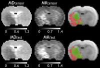
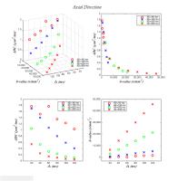
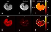
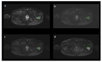
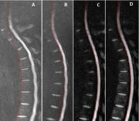
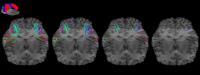
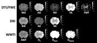
A rat model of graded spinal cord injury was used to evaluate several diffusion models for the ability to detect injury at acute and chronic time points.  Parameters from diffusion tensor imaging, free water estimation, diffusion kurtosis imaging, and white matter tract integrity models demonstrated that higher order modeling showed better separation of injury severity, especially in the chronic time point.  Furthermore, parameters sensitive to volume changes associated with edema and inflammation demonstrated the greatest separation of these injury groups, indicating the importance of these processes in altering diffusion characteristics in spinal cord injury.


3079.   
74 Intracellular metabolites exhibit non-Gaussian diffusion in the healthy human brain using magnetic resonance spectroscopy at 7 Tesla
Carson Ingo1, Wyger M. Brink1, Ece Ercan1, Andrew G. Webb1, and Itamar Ronen1
1C.J. Gorter Center for High Field MRI, Department of Radiology, Leiden University Medical Center, Leiden, Netherlands
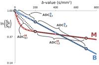
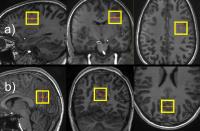
Since choline mostly resides in astrocytes, N-acetyl-aspartate mostly presides in axons, and creatine is distributed between both neural cell types, these intracellular metabolites can provide more specific microstructural compartment information compared to water. In this study, we apply diffusion-weighted spectroscopy to analyze axonal and glial structures by identifying non-Gaussian movement of intracellular metabolites in both white and gray matter of the healthy human brain at b-values up to ~17,000 s/mm2. We establish that all measured metabolites exhibited non-Gaussian subdiffusion in both tissue types with the gray matter intracellular space appearing more heterogeneous than white matter, opposite to water diffusion dynamics.


3080.   
75 
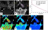
Relations between the stretched exponential DWI model and tumor malignancy related microstructural changes
Chu-Yu Lee1, Kevin M Bennett2, Josef P Debbins3, In-Young Choi1,4,5, and Phil Lee1,5
1Hoglund Brain Imaging Center, University of Kansas Medical Center, Kansas city, KS, United States, 2Department of Biology, University of Hawaii, Manoa, HI, United States, 3Neuroimaging research, Barrow Neurological Institute, Phoenix, AZ, United States, 4Department of Neurology, University of Kansas Medical Center, Kansas City, KS, United States, 5Department of Molecular & Integrative Physiology, University of Kansas Medical Center, Kansas City, KS, United States
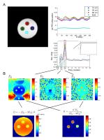
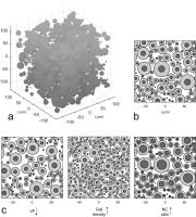
Diffusion weighting imaging (DWI) has been shown to be useful in differentiating low- and high-grade tumors in the brain. The decreased apparent diffusion coefficient (ADC) has been associated with increased tumor cellularity. However, tumor malignancy involves multiple microstructural changes that may also affect changes in the ADC. The alternative way to assess water diffusion in the complex microstructure is through the diffusion heterogeneity measured by the stretched exponential model (α-DWI). Recent studies using the α-DWI model have shown the increased diffusion heterogeneity in high-grade tumors. However, it remains unclear about the microstructural information provided by the α. The purpose of this study was to investigate how the α-DWI model responds to tumor malignancy related microstructural changes. We simulated a 3-D microenvironment in tumors and a DWI experiment. We studied how ADC and the fitted parameters of the α-DWI model responded to microstructural changes related to tumor malignancy.


3081.   
76 Validation and comparison of diffusion MR methods measuring transcytolemmal water exchange rate - Video Not Available
Xin Tian1,2, Hua Li1, Xiaoyu Jiang1, Jingping Xie1, John C Gore1, and Junzhong Xu1
1Radiology and Radiological Sciences, Vanderbilt University, Nashville, TN, United States, 2Radiology, The Second Hospital of Hebei Medical University, Shijiazhuang, China, People's Republic of
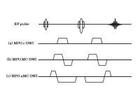
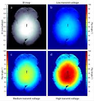
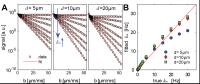
Two diffusion-based method, the CG (constant gradient) and FEXI (filtered exchange imaging) methods, have been developed to provide a flexible and safer means to measure transcytolemmal water exchange rate $$$k_{in}$$$ non-invasively in vivo. However, neither methods have been fully validated up to date. In the present work, computer simulations and in vitro experiments with well-controlled cultured cells with different sizes and permeabilities were performed to evaluate the accuracy of the CG and FEXI methods. The results suggest that $$$k_{in}$$$ can be accurately estimated when $$$k_{in}$$$ < 10 Hz. Although the FEXI method provides less accurate results, the linear dependence of AXR on $$$k_{in}$$$ suggesting it is still a reliable method.


3082.   
77 
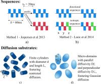
Metrics of microscopic anisotropy: a comparison study
Andrada Ianu?1, Noam Shemesh2, Daniel C. Alexander1, and Ivana Drobnjak1
1CMIC, University College London, London, United Kingdom, 2Champalimaud Neuroscience Programme, Champalimaud Centre for the Unknown, Lisbon, Portugal
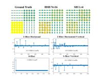
Microscopic anisotropy disentangles the effects of pore shape from orientation distribution, and thus can serve as a valuable metric for underlying microstructural configurations. Recent developments in diffusion MRI proposed different approaches to acquire and analyse data for extracting information regarding microscopic anisotropy. This work compares in simulation two recently introduced metrics of microscopic anisotropy: fractional eccentricity (FE), derived from double-diffusion-encoding (DDE) sequences and microscopic fractional anisotropy (μFA), derived from a combination of sequences with isotropic and directional diffusion weighting. We find that DDE-derived metrics are more reliable for quantifying underlying microstructures if diffusion is restricted, while μFA is closer to the ground truth values when individual micro-domains feature Gaussian diffusion. 


3083.   
78 
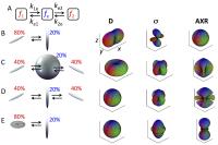
Apparent Exchange Rate in Multi-compartment Anisotropic Tissue - Permission Withheld
Samo Lasic1, Sune N. Jespersen2,3, Henrik Lundell4, Markus Nilsson5, Tim B. Dyrby4, and Daniel Topgaard6
1CR Development, AB, Lund, Sweden, 2CFIN/MINDLab, Department of Clinical Medicine, Aarhus University, Arhus, Denmark, 3Department of Physics and Astronomy, Aarhus University, Arhus, Denmark,4Danish Research Centre for Magnetic Resonance, Copenhagen University Hospital, Hvidovre, Copenhagen, Denmark, 5Lund University Bioimaging Center, Lund University, Lund, Sweden, 6Physical Chemistry, Lund University, Lund, Sweden
Filter exchange imaging (FEXI) is a noninvasive method to probe Apparent Exchange Rate (AXR). Understanding how diffusion anisotropy affects AXR is fundamental in experimental design and interpretation of results. In case of only two compartments, AXR is isotropic regardless of diffusion anisotropy. The key finding of this work is that AXR is anisotropic even in systems with a single exchange rate if there are more than two orientationally dispersed compartments. These findings may guide identification of different fiber populations and their directions and could be useful for analysis of fiber-specific characteristics.  


3084.   
79 
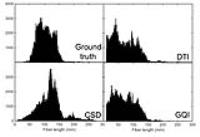
Simulating axon packing for investigating white matter tissue characteristics with diffusion MRI
Hamed Y. Mesri1, Dmitry S. Novikov2, Max A. Viergever1, and Alexander Leemans1
1Image Sciences Institute, University Medical Center Utrecht, Utrecht, Netherlands, 2Bernard and Irene Schwartz Center for Biomedical Imaging, Department of Radiology, New York University School of Medicine, New York, NY, United States
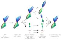
A novel algorithm for simulating axon packing in nerve bundles is proposed. Statistical analysis of the results demonstrates that, in contrast to conventional methods, the proposed method eliminates the bias in the estimated distribution and achieves higher packing densities, while preserving the random nature of the axon packing structure. The resultant tissue models can be used subsequently to study the Brownian motion of water molecules within nerve bundles. With our novel axon packing simulation framework, the effect of axon properties on the derived diffusion-weighted MR signal can be investigated more reliably now.


3085.   
80 Magnetic Resonance Diffusion Pore Imaging on Preclinical 9.4T-Animal-Scanner
Marco Bertleff1, Sebastian Domsch1, Frederik Laun2, Tristan Kuder2, and Lothar Schad1
1Computer Assisted Clinical Medicine, Heidelberg University, Medical Faculty Mannheim, Mannheim, Germany, 2Department of Medical Physics in Radiology, German Cancer Research Center (DKFZ), Heidelberg, Germany
The study of porous microstructures is of high interest in medical imaging. Diffusion pore imaging (DPI) has recently been proposed as a means to acquire images of the average cell shape in a voxel or region of interest. In this work, we present the feasibility of DPI phantom measurements on a preclinical 9.4T animal scanner for the first time and preliminarily compare two different sequence implementations. The shown feasibility on a preclinical system opens the possibility of a potential in-vivo measurement realization.


3086.   
81 Diffusion microstructure in the population: variability and effect size of biophysical compartment model parameters over 100 subjects
Robbert Harms1, Rainer Goebel1, and Alard Roebroeck1
1Maastricht University, Maastricht, Netherlands
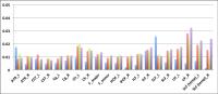
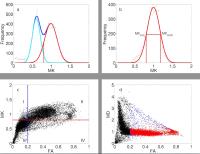
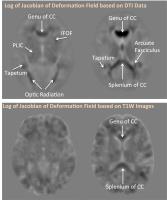
Statistical power in neuroscience studies is often limited, leading to, among others, low reproducibility of results[1]. Robust effect size estimates over a large subject group are crucial for power assessments. Here, we computed these estimates for microstructural differences in splenium, body and genu of the Corpus Callosum (CC) using diffusion MRI microstructure modeling over 100 subjects. We fitted Tensor, Ball&Stick, NODDI and CHARMED using GPU-accelerated software (MDT) and extracted subject specific parameters for the CC for each model. We observe medium to large effect sizes (Cohen’s d=1-3) for dMRI microstructure measures, promising for power and reproducibility of dMRI microstructure studies.


3087.   
 
82 Obtaining geometrical information from the time-dependent apparent diffusion coefficient
Simona Schiavi1, Houssem Haddar1, and Jing-Rebecca Li1
1CMAP, INRIA, Ecole Polytechnique, Palaiseau Cedex, France
Diffusion MRI (dMRI) has been established as a useful tool to obtain voxel-level information on the tissue micro-structure. An important quantity measured in dMRI is the apparent diffusion coefficient (ADC), and it has been well established by in-vivo brain imaging experiments that the ADC depends significantly on the diffusion time.  We derive an explicit formula for the time-dependent ADC, and, using the ADCs at multiple diffusion times and gradient directions, we estimate the surface to volume ratio, the eigenvalues and the first moment of the dominant eigen-functions associated to the geometry of the biological cells.


3088.   
83 Precise Inference of Cellular and Axonal Structural Organization (PICASO) using diffusion MRI
Lipeng Ning1,2, Carl-Fredrik Westin1,2, and Yogesh Rathi1,2
1Brigham and Women's Hospital, Boston, MA, United States, 2Harvard Medical School, Boston, MA, United States
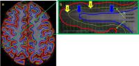
We propose a novel model termed PICASO for investigating the microstructural layout of brain tissue using in vivo diffusion MRI (dMRI) measurements. Our method provides a direct connection between the structural organization of biological tissue and a function representing the disorder in the evolution of magnetization density. This is achieved by extending the Bloch-Torrey equation to include variability in diffusivity due to restrictions and hindrances. Using in vivo data from the Humman Connectome Project (HCP), we show that the PICASO model can provide novel information about the microstructural layout of the axonal packing in human brain. Thus, our method can be applied in clinical settings to investigate brain abnormalities.


3089.   
84 Simulation study investigating the effect of diffusion, susceptibility, and vessel topology in characterizing normal and tumorous vasculature using R2*
Mohammed Salman Shazeeb1,2, Jayashree Kalpathy-Cramer1, and Bashar Issa2
1Athinoula A. Martinos Center for Biomedical Imaging, Massachusetts General Hospital and Harvard Medical School, Boston, MA, United States, 2Department of Physics, UAE University, Al-Ain, Abu Dhabi, United Arab Emirates
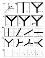
Brain vasculature is conventionally represented as straight cylinders when simulating BOLD contrast effects in fMRI. In reality, the vasculature is more complicated with branching and coiling especially in tumors. We applied a cylinder fork model to reflect the bifurcation, tortuosity, and size of vessels and performed simulations to study the effect of the rotation angle (?) on R2* at different bifurcation angles (β), vessel diameters, diffusion constants, and susceptibility values. This model clearly showed an R2* dependence on ?, which could potentially be used as a tool to differentiate between normal and tumor vessels.


3090.   
85 Anomalous diffusion parameters are sensible to microstructural variations in brain due to aging
Michele Guerreri1,2, Alessandra Caporale1,2, Marco Palombo1,3, Ivan De Berardinis1, Emiliano Macaluso4, Marco Bozzali4, and Silvia Capuani1
1Department of physics, CNR ISC UOS Roma Sapienza, Rome, Italy, 2Department of anatomical, histological, forensic and of the locomotor system science, Morphogenesis & Tissue Homeostasis, Sapienza University, Rome, Italy, 3MIRCen, CEA/DSV/I2BM, Fontenay-Aux Roses, France, 4Neuroimaging Laboratory, Santa Lucia Foundation, Rome, Italy
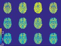
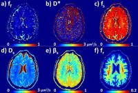
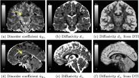
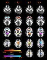
We investigated the anomalous diffusion (AD) stretched exponential γ-imaging model to overcome the sensitivity limitations of conventional DTI approach based on the assumption of the Gaussian model with regard of displacements of water molecules in tissues. The benefits of this approach are illustrated with an in-vivo diffusion study of the human brain performed on 18 healthy volunteers in the age range (23-70 years). Mean γ (Mγ) and anisotropic γ (Aγ) maps are obtained and compared with DTI maps. The current study suggests that Mγ and Aγ are more sensitive to micro-structural changes caused by normal aging, compared to DTI metrics. 


3091.   
86 Diffusion Microstructure Imaging With High-Performance Head-Only Gradient: Preliminary Results
Ek T Tan1, Jonathan I Sperl2, Miguel Molina Romero2,3, Seung-Kyun Lee1, Matt A Bernstein4, and Thomas KF Foo1
1GE Global Research, Niskayuna, NY, United States, 2GE Global Research, Munich, Germany, 3Technical University of Munich, Munich, Germany, 4Mayo Clinic, Rochester, MN, United States
A high-performance head-only gradient coil (Gmax=80 mT/m, SR=700 T/m/s) allows diffusion imaging at substantially shorter echo-time and echo-spacing than conventional whole-body gradient coil systems. This greatly benefits microstructure imaging with diffusion EPI, providing reduced echo spacing by up-to two-fold and shorter TE by up-to 30%. Imaging results demonstrate reduced distortion and improved white matter SNR. Preliminary results on axonal radius mapping with high b-value imaging (up-to b=12,000 s/mm2) demonstrate the feasibility of 2 mm-isotropic imaging with the head-gradient.


3092.   
 
87 Quantifying White Matter Microstructure with a Unified Spatio-Temporal Diffusion Weighted MRI Continuous Representation
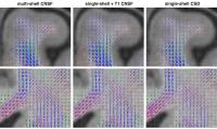
Demian Wassermann1, Alexandra Petiet2, Rutger Fick1, Mathieu Santin2, Anne-Charlotte Philippe 2, Stephane Lehericy2, and Rachid Deriche1
1Athena, Inria, Sophia-Antipolis, France, 2CENIR, Brain and Spine Institute, Paris, France
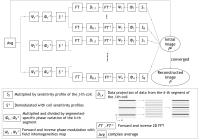
A current problem Diffusion MRI (dMRI) based microscopy faces under the narrow pulse approximation is how to best exploit the 4D (q-space + diffusion time) nature of the signal. Assaf et al. showed that exploring the dMRI attenuation at different diffusion times provides information on the apparent distribution of axonal diameters within a voxel in their seminal work: AxCaliber1. However, AxCaliber requires knowing beforehand the predominant orientation of the axons within the analyzed volume to adjust the q-space sampling accordingly. In this work, we show that our novel sparse representation of the 3D+t dMRI signal2 enables the recovery of axonal diameter distribution parameters with two main advantages. First,  it doesn't require knowledge of the predominant axonal direction at acquisition time. Second, using the hypothesised dMRI signal symmetry, it allows computing the average attenuation on the plane perpendicular to the predominant axonal direction analytically. Hence, it takes advantage of the full 3D+t signal information to fit the AxCaliber model.


3093.   
88 Cytoarchitectonic abnormalities along white matter pathways in temporal lobe epilepsy: Combining diffusional kurtosis imaging and automated fiber quantification - Permission Withheld
Russell Glenn1, Jens H Jensen1, Simon S Keller2, Joseph A Helpern1, and Leonardo Bonilha3
1Medical University of South Carolina, Charleston, SC, United States, 2University of Liverpool, Liverpool, United Kingdom, 3Charleston, SC, United States
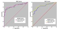
Temporal lobe epilepsy (TLE) is the most common form of medically refractory epilepsy and is associated with focal brain abnormalities causing recurrent, unprovoked seizures originating from the temporal lobe. However, cytoarchitectronic changes can be detected outside of the temporal lobe and may be associated with the clinical course of the disease. We implement a novel neuroimaging approach which combines the strengths of diffusional kurtosis imaging and automated fiber quantification for the non-invasive characterization of white matter pathways and demonstrate its sensitivity to detect pathological alterations associated with TLE. The proposed technique may provide further insights into the clinicopathology of TLE.


3094.   
89 
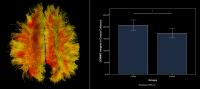
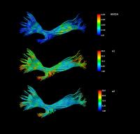
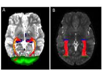
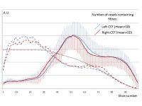
Short-term mindfulness-based stress reduction training increases tract integrity in right auditory radiation and anterior and posterior commissures
Chang-Le Chen1, Yao-Chia Shih2, Tzung-Kuen Wen3, Shih-Chin Fang4, Da-Lun Tang5, Si-Chen Lee6, and Wen-Yih Isaac Tseng1,7,8
1Graduate Institute of Brain and Mind Sciences, National Taiwan University College of Medicine, Taipei, Taiwan, 2Institute of Biomedical Engineering, National Taiwan University, Taipei, Taiwan,3Department of Buddhist Studies, Dharma Drum Institute of Liberal Arts, New Taipei City, Taiwan, 4Department of Neurology, Cardinal Tien Hospital Yonghe Branch, New Taipei City, Taiwan, 5Department of Mass Communication, Tamkang University, Taipei, Taiwan, 6Department of Electrical Engineering, National Taiwan University, Taipei, Taiwan, 7Institute of Medical Device and Image, National Taiwan University College of Medicine, Taipei, Taiwan, 8Molecular Imaging Center, National Taiwan University, Taipei, Taiwan
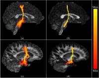
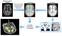
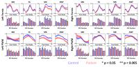
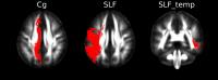
Mindfulness-based stress reduction (MBSR) is an 8-week mindfulness meditation training which exerts beneficial effects on physical and mental health. Many researches showed that the changes in brain structure were related to mindfulness meditation. However, few studies have investigated the relationships between short-term mindfulness meditation and altered white matter tracts. Therefore, a longitudinal study was designed in this study to identify the effects of 8-week MBSR program on white matter tract integrity. We found that there was significant difference in three white matter tracts, right auditory radiation, anterior commissure and posterior commissure, in the novice practitioners.


3095.   
90 A physically-constrained model for diffusion kurtosis imaging
Darryl McClymont1, Irvin Teh1, Hannah Whittington1, Vicente Grau2, and Jurgen Schneider1
1Division of Cardiovascular Medicine, University of Oxford, Oxford, United Kingdom, 2Department of Engineering Science, University of Oxford, Oxford, United Kingdom
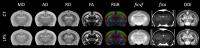
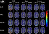
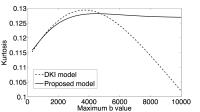
Diffusion kurtosis imaging provides higher-order information about diffusion. However, the quadratic term in the diffusion kurtosis model produces undesirable behaviour at high b-values as a result of the negative tails of the diffusivity distribution. A truncated normal distribution has been proposed to address this in one dimension. This work extends this concept to a multivariate truncated normal distribution, and extends the range of b-values over which kurtosis can be estimated. The proposed model is fit to diffusion data from rat hearts, and yields kurtosis values that are consistent with the DKI model.


3096.   
91 Intravoxel Incoherent Motion in Normal Pituitary Gland: Initial Study with Turbo Spin-echo Diffusion-weighted Imaging - Permission Withheld
Kiyohisa Kamimura1, Masanori Nakajo1, Yoshihiko Fukukura1, Takashi Iwanaga2, Tomonori Saito2, Masashi Sasaki2, Takuro Fujisaki2, Atsushi Takemura3, Tomoyuki Okuaki 3, and Takashi Yoshiura1
1Radiology, Kagoshima University Medical and Dental Hospital, Kagoshima, Japan, 2Clinical Engineering Department Radiation Section, Kagoshima University Hospital, Kagoshima, Japan, 3Philips Electronics Japan, Tokyo, Japan
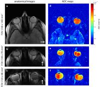
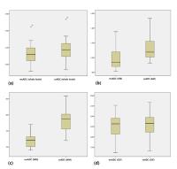
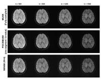
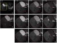
Our purpose was to evaluate the feasibility of intravoxel incoherent motion (IVIM) assessment based on turbo spin-echo diffusion-weighted imaging (TSE-DWI) in the normal pituitary gland. In a validation study using normal brain white matter (WM), Bland-Altman analyses revealed fair to good agreement with conventional echo-planar-based DWI (EP-DWI) in the true diffusion coefficient (D) and perfusion fraction (f). In 7 volunteers, both D and f in the anterior pituitary lobe were significantly higher than those in WM, being consistent with high microvascular density in the pituitary gland. Results demonstrated that IVIM assessment based on TSE-DWI in the pituitary gland is feasible.


3097.   
92 White Matter Asymmetry During Development Using Diffusion Kurtosis Imaging - Permission Withheld
Xiang Gao1, Farida Grinberg1,2, Ezequiel Farrher1, Fei Li1, Eileen Oberwelland3,4, Irene Neuner1,5,6, Kerstin Konrad4,6,7, and N.Jon. Shah1,2,6
1Institute of Neuroscience and Medicine - 4, Forschungszentrum Juelich GmbH, Juelich, Germany, 2Department of Neurology, Faculty of Medicine, RWTH Aachen University, Aachen, Germany,3Translational Brain Research in Psychiatry and Neurology, Department of Child and Adolescent Psychiatry, Psychosomatics and Psychotherapy, RWTH Aachen University, Aachen, Germany, 4Institute of Neuroscience and Medicine - 3, Forschungszentrum Juelich GmbH, Juelich, Germany, 5Department of Psychiatry, Psychotherapy and Psychosomatics, RWTH Aachen University, Aachen, Germany, 6JARA - BRAIN, Translational Medicine, Juelich, Germany, 7Child Neuropsychology Section, Department of Child and Adolescent Psychiatry, Psychosomatics and Psychotherapy, RWTH Aachen University, Aachen, Germany
We compare changes in the white matter asymmetry index in conventional fractional anisotropy (FA) and other diffusion kurtosis imaging (DKI) metrics in adults and children. For some fibres, such as cingulate gyrus, hippocampus and superior longitudinal fasciculus, other DKI parameters show significant asymmetry where FA fails. When compared to adults, children showed more laterality in cingulate gyrus, superior longitudinal fasciculus and superior longitudinal fasciculus in temporal parts, which indicate that the degree of asymmetry in these fibres is higher during childhood.


3098.   
93 
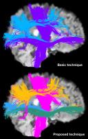
Anisotropy measure from High angular resolution diffusion imaging Data Using Higher Order Diffusion Tensor model
Getaneh Bayu Tefera1 and Ponnada A. Narayana1
1Diagnostic & Interventional Imaging, University of Texas at Houston, Houston, TX, United States
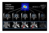
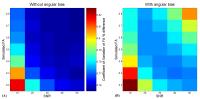
Different anisotropy indices such as generalized anisotropy (GA) and generalized fractional anisotropy (GFA) for HARDI data have been reported, but they have their own limitations. Here we propose a new anisotropy measure (HFA) for the HARDI data that is rotationally invariant.  The new proposed measure is compared with GA and GFA using the contrast-to-noise ratio and coefficient of variation as the metrics for three white matter regions. HFA and GFA have shown better CNR than FA and GA in two and three crossing regions. The results described above were very similar across all the five subjects.


3099.   
94 Tract Orientation and Angular Dispersion Deviation Indicator (TOADDI): A framework for single-subject analysis in diffusion tensor imaging
Cheng G. Koay1,2, Ping-Hong Yeh2,3, John M. Ollinger2, M. Okan Irfanoglu1,3, Carlo Pierpaoli1, Peter J. Basser1, Terrence R. Oakes2, and Gerard Riedy2
1Eunice Kennedy Shriver National Institute of Child Health and Human Development, National Institutes of Health, Bethesda, MD, United States, 2National Intrepid Center of Excellence, Walter Reed National Military Medical Center, Bethesda, MD, United States, 3The Henry M. Jackson Foundation for the Advancement of Military Medicine, Bethesda, MD, United States
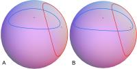
The purpose of the proposed framework is to carry out single-subject analysis of diffusion tensor imaging (DTI) data. This framework is termed Tract Orientation and Angular Dispersion DeviationIndicator (TOADDI). It is capable of testing whether an individual tract as represented by the major eigenvector of the diffusion tensor and its corresponding angular dispersion are significantly different from a group of tracts on a voxel-by-voxel basis. This work develops two complementary statistical tests (orientation and shape tests) based on the elliptical cone of uncertainty, which is a model of uncertainty or dispersion of the major eigenvector of the diffusion tensor.


3100.   
95 Assessment of brain structural abnormalities and the correlation with inhibitory control in betel nut chewers with DTI
Te-Wei Kao1, Ming-Chou Ho2, and Jun-Cheng Weng1,3
1Department of Medical Imaging and Radiological Sciences, Chung Shan Medical University, Taichung, Taiwan, 2Department of Psychology, Chung Shan Medical University, Taichung, Taiwan,3Department of Medical Imaging, Chung Shan Medical University Hospital, Taichung, Taiwan
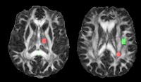
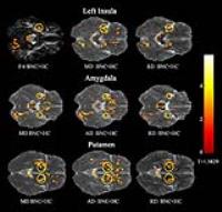
Betel nut is one of the common addictive substances in many countries. The brain influence of cocaine, alcohol, and tobacco cigarette have been studied by several studies. However, only few studies focused on the brain influence of betel nut, and most of them used fMRI or PET. Thus, our study aim was to use diffusion tensor imaging (DTI) to evaluate the impact of neurological structure of white matter caused by betel nut. The brain structural differences between the betel nut chewers and healthy controls and the correlation with inhibitory control were also discussed. Our results pointed out the significant neurological structural differences in the insula, amygdala and putamen of DTI indices between the betel nut chewers and healthy controls.


3101.   
96 Assessment of pharmacotherapy effects on APP/PS1 mice brain by Diffusion Spectrum Imaging
Chih-Hsien Tseng1,2, Yu-Jen Chen1, and Wen-Yih Isaac Tseng1,2,3,4
1Institute of Medical Device and Imaging, National Taiwan University College of Medicine, Taipei, Taiwan, 2Institute of Biomedical Engineering, National Taiwan University College of Medicine, Taipei, Taiwan, 3Graduate Institute of Brain and Mind Sciences, National Taiwan University College of Medicine, Taipei, Taiwan, 4Molecular Imaging Center, National Taiwan University, Taipei, Taiwan
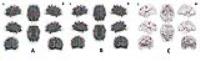
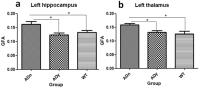
To determine the effects of a novel neuroprotective drug on white matter integrity in Alzheimer’s disease (AD), generalized fractional anisotropy (GFA) was assessed in mice brains using diffusion spectrum imaging (DSI). The mice included 5 AD mice without medication, 4 AD mice with medication, and 5 control mice. Comparing with the control mice, the AD mice without medication showed significantly increased GFA in the hippocampus and thalamus, whereas the AD mice with medication showed no significant difference. Our findings imply that DSI can be used to monitor the drug effects in AD mice.
 

The International Society for Magnetic Resonance in Medicine is accredited by the Accreditation Council for
Continuing Medical Education to provide continuing medical education for physicians.