|
Exhibition Hall 17:30 - 18:30 |
|
|
|
Computer # |
|
3123.
 |
25 |
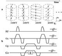
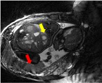
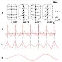
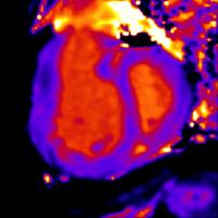
Short-breath hold cine DENSE 
Andrew David Scott1,2, Upasana Tayal1,2,
Sonia Nielles-Vallespin1,3, Pedro Ferreira1,2,
Xiaodong Zhong4, Frederick Epstein5,
Sanjay Prasad1,2, and David Firmin1,2
1NIHR funded Cardiovascular Biomedical Research
Unit, Royal Brompton Hospital, London, United Kingdom, 2National
Heart and Lung Institute, Imperial College London, London,
United Kingdom,3National Heart Lung and Blood
Institute, National Institutes of Health, Bethesda, MD,
United States, 4MR
R&D Collaborations, Siemens Healthcare, Atlanta, GA, United
States, 5University
of Virginia, Department of Biomedical Engineering,
Charlottesville, VA, United States
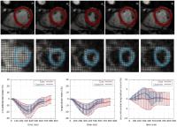
Displacement encoding with stimulated echoes (DENSE) can
provide valuable strain information, but acquisitions are
typically too long for patient cohorts who have difficulty
breath holding. In this work we accelerate 2D cine spiral
DENSE acquisitions by selectively exciting a small field of
view around the heart. We compare strain data derived from
DENSE acquired with unaccelerated and up to 2.5x
acceleration in a cohort of healthy subjects and show
minimal differences when the acquisition is accelerated. We
also show an example from a patient with a myocardial
infarction where the accelerated DENSE data shows abnormal
strain in the infarcted regions.
|
|
3124.
 |
26 |
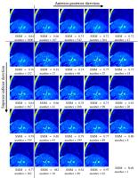
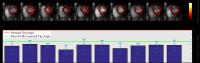
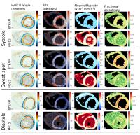
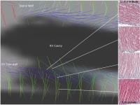
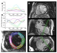
Biventricular Cardiac Mechanics in Healthy Subjects using 3D
Spiral Cine DENSE and Mesh-Free Strain Analysis
Jonathan D Suever1,2, Gregory J Wehner3,
Christopher M Haggerty1,2, Linyuan Jing1,2,
David K Powell3, Sean M Hamlet4,
Jonathan D Grabau2, Dimitri Mojsejenko2,
and Brandon K Fornwalt1,2,3
1Institute for Advanced Application, Geisinger
Health System, Danville, PA, United States, 2Pediatrics,
University of Kentucky, Lexington, KY, United States, 3Biomedical
Engineering, University of Kentucky, Lexington, KY, United
States, 4Electrical
Engineering, University of Kentucky, Lexington, KY, United
States
Cardiac mechanics have been extensively characterized in the
left ventricle (LV). However, the right ventricle (RV) is
rarely studied due to both acquisition and post-processing
challenges. In this study, we combined 3D
displacement-encoded (DENSE) imaging with custom
post-processing that utilizes a local coordinate system to
extract advanced measures of cardiac mechanics in an effort
to characterize healthy biventricular function. We found
that torsion as well as circumferential and longitudinal
strain vary throughout the RV, but globally were comparable
to their LV counterparts. This data can be used to better
understand how biventricular function is disrupted by
disease.
|
|
3125.
 |
27 |
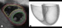
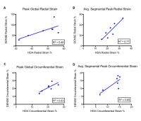
Myocardial Strain Analysis With CMR in Breast Cancer Patients
with Iatrogenic Cardiotoxicity Using Heart Deformation Analysis:
Comparison to DENSE
Abraham Bogachkov1, Kai Lin2,
Ahmadreza Ghasemiesfe2, Amir Ali Rahsepar3,
Bruce Spottiswoode3, Ben Freed4,
Michael Markl2, James Carr2, and
Jeremy Collins2
1Northwestern University Feinberg School of
Medicine, Chicago, IL, United States, 2Radiology,
Northwestern University, Chicago, IL, United States, 3Cardiovascular
MR R&D, Siemens Healthcare, Chicago, IL, United States, 4Cardiology,
Northwestern University, Chicago, IL, United States
Strain imaging at cardiac MR has been shown to be a powerful
tool in the pre-clinical detection of early cardiac
dysfunction in the heart failure population, but has been
only minimally studied in cardiotoxicity patients. This
study evaluated a semi-automatic heart deformation analysis
(HDA) tool in the assessment of left ventricular myocardial
strain in patients with known cardiotoxicity, and found very
good to excellent agreement with global strain values
calculated using displacement encoding with stimulated
echoes (DENSE). HDA analysis of conventional cine sequences
has the potential to play a significant role in the
evaluation of patients at risk for cardiotoxicity.
|
|
3126.
 |
28 |
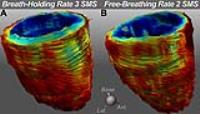
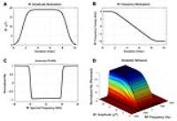
Free-breathing 2D cine DENSE MRI using localized signal
generation, image-based navigators, motion compensation and
compressed sensing
Xiaoying Cai1, Xiao Chen2, Yang Yang1,
Michael Salerno3, Daniel S. Weller4,
Craig H. Meyer1, and Frederick H. Epstein1
1Biomedical Engineering, University of Virginia,
Charlottesville, VA, United States, 2Medical
Imaging Technologies, Siemens Healthcare, Princeton, NJ,
United States, 3University
of Virginia, Charlottesville, VA, United States, 4Electrical
and Computer Engineering, University of Virginia,
Charlottesville, VA, United States
Current cine DENSE protocols require breath-holding, which
limits the use of this technique to patients with good
breath-holding capabilities and excludes many pediatric and
heart failure patients. To accomplish free-breathing scans
with high efficiency and quality, we developed a 2D cine
DENSE acquisition and reconstruction framework that utilizes
localized signal generation, image-based self-navigated
motion estimation, k-space motion correction and compressed
sensing. Reconstructions and Bland-Altman analysis from 5
volunteers demonstrated that the proposed method recovered
high-quality images and strain data from free-breathing
data, showing better agreement than conventional
reconstructions of the same data with breath-holding scans.
|
|
3127.
 |
29 |
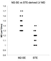
Effect of Respiratory Suspension on the Computation of Left
Ventricular (LV) Volume and Rate of Volume Change (dV/dt)-based
Diastolic Indices with Echocardiography as a Reference
Amol Pednekar1, Jiming Zhang2, Debra
Dees3, Benjamin Y Cheong3, and Raja
Muthupillai3
1Phillips Healthcare, Cleveland, OH, United
States, 2Diagnostic
and interventional Radiology, CHI St Luke's Health, Houston,
TX, United States, 3Diagnostic
and Interventional Radiology, CHI St Luke's Health, Houston,
TX, United States
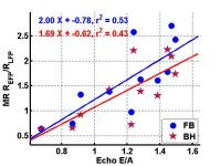
Diastolic functional indices based on trans-mitral blood
flow velocities are pre-load dependent and early diastolic
filling can be diminished by activities such as inspiration
or Valsalva maneuver. Cardiac cine MR images are typically
acquired during suspended respiration and thus could induce
systemic bias. In this study, we evaluate the impact of
respiratory suspension on the computation of volume-based
diastolic indices using peak velocity-based Doppler echo
measurements as the reference. The volume based diastolic
indices derived from high temporal resolution cine MR
correlated well with velocity based E/A ratio from echo
while indicating the direct impact of respiratory
suspension.
|
|
3128.
 |
30 |
Peak Filling Rates assessed by Cardiac Magnetic Resonance
Imaging indicate Diastolic Dysfunction from Myocardial Iron
Toxicity
Jin Yamamura1, Sarah Keller1, Roland
Fischer2,3, Regine Grosse4, Gregory
Kurio3, Gunnar Lund1, Joachim
Graessner5, Gerhard Adam1, and Bjoern
Schoennagel1
1Diagnostic and Interventional Radiology,
University Medical Center Hamburg-Eppendorf, Hamburg,
Germany, 2Biochemistry,
University Medical Center Hamburg-Eppendorf, Hamburg,
Germany,3Department of Radiology, UCSF Benioff
Children's Hospital Oakland, Oakland, CA, United States, 4Department
of Pediatric Hematology/Oncology, University Medical Center
Hamburg-Eppendorf, Hamburg, Germany, 5Siemens
Healthcare, Hamburg, Germany
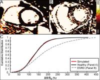
The diastolic peak filling rate ratio (PFRR) is a sensitive
marker to indicate diastolic dysfunction from myocardial
iron toxicity in patients with systemic iron overload
disease. Precise assessment of the PFRR by CMR requires a
volumetric approach with exclusion of trabeculae and
papillary muscles from the LV cavity. The PFRR assessed by
CMR may be a valuable parameter for the screening and
monitoring of myocardial iron toxicity due to iron
deposition in patients with preserved systolic function.
|
|
3129.
 |
31 |
Exercise stress cardiac MR assessment of diastolic function in
healthy volunteers and pulmonary hypertension
Thomas Kennedy1, Omid Forouzan2,
Oliver Wieben1,3, Naomi C Chesler2,
Jacob Macdonald3, and Christopher J Francois1
1Radiology, University of Wisconsin- Madison,
Madison, WI, United States, 2Biomedical
Engineering, University of Wisconsin- Madison, Madison, WI,
United States, 3Medical
Physics, University of Wisconsin- Madison, Madison, WI,
United States
Dyspnea on exertion is a common manifestation of systolic
and diastolic heart failure. Using an MRI-compatible
exercise device allowing subjects to exercise while in the
bore of the scanner, we assessed exercise-induced changes in
diastolic transmitral flow in younger and older healthy
volunteers and subjects with pulmonary hypertension. The
measurements we obtained demonstrated decreased E/A ratios
for older healthy volunteers and PH subjects when compared
to younger healthy volunteers, however these differences
were not statistically significant.
|
|
3130.
 |
32 |
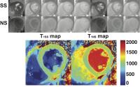
Quantitative, Time-efficient, Heart-Rate Independent Myocardial
BOLD MRI with Whole-heart Coverage at 3T in a Canine Model of
Coronary Stenosis with Simultaneous 13N-Ammonia PET Validation
Hsin-Jung Yang1, Damini Dey1, Jane
Sykes2, John Butler2, Xiaoming Bi3,
Behzad Sharif1, Sotirios Tsaftaris4,
Debiao Li1, Piotr Slomka1, Frank Prato2,
and Rohan Dharmakumar1
1Cedars Sinai Medical Center, Los Angeles, CA,
United States, 2Lawson
Health Research Institute, london, ON, Canada, 3Siemens
Healthcare, Los Angeles, CA, United States, 4IMT
Institute for Advanced Studies Lucca, Lucca, Italy
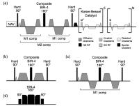
Current myocardial BOLD MR methods are limited by: (a) poor
spatial coverage and imaging speed; (b) imaging confounders;
and (c) imaging artifacts, particularly at 3T. To address
these limitations, we developed a heart-rate independent,
free-breathing 3D T2 mapping technique at 3T that utilizes
near 100% imaging efficiency, which can be completed in 3
minutes with full LV coverage. We tested our method in a
canine model of coronary stenosis and validated our findings
with simultaneously acquired13N-ammonia PET perfusion data
in a whole-body PET/MR system.
|
|
3131.
 |
33 |
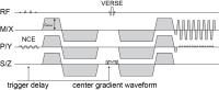
Spiral SPIRIT Tissue Phase Mapping enables the acquisition of
myocardial motion with high temporal and spatial resolution
during breath-hold
Marius Menza1, Daniela Föll2, Jürgen
Hennig1, and Bernd Jung3
1University Medical Center Freiburg, Dept. of
Radiology - Medical Physics, Freiburg, Germany, 2University-Heart
Center Freiburg, Cardiology und Angiology I, Freiburg,
Germany, 3University
Hospital Bern, Institute of Diagnostic, Interventional and
Pediatric Radiology, Bern, Switzerland
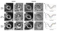
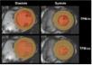
MR Tissue Phase Mapping (TPM) is a powerful approach to
assess left ventricular (LV) function. Conventional
Cartesian acquisition-strategies with k-t-based parallel
imaging acceleration allow the acquisition of a single slice
within a breath-hold, but suffer from low spatial
resolution. In this work a comparison with undersampled
high-resolution spiral SPIRIT TPM for different trajectory
designs within one breath-hold and free breathing Cartesian
k-t-accelerated PEAK TPM is presented. High image quality,
comparable peak velocity values and time to peaks of spiral
SPIRIT TPM for high resolution within a breath-hold might
enhance myocardial functional analysis.
|
|
3132.
 |
34 |
Comparison Between Radial and Cartesian Sampling Patterns in
Accelerated Real-Time Cardiac Cine MRI
Elwin Bassett1, Ganesh Adluru2, Brent
D. Wilson3, Cory Nitzel3, Tobias Block4,
Hassan Haji-Valizadeh5, Edward VR DiBella2,
and Daniel Kim2
1Physics, University of Utah, Salt Lake City, UT,
United States, 2Radiology,
UCAIR, University of Utah, Salt Lake City, UT, United
States, 3Internal
Medicine, Division of Cardiology, University of Utah, Salt
Lake City, UT, United States, 4School
of Medicine, Radiology, New York University, New York, NY,
United States, 5Bioengineering,
University of Utah, Salt Lake City, UT, United States
To date, no study has compared 12-fold accelerated real-time
cine MRI with compressed sensing (CS) between Cartesian and
radial k-space sampling schemes. We sought to compare their
performance in patients and volunteers. We compared point
spread functions (PSF) to determine which sampling pattern
generates more incoherent aliasing artifacts. We also
compared their performance in a group of 15 patients and one
volunteer, where 3-fold accelerated product real-time MRI
was used as reference. Two cardiologists independently
graded images from each subject. PSF analysis showed that
radial produces more incoherent aliasing artifacts. Image
quality was better for radial than Cartesian sampling
schemes.
|
|
3133.
 |
35 |
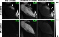
Wideband Cardiac MR Perfusion Pulse Sequence for Imaging
Patients with implantable cardioverter defibrillator
KyungPyo Hong1,2 and
Daniel Kim1
1Radiology, UCAIR, University of Utah, Salt Lake
City, UT, United States, 2Bioengineering,
University of Utah, Salt Lake City, UT, United States
Patients with end-stage heart failure (HF) often require
advanced therapeutics, but current clinical profiles and
biomarkers are not adequate for predicting outcomes.
Myocardial perfusion reserve may be an important predictor,
but it is technically challenging to perform perfusion MRI
in patients with end-stage HF because they often have
implantable cardioverter defibrillator (ICD), which
generates significant image artifacts. We developed a novel
cardiac perfusion pulse sequence using a wideband saturation
pulse. Compared with standard perfusion MRI, wideband
perfusion MRI suppresses image artifacts induced by ICD.
|
|
3134.
 |
36 |
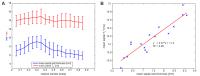
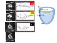
Feature tracking imaging (FTI) for right ventricular strain
assessment in patients with chronic thromboembolic pulmonary
hypertension (CTEPH)
Yoshiaki Morita1, Naoaki Yamada1,
Makoto Amaki2, Emi Tateishi2, Asuka
Yamamoto2, Masahiro Higashi1, and
Hiroaki Naito1
1Department of Radiology, National Cerebral and
Cardiovascular Center, Suita, Osaka, Japan, 2Division
of Cardiology, National Cerebral and Cardiovascular Center,
Suita, Osaka, Japan
Right ventricular (RV) function has a significant impact on
the prognosis of chronic thromboembolic pulmonary
hypertension (CTEPH), as it does with other forms of
pulmonary arterial hypertension (PH). In this study, we
demonstrated that feature tracking imaging (FTI) is fast,
simple, and has potential for clinical use for assessing RV
strain in CTEPH. The global longitudinal strain (GLS) showed
better correlation with the RV ejection fraction (RVEF) and
mean pulmonary artery pressure (mPAP). FTI-derived strain
measurement might offer a modality for good detection of RV
dysfunction and repeatable monitoring after therapeutic
intervention.
|
|
3135.
 |
37 |
Cardiac MR Assessment of Diastolic Function
Thomas Kennedy1, Niti Aggarwal2,
Christopher Francois1, Mark Schiebler1,
and Jeremy Collins3
1Radiology, University of Wisconsin- Madison,
Madison, WI, United States, 2Cardiology,
Univesity of Wisconsin-Madison, Madison, WI, United States, 3Radiology,
Northwestern University, Chicago, IL, United States
Diastolic dysfunction is the primary cause of CHF in 40-60%
of patients with heart failure in the United States and has
been shown to lead to poor outcomes. Early diagnosis and
treatment of the causes of diastolic dysfunction is
effective in relieving symptoms and reducing mortality. The
non- invasive methods which can be used to assess diastolic
function include cardiac magnetic resonance (CMR) imaging.
The purpose of this educational poster is to describe the
CMR techniques which can be used to evaluate diastolic
function and review the CMR findings of this disorder.
|
|
3136.
 |
38 |
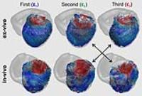
Finite Element Digital Image Correlation for Cardiac Strain
Analysis from 3D Whole-Heart Tagging
Martin Genet1,2, Christian T Stoeck3,4,
Constantin von Deuster3,4, Lik Chuan Lee5,
Julius M Guccione6, and Sebastian Kozerke3,4
1École Polytechnique, Palaiseau, France, 2Institute
for Biomedical Engineering, ETHZ, Zurich, Switzerland, 3KCL,
London, United Kingdom, 4ETHZ,
Zurich, Switzerland, 5MSU,
East Lansing, MI, United States, 6UCSF,
San Francisco, CA, United States
The objective of the present work was to develop, validate
and analyze a finite element digital image correlation
approach of extracting ventricular strain data from MR
images, which can be applied to both 3D CSPAMM images and
conventional multi-slice cine images. Cine and 3D CSPAMM
data was acquired on a normal human volunteer, and analyzed.
The proposed method provided similar circumferential strain
data compared to already validated SinMod method. In
contrast to strain mapping from cine images, strain mapping
from 3D CSPAMM images captures ventricular twist and torsion
in agreement with physiological values, and is less
sensitive to image misregistration.
|
|
3137.
 |
39 |
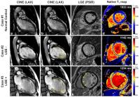
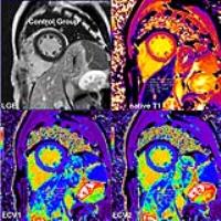
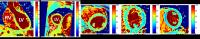
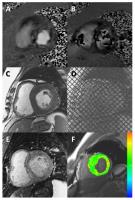
A data analysis framework to study remodeling after myocardial
infarction
Freddy Odille1,2,3, Lin Zhang1,2,
Bailiang Chen1,2,3, Jacques Felblinger1,2,3,4,5,
Damien Mandry1,2,4, and Marine Beaumont3,5
1U947, Inserm, Nancy, France, 2IADI,
Université de Lorraine, Nancy, France, 3CIC-IT
1433, Inserm, Nancy, France, 4Pôle
imagerie, CHRU de Nancy, Nancy, France, 5Pôle
S2R, CHRU de Nancy, Nancy, France
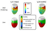
A data analysis framework is proposed to study the relation
between scar severity and regional myocardial function
during the process of remodeling after acute myocardial
infarction (MI). The framework includes registration steps
to correct for slice-to-slice inconsistencies, to align cine
with late gadolinium enhancement (LGE) data and to align
data from follow-up scans. The framework was evaluated in
114 patients with CMR scans within 3 days after MI and at 6
months. Registration accuracy was below 3 mm. Results show
that function at 6 months was inversely associated with scar
transmuraltiy at both 3 days and 6 months.
|
|
3138.
 |
40 |
High-resolution MR Imaging of Left-ventricular Function in
Newborn Mice
Mahon L Maguire1, Mala Rohling2, Megan
Masters2, Debra McAndrew1, Paul Riley2,
and Jurgen E Schneider1
1Radcliffe Department of Medicine, University of
Oxford, Oxford, United Kingdom, 2Department
of Physiology, Anatomy, and Genetics, University of Oxford,
Oxford, United Kingdom
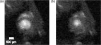
The neonatal mouse heart has been reported to regenerate
following myocardial injury during the first days of life.
Research into this regenerative capability is being actively
pursued for translation into the clinic. This study
presents cardiac cine imaging of one-day old mice using
retrospectively gated, accelerated MR imaging with
recovery. Images were acquired with 78x78x500 μm
resolution. Left ventricular functional parameters were
derived from the images and are presented. This proof of
concept study demonstrates that cardiac functional MR
imaging with recovery in newborn mice is practical. It also
allows the investigation of myocardial regeneration during
the first days of life.
|
|
3139.
 |
41 |
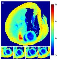
Fast myocardial perfusion mapping in mice using heart cycle
dependent data weighting
Fabian Tobias Gutjahr1, Thomas Kampf1,
Stephan Michael Guenster1, Volker Herold1,
Patrick Winter1, Xavier Helluy2,
Wolfgang Bauer3, and Peter Jakob1
1Experimental Physics V, University of Wuerzburg,
Wuerzburg, Germany, 2NeuroImaging
Centre, Ruhr University, Bochum, Germany, 3Department
of Internal Medicine 1, Universitaetsklinikum Würzburg,
Wuerzburg, Germany
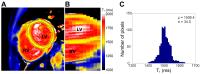
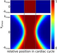
A fast method for the measurement of myocardial perfusion in
mice is presented. Using an efficient retrospective data
selection and weighting process in combination with a model
based reconstruction perfusion maps can be acquired within
3.5min.
|
|
3140.
 |
42 |
A preliminary study of Intravoxel Incoherent Motion MR for
quantitative evaluation of myocardial perfusion in diabetes
and/or hypertension
Anna Mou1, Zhiyong Li1, Mengying Li2,
Qingwei Song2, Chen Zhang2, and Ailian
Liu2
1Radiology, The First Affiliated Hospital of
Dalian Medical University, Dalian, China, People's Republic
of, 2Dalian,
China, People's Republic of
Myocardial microcirculation perfusion dysfunction plays an
important role in assessment cardiac disease especially
diabetes and hypertension because of their high incidence in
the world. We preliminarily investigated the difference of
myocardial micro-vascular perfusion between patients with
diabetes/hypertension and normal volunteers with IVIM (0,
20, 50, 80, 120, 150, 200, 300, 500 s/mm2) diffusion
weighted imaging. We found that Fast ADC values
in patients were significant lower than in healthy
volunteers. We concluded that IVIM CMR could quantitatively
and noninvasively evaluate perfusion status in patients with
diabetes and/or hypertension.
|
|
3141.
 |
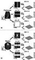
43 |
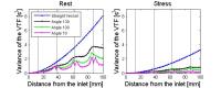
A Computational Fluid Dynamics Simulation Study on the Influence
of the Tortuosity of the Coronary Arteries on Contrast Agent
Bolus Dispersion in Contrast-Enhanced Myocardial Perfusion MRI
Regine Schmidt1, Hanns-Christian Breit1,
and Laura Maria Schreiber1,2
1Section of Medical Physics, Department of
Radiology, Johannes Gutenberg University Medical Center,
Mainz, Germany, 2Department
of Cellular and Molecular Imaging, Comprehensive Heart
Failure Center (CHFC), Wuerzburg, Germany
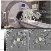
The dispersion of the contrast agent bolus at T1-weighted
contrast-enhanced first-pass myocardial perfusion MRI was
examined by means of computational fluid dynamics
simulations. In this study simulations in idealized coronary
artery geometries with different extent of vessel tortuosity
and in a straight reference vessel geometry have been
performed for the condition of rest and stress. The contrast
agent bolus dispersion was larger at rest compared to
stress. Furthermore, a negative correlation between the
extent of tortuosity and the contrast agent bolus dispersion
was found.
|
|
3142.
 |
44 |
Myocardial ASL Perfusion Imaging using MOLLI
Hung Phi Do1 and
Krishna S Nayak2
1Department of Physics and Astronomy, University
of Southern California, Los Angeles, CA, United States, 2Ming
Hsieh Department of Electrical Engineering, University of
Southern California, Los Angeles, CA, United States
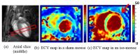
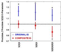
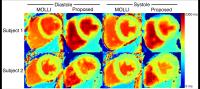
Modi?ed Look-Locker Inversion Recovery (MOLLI) provides the
highest precision and reproducibility for myocardial T1 mapping,
and extracellular volume (ECV) mapping. In this work, we
determine its effectiveness for measuring myocardial blood
flow (MBF), based on apparent-T1 mapping
under two conditions, slice-selective inversion and
non-selective inversion. We demonstrate that MOLLI provides
measured MBF comparable to the reference FAIR-SSFP ASL
method.
|
|
3143.
 |
45 |
High-resolution myocardial perfusion imaging with radial
simultaneous multi-slice imaging and constrained reconstruction
Ganesh Adluru1, Chris Welsh1, John
Roberts1, and Edward DiBella1
1Radiology, University of Utah, Salt Lake City,
UT, United States
High-resolution myocardial perfusion imaging offers improved
delineation of subendocardial ischemic regions and can lead
to improved diagnosis. Here we use undersampled radial
simultaneous multi-slice (SMS) acquisitions in conjunction
with constrained reconstruction with temporal total
variation and spatial block-matching 3D (BM3D) constraints
to obtain high in-plane spatial resolution perfusion
imaging. Promising results are shown with two types of
myocardial perfusion acquisitions (i) a set of 3
simultaneous slices after a saturation pulse, repeated
several times per beat at different cardiac phases, and (ii)
a ‘hybrid’ perfusion acquisition with one saturation pulse
per beat and the reconstructed cardiac phase of the 3 slices
chosen retrospectively.
|
|
3144.
 |
46 |
3D first-pass myocardial perfusion stack-of-stars imaging using
balanced steady state free precession
Merlin J Fair1,2, Peter D Gatehouse1,2,
Liyong Chen3,4, Ricardo Wage2, Edward
VR DiBella5, and David N Firmin1,2
1NHLI, Imperial College London, London, United
Kingdom, 2NIHR
Cardiovascular BRU, Royal Brompton Hospital, London, United
Kingdom, 3UC
Berkeley, Berkeley, CA, United States, 4Advanced
MRI Technologies, Sebastopol, CA, United States, 5UCAIR,
University of Utah, Salt Lake City, UT, United States
A method for enabling a balanced steady-state free
precession 3D stack-of-stars approach to whole-heart
first-pass myocardial perfusion imaging is investigated.
Consideration is made of the impact of potential
off-resonance effects at 3T and sequence-based modifications
to rectify this are examined. Demonstration of the
feasibility of this approach is then performed in-vivo.
|
|
3145.
 |
47 |
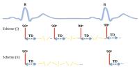
Double-gated Myocardial ASL Perfusion Imaging provides
Insensitivity to Heart Rate Variation
Hung Phi Do1, Andrew J Yoon2, Michael
W Fong2, Farhood Saremi3, Mark L Barr4,
and Krishna S Nayak5
1Department of Physics and Astronomy, University
of Southern California, Los Angeles, CA, United States, 2Department
of Medicine, Divison of Cardiology, Keck School of Medicine
of USC, University of Southern California, Los Angeles, CA,
United States, 3Department
of Radiology, Keck School of Medicine of USC, University of
Southern California, Los Angeles, CA, United States, 4Department
of Cardiothoracic Surgery, Keck School of Medicine of USC,
University of Southern California, Los Angeles, CA, United
States, 5Ming
Hsieh Department of Electrical Engineering, University of
Southern California, Los Angeles, CA, United States
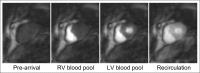
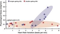
Double-gating in myocardial ASL allows for variations in the
post-labeling delay in order to ensure that both labeling
and imaging occur in the same cardiac phase. Originally
proposed by Poncelet et al. in 1999, this was believed to
provide insensitivity to heart rate variation. Despite this,
most groups have utilized single-gating with a fixed
post-labeling delay for pairs of control and tagged images,
since this allows for simpler quantification of myocardial
blood flow. In this study, we demonstrate that the
double-gating is indeed more robust to heart rate variation
compared to single-gating for myocardial ASL, based on
experiments in healthy volunteers and heart transplant
recipients.
|
|
3146.
 |
48 |
Demonstration of Velocity Selective Myocardial Arterial Spin
Labeling
Terrence Jao1 and
Krishna Nayak2
1Biomedical Engineering, University of Southern
California, Los Angeles, CA, United States, 2Electrical
Engineering, University of Southern California, Los Angeles,
CA, United States
Arterial spin labeled CMR is a non-contrast myocardial
perfusion imaging technique capable of assessing coronary
artery disease. A limitation of current methods is potential
underestimation of blood flow to myocardial segments that
have coronary arterial transit time longer than 1 R-R, which
are found in regions with significant collateral development
from chronic myocardial ischemia. In this work, we
demonstrate the feasibility of a velocity selective labeling
scheme for ASL-CMR that is insensitive to arterial transit
time.
|
|