ISMRM 24th Annual Meeting & Exhibition • 07-13 May 2016 • Singapore

Electronic Poster Session: Engineering

3500 -3523 RF Coils & Arrays
3524 -3547 UHF Applications & Modeling
3548 -3571 Gradients & Systems, MR Electronics, MR/PET
3620 -3643 MR Engineering Beyond RF Coils

Exhibition Hall 

16:00 - 17:00

    Computer #

 
3500.   
1 7 Tesla dual element 31P TxRx/1H Rx endorectal coil combined with an 8-channel 1H-TxRx body coil
Mark J. van Uden1, Bart Philips1, Miriam Lagemaat1, and T.W.J. Scheenen1,2
1Radiology and Nuclear Medicine, Radboud university medical center, Nijmegen, Netherlands, 2Erwin L. Hahn Institute for Magnetic Resonance Imaging, Essen, Germany
Multiparametric MRI (mpMRI) is a valuable tool to assess aggressiveness and stage of localized prostate cancer. When combined with metabolic information from 1H and 31P spectra valuable information on the metabolism of the disease can be derived. Here we demonstrate a dual element endorectal coil with 1H Rx and 31P TxRx capabilities combined with an external 8-channel transmit-receive body array. One patient (Gleason score 4+4) was measured. High quality proton spectra show elevated total choline in tumor tissue, which corresponded with increased glycerophosphocholine, glycerophosphoethanolamine and phosphocholine in the 31P spectra of the tumor.


3501.   
2 Prostate MRI at 7.0 Tesla using an actively-tuned endorectal coil
M. Arcan Erturk1 and Gregory J Metzger1
1Center for Magnetic Resonance Research, University of Minnesota, Minneapolis, MN, United States
Using endorectal coils (ERC) in combination with external surface arrays (ESA) can improve the SNR inside the prostate at 7.0T. When used in transmit, ERC can provide higher B1+ levels at the expense of field uniformity. Here, we develop a novel actively-tuned ERC that can be used both in “transmit” and “receive-only” modes utilizing the high B1+ of the ERC and field uniformity of the ESA inside the prostate. Transmit capabilities of this approach are investigated using simulations in a human model. Prostate MRI is acquired using the combined ESA+ERC approach to demonstrate the feasibility for translational studies at 7.0T.


3502.   
3 The Trellis Coil: A Morphing, Size Adaptable Array Coil
Graham Wiggins1,2 and August Frank3
1The Center for Advanced Imaging Innovation and Research (CAI2R), Department of Radiology, New York University School of Medicine, New York, NY, United States, 2The Bernard and Irene Schwartz Center for Biomedical Imaging, Department of Radiology, New York University School of Medicine, New York, NY, United States, 3University of Pennsylvania, Philadelphia, PA, United States
The sensitivity of surface coil arrays can be maximized by conforming them closely to the human body, maximizing coil loading and minimizing the contribution of coil noise. There is, however, great diversity in body shape and size. Rigid coils must adopt a compromise, allowing for the largest cases while not giving up too much sensitivity when imaging smaller examples. Flexible arrays can be wrapped tightly around the body, but if the overall array dimensions are fixed there will always be gaps or undesired overlaps depending on body habitus. We present here a geometrical approach based on a trellis-like lattice of interlinked slats which allows a surface coil array to be re-shaped while maintaining coil tuning, coil loading and decoupling of neighboring coils.


3503.   
4 Liquid metal based deformable transmitter for MR imaging: A feasibility study
Qi Duan1, Hai Lu2,3, Chris Cooper4, Xiaopeng Zong3, Jeff H. Duyn1, Michael D. Dickey4, and Shumin Wang2,3
1Laboratory of Functional and Molecular Imaging, National Institute of Neurological Disorders and Stroke, National Institutes of Health, Bethesda, MD, United States, 2UNC/NCSU Joint Department of Biomedical Engineering, University of North Carolina of Chapel Hill, Chapel Hill, NC, United States, 3Biomedical Research Imaging Center, University of North Carolina of Chapel Hill, Chapel Hill, NC, United States, 4Department of Chemical & Biomolecular Engineering, NC State University, Raleigh, NC, United States
Recent advances of liquid metal technology have made it possible to build deformable RF transmitters. Such coils have many attractive features for MR, such as easy reconfiguration, flexibility, and self-healing capabilities. However, given that the conductivity of liquid metal is more than an order of magnitude lower than the copper, it is unclear whether it is suitable to be used as a RF transmitter in MR applications. In this work, such feasibility was demonstrated by comparing the performance of two electric dipole antennas with similar size, one made of liquid metal and the other one made of copper, on a 7T scanner.


3504.   
5 RF coil design for multi-nuclear lung MRI of 19F fluorinated gases and 1H using MEMS
Adam Maunder1, Madhwesha Rao1, Fraser Robb2, and Jim Wild1
1Unit of Academic Radiology, University of Sheffield, Sheffield, United Kingdom, 2GE Healthcare, Aurora, OH, United States
Mutli-nuclear lung MRI using inhaled inert fluorinated 19F gas and  1H provide complementary structural-functional information. The close MR resonance frequencies of the two nuclei preclude the use of dual-tuned coils using trap circuits. Thus, we introduce the use of Micro-electromechanical systems (MEMS) in a quadrature transmit-receive RF coil to switch between two resonance frequencies of19F and 1H at 1.5T. Characterization of the additional loss and co-registered imaging of 19F and 1H with a custom-built body sized phantom is demonstrated.


3505.   
6 Performance of 7 Tesla Normal Metal and Superconducting Cryo-coils for MRI of Rat Brain - Permission Withheld
Jarek Wosik1,2, Kurt Bockhorst3, Tan I-Chih4, Kuang Qin1, Krzysztof Nesteruk5, and Ponnada A Narayana3
1Electrical and Computer Engineering, University of Houston, Houston, TX, United States, 2Texas Center for Superconductivity, University of Houston, Houston, TX, United States, 3Radiology, The University of Texas Health Science Center, Houston, TX, United States, 4The Brown Foundation Institute of Molecular Medicine, The University of Texas Health Science Center, Houston, TX, United States,5Institute of Physics of Polish Academy of Science, Warsaw, Poland
 We report on the performance of receive only 300-MHz planar cryogenic (both copper and superconducting) coils for MRI of rat brain at 7 T. The double-sided coils were fabricated by patterning Cu and YBCO on laminate and 0.33 mm thick sapphire substrate, respectively. Practical limits of the performance of the cryocoils (20 mm Cu and 17 mm superconducting) were tested using 3D-RARE, 2D-RARE, and RGB-FA maps recorded with EPI_DTI protocols. Both in-vivo and ex-vivo images were acquired. Twofold SNR gain was achieved at 65K. Cryo-system stability tests with over 11 hours of DTI ex-vivo scanning are presented.


3506.   
7 8CH 19F/1H Transceiver Array for Lung Imaging at 7T (pTX)
Helmar Waiczies1, Andre Kuehne1, Armin M. Nagel2,3, Dennis Schuchardt1, Darius Lysiak1, Jan Rieger1, and Thoralf Niendorf1,4
1MRI.Tools GmbH, Berlin, Germany, 2Division of Medical Physics in Radiology, Cancer Research Center (DKFZ), Heidelberg, Germany, 3Diagnostic and Interventional Radiology, University Medical Center Ulm, Ulm, Germany, 4Berlin Ultrahigh FieldFacility (B.U.F.F.), Max Delbrück Center for Molecular Medicine, Berlin, Germany
Lung MR-Imaging is challenging due to low proton density in the lung. Ventilation can only be visualized directly using exogenous contras agents such as hyperpolarized noble gases or 19F-containing gasses as a “contrast agents”. This work proposes a multi-channel transmit and receive (TX/RX) radiofrequency (RF) coil that supports eight TX/RX channels (4 loop- and 4 dipole-elements) for 19F and 1H lung imaging at 7.0T using a pTX-array.


3507.   
8 Comparison between commercial RF head coils and a new hybrid transmit-array coil based on 12 transmit elements and 22 receive elements for an 8-channel transmission system at 7 T
Alexis Amadon1, Guillaume Ferrand2, Elodie Georget1, Eric Giacomini1, Edouard Chazel1, Marie-France Hang1, Jeremy Bernard1, Nicolas Boulant1, Vincent Gras1, Alexandre Vignaud1, and Michel Luong2
1CEA, DSV, I2BM, NeuroSpin, UNIRS, Gif-sur-Yvette, France, 2CEA, DSM, IRFU, SACM, LISAH, Gif-sur-Yvette, France
This work compares the performance of two commercial RF coils with a new transmit-array hybrid coil developed at NeuroSpin for the human head at 7T. This z-segmented coil uses 11 transceiver dipoles connected to 7 power channels via an SVD-box, one transceiver patch to cover the top of the head, and 10 receive-only 3D loops in-between the dipoles. Its performance outbeats that of the commercial coils  both in terms of signal-to-noise ratio and dynamic RF shimming efficiency.


3508.   
9 A size-adaptable electric dipole array for 7T body imaging
Bei Zhang1, Martijn Cloos1, Gang Chen1, and Graham C. Wiggins1
1Department of Radiology, The Bernard and Irene Schwartz Center for Biomedical Imaging, New York, NY, United States
A major challenge in 7T coil array design for body imaging is to achieve optimum distribution in different body sizes with only one coil array. In this work, we propose a flexible 8 channel dipole array with trellis-like substrate which can expand and contract to fit various body sizes and hold electric dipole antenna elements in the optimum distribution around the body for all subjects. The performance of the trellis array was compared with a flexible yet fixed-element-spaced folded dipole body array [1] in terms of flip angle maps and SNR on two different sizes of body phantoms. Both phantom and invivo images show that the trellis array does not have common nulls between the circular polarized (CP) and gradient modes.


3509.   
10 13C MRS in human calf muscles at 7T using a double tuned 4 channel 13C-4 channel 1H transceiver phased array.
Guillaume Donati1, Eulalia Serés Roig1, and Rolf Gruetter1,2
1Laboratory of Functional and Metabolic Imaging, École Polytechnique Fédérale de Lausanne, Lausanne, Switzerland, 2Department of Radiology, Universities of Lausanne and Geneva, Lausanne, Geneva, Switzerland
The inherent low sensitivity of 13C MRS makes direct 13C detection challenging. Moreover, the 13C-1H heteronuclear J coupling requires double tuned 13C-1H coils to perform proton decoupling during13C signal acquisition. High sensitivity can be achieved over a large field-of-view using array coils. We aimed to design and build a 4 channel 13C-4 channel 1H transceiver phased-array coil for 13C-MRS studies in humans at ultra-high field. Electromagnetic performance of the coil was evaluated by FDTD simulations in calf muscles at both frequencies. Finally, 1H-decoupled 13C resonance of Glycogen C1was successfully detected in volunteer calf muscles at 7T.


3510.   
11 Use of the ultimate signal-to-noise ratio to assess the impact of coil coverage for brain - Video Not Available
Bastien Guerin1, Jorge F. Villena2, Athanasios G. Polimeridis3, Elfar Adalsteinsson4,5,6, Luca Daniel4, Jacob K. White4, and Lawrence L. Wald1,5
1A. A. Martinos Center for Biomedical Imaging, Massachusetts General Hospital, Charlestown, MA, United States, 2Cadence Design Systems, Feldkirchen, Germany, 3Skolkovo Institute of Science and Technology, Moscow, Russian Federation, 4Department of Electrical Engineering and Computer Science, Massachusetts Institute of Technology, Cambridge, MA, United States, 5Harvard-MIT Division of Health Sciences Technology, Cambridge, MA, United States, 6Institute for Medical Engineering and Science, Massachusetts Institute of Technology, Cambridge, MA, United States
We assess the impact of coil coverage for brain imaging at 3 T and 7 T using the ultimate SNR (uSNR). We simulate three coil configurations that cover (i) the whole head (ultimate), (ii) the whole head but the neck (realistic ultimate) and (iii) the head except the neck and face (helmet geometry). We compute the maximum SNR (unaccelerated and accelerated) achievable by any coils with these head coverage using random excitations of a dense dipole cloud placed around the head. Not covering the face (eye/nose/mouth) and neck has little impact on unaccelerated and accelerated SNR compared to the ultimate.


3511.   
12 A multisegment detunable HEM-mode dielectric resonator for increased patient comfort at 7 T
Rita Schmidt1, Wouter M. Teeuwisse1, and Andrew Webb1
1Radiology, Leiden University Medical Center, Leiden, Netherlands
At ultrahigh field (≥7T) cavity- and waveguide-based RF coils become possible to construct, and have the advantage that simple designs can be used. One such design uses materials with a high dielectric constant. So far only completely circular structures have been produced; to improve the usability of such a dielectric resonator, it is important to be able to design a splittable and easily detunable resonator. In this work, we tested designs that allow splitting of the cylinder to sections, while maintaining the circularly polarized RF field. In-vivo measurements of the knee at 7 Tesla showed the feasibility of this approach. 


3512.   
13 Segmented Dipole: A Remotely Reconfigurable Near-Field Dipole Antenna Transmitter for Optimized 7 T Spine Imaging
Qi Duan1, Jeff H. Duyn1, and Hellmut Merkle1
1Laboratory of Functional and Molecular Imaging, National Institute of Neurological Disorders and Stroke, National Institutes of Health, Bethesda, MD, United States
7T spine array based on electric dipole antenna starts to gain attention recently. Nevertheless, the transmit efficiency of the dipole has a natural decay from the feeding point, which does not necessarily corresponding to the target imaging area. In addition, a variety of patient size requires the dipole to be reconfigured for optimum performance. In this work, we propose a concept of “segmented dipole” that can be remotely reconfigured for transmit field-of-view shifting, tuning, and impedance matching characteristics without the need of a parallel-transmit system. A prototype transmitter was built and the reconfiguration capability was demonstrated at a 7T scanner.


3513.   
14 Dedicated surface coils for MR studies in the temporal and the frontal lobes of the human brain at 7T
Jérémie Daniel Clément1, Lijing Xin2, Rolf Gruetter3,4,5, and Özlem Ipek2
1CIBM-LIFMET, Ecole Polytechnique Fédérale de Lausanne, Lausanne, Switzerland, 2CIBM-AIT, Ecole Polytechnique Fédérale de Lausanne, Lausanne, Switzerland, 3LIFMET, Ecole Polytechnique Fédérale de Lausanne, Lausanne, Switzerland, 4Department of Radiology, University of Geneva, Geneva, Switzerland, 5Department of Radiology, University of Lausanne, Lausanne, Switzerland
The purpose of the study was to build dedicated surface coils for the temporal and the frontal lobes of the human brain at 7T. Their transmit field efficiency and single-voxel spectroscopy performances were compared with a birdcage coil with a dielectric pad. An increased B1+-field for the brain regions was measured with the surface coils compared to the birdcage with the pad, which allows single-voxel SPECIAL spectroscopy and anatomical image acquisitions in the peripheral temporal and frontal lobes.


3514.   
15 A combined microcoil and microdialysis approach to measure metabolic response in real-time. - Permission Withheld
Stefan Glöggler1, Silvia Rizzitelli2, Noel Pinaud3, Gérard Raffard2, Véronique Bouchaud2, Stéphane Sanchez2, Alan Wong4, and Yannick Crémillieux2
1University of Southampton, Southampton, United Kingdom, 2CRMSB, Université Bordeaux, Bordeaux, France, 3Université Bordeaux, Bordeaux, France, 4CEA Saclay, Gif-sur-Yvette, France
We present two highly sensitive microcoil probes (1H and 13C) that are connected to microdialysis probes inside a MRI scanner. Under continuous flow conditions the metabolic response can be monitored in real-time in vitro and in vivo.


3515.   
16 High Spatial Resolution Functional Imaging of Human Temporal Lobe Cortical Layers at 3T
Pu-Yeh Wu1, Ying-Hua Chu1, Shang-Yueh Tsai2, Wen-Jui Kuo3, and Fa-Hsuan Lin1
1Institute of Biomedical Engineering, National Taiwan University, Taipei, Taiwan, 2Institute of Applied Physics, National Chengchi University, Taipei, Taiwan, 3Institute of Neuroscience, National Yang Ming University, Taipei, Taiwan
By using a dedicated 24-channel coil array and tailored sequence, we achieved human temporal lobe functional imaging with 1.5 mm isotropic resolution. Our coil array had two-fold SNR and tSNR improvement in the temporal lobe compared to a 32-channel whole-head coil array. Functional MRI showed more robust BOLD signals elicited by music stimuli in the primary auditory cortex. Tonotopic mapping revealed the clearest frequency progressions in the middle layer of auditory cortex gray matter, which was consistent with the result acquired by electrophysiological recordings on rodents.


3516.   
17 A Slot Antenna Concept for High Fidelity Body Imaging at Ultra High Field - Permission Withheld
Leeor Alon1,2,3,4, Cem M. Deniz1,2,3,4, Ryan Brown1,2, Daniel Sodickson1,2,3, and Christopher M. Collins1,2,3
1Center for Advanced Imaging Innovation and Research (CAI2R), New York University School of Medicine, New York, NY, United States, 2Center for Biomedical Imaging, New York University School of Medicine, New York, NY, United States, 3NYU Wireless, NYU-Poly, New York, NY, United States, 4RF Test Labs, New York, NY, United States
In recent years dipoles and other radiative antenna structures have become popular for ultra high field MR imaging. In this work, we introduce the radiative slot antenna, which generates a horizontally polarized E field showing promise as a simple coil structure for high fidelity axial imaging.


3517.   
18 A Novel 8-channel transceive open knee coil for dynamic musculoskeletal imaging at 7 Tesla
Bassem Henin1, Ewald Weber1, and Stuart Crozier1
1ITEE, The University of Queensland, Brisbane, Australia
Magnetic resonance imaging (MRI) is a very important non-invasive modality for the diagnosis of knee anatomy and pathology. The use of 7 Tesla systems for knee imaging can provide higher signal-to-noise ratios (SNR) than lower field systems. In this work, a novel 8-channel transceive open knee coil for dynamic musculoskeletal (MSK) MR imaging of the knee is simulated and constructed. The open design concept will facilitate the diagnostic and functional assessment of knee injuries and pathology, and allow for imaging of a moving knee.


3518.   
19 A 4-element dual-tuned 1H/31P coil array for MR imaging and spectroscopy of the human heart at 3 Tesla - Permission Withheld
Adrianus J. Bakermans1, Bart-Jan van den Berg2, Gustav J. Strijkers3, Maarten J. Versluis4, Dennis W.J. Klomp2,5, Aart J. Nederveen1, and Jeroen A.L. Jeneson1,6
1Department of Radiology, Academic Medical Center, Amsterdam, Netherlands, 2MR Coils B.V., Drunen, Netherlands, 3Department of Biomedical Engineering and Physics, Academic Medical Center, Amsterdam, Netherlands, 4Philips Healthcare Benelux, Eindhoven, Netherlands, 5Department of Radiology, University Medical Center Utrecht, Utrecht, Netherlands, 6Neuroimaging Center, University Medical Center Groningen, Groningen, Netherlands
Maximizing the signal-to-noise ratio (SNR) in in vivo cardiac phosphorus-31 magnetic resonance spectroscopy (31P-MRS) remains a major challenge in investigations of human myocardial energy metabolism. Here, we demonstrate that with a dual-tuned 1H/31P coil array with 4 elements, it is feasible to achieve a higher SNR and a more homogeneous receive sensitivity distribution over the region of interest for cardiac 31P-MRS at 3 Tesla than with a standard linear single-turn 31P surface coil. Furthermore, the 4 channels available for 1H signal reception allow for cardiac cine 1H-MRI with parallel imaging. 


3519.   
20 Assessment of coil arrays with small loop diameter at 7T for micron-scale resolution fMRI of human neocortex.
Alexander Beckett1, An T Vu1,2, Boris Keil3, Kawin Setsompop3, Lawrence L Wald3, Scott Schillak4, and David A Feinberg1,2
1Helen Wills Neuroscience Institute, University of California, Berkeley, CA, United States, 2Advanced MRI Technologies, Sebastopol, CA, United States, 3Martinos Center for Biomedical Imaging, Charlestown, MA, United States, 4Virtumed, LLC, Minneapolis, MN, United States
The use of array coils with smaller loop sizes allows increased signal to noise ratio close to the coils with decreased coverage of the brain. By focusing on cortical imaging (MR Corticography, MRCoG), this increased SNR can be leveraged to allow ultra-high resolution neuroimaging and fMRI. We assess array coils with varying loop sizes (50mm-20mm) in phantom and with 8-channel arrays  (70mm, 40mm, 30mm) in brain at ultra-high resolution (≤ 500 micron isotropic voxels), and show increases in SNR and BOLD contrast in the human cortex as loop size decreases.


3520.   
21 A larger subject Field-of-View and denser coil arrays at UHF: What do we gain?
Shubharthi Sengupta1, Gregor Adriany2, Valentin G Kemper1, Rainer Goebel1, and Alard Roebroeck1
1Dept. of Cognitive Neuroscience, Faculty of Psychology and Neuroscience, Maastricht University, Maastricht, Netherlands, 2Dept. of Radiology, Center for Magnetic Resonance Research, University of Minnesota, Minneapolis, MN, United States
Functional magnetic resonance imaging (fMRI) of the human brain has now become the preferred method for mapping functional pathways in the human visual cortex, among other brain regions. At ultra-high fields of 7 Tesla and above, the increased Contrast-to-Noise ratio for blood oxygenation level dependent (BOLD) techniques further facilitates this acquisition methodology. We designed and constructed a frontally open RF coil with a phased array transmit and a dense receiver array layout for human visual cortical imaging at 7 Tesla, specifically for visual fMRI experiments at sub-millimeter resolutions. We compared the coil's performance to that of a standard 32-channel whole-brain volume coil to inquire into its efficacy as a suitable substitute for the whole-brain coil for high resolution fMRI.


3521.   
22 An investigation of coupling and loading effects for adaptive coil design
Barbara Dornberger1, Markus Vester1, Robert Rehner1, Michael Zenge2, Riccardo Lattanzi3, and Graham Wiggins3
1Siemens Healthcare GmbH, Erlangen, Germany, 2Siemens Medical Solutions USA, Inc., Malvern, PA, United States, 3Department of Radiology, New York University School of Medicine, Center for Advanced Imaging Innovation and Research (CAI2R) and Center for Biomedical Imaging, New York, NY, United States
This abstract examines the feasibility of an adaptive coil design to overcome the disadvantage of poorly loaded and non-fitting local coils. The coil adjusts to the circumference of e.g. a knee by changing the overlap between coil elements. Through bench measurements and simulation we investigated the tradeoff between maintaining optimal coil loading and signal-to-noise ratio (SNR) losses due to coupling. Broadband matching was considered to reduce SNR degradation. Although results show that an adaptive coil can minimize coil losses, the non-ideal overlap of this design leads to a larger SNR degradation than the coil losses of a non-perfect fitting coil.


3522.   
23 Decoupling between coils in a flexible phased-array using stacked circumferential shielding
Jhy-Neng Tasso Yeh1 and Fa-Hsuan Lin1
1Institute of Biomedical Engineering, National Taiwan University, Taipei, Taiwan
We propose a flexible coil array design consisting of circumferential shielding stacked on circular coils to achieve robust decoupling between coil when the array is either bended or on a flat plane. Two types of circumferential shielding were tested through numerical simulation. The results demonstrated that both types can have good decoupling on a curved surface with S21 < -17.1 dB. The maximum |B1| for the array coil with Type-II (two-parallel-ring) shielding was 63% larger than that with Type-I (single-strip) shielding. Future work will empirically construct these coils to compensate simulation inaccuracies and validate their performance In Vivo.


3523.   
24 Sensitive detection of hemodynamic responses to TMS pulse at human sensorimotor cortex
Pu-Yeh Wu1, Ying-Hua Chu1, Aapo Nummenmaa2, Thomas Witzel2, Shang-Yueh Tsai3, Wen-Jui Kuo4, and Fa-Hsuan Lin1
1Institute of Biomedical Engineering, National Taiwan University, Taipei, Taiwan, 2Athinoula A. Martinos Center for Biomedical Imaging, Massachusetts General Hospital, Charlestown, MA, United States,3Institute of Applied Physics, National Chengchi University, Taipei, Taiwan, 4Institute of Neuroscience, National Yang Ming University, Taipei, Taiwan
We tested how TMS pulses and the separation between TMS pulse and RF excitation affect MRI SNR and tSNR using our tailored 8-channel TMS-compatible MRI coil array. Result from an in vivo experiment using this integrated system to record the BOLD signal elicited by TMS pulses at human primary motor cortex is also reported.
Exhibition Hall 

16:00 - 17:00

    Computer #

 
3524.   
49 
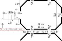
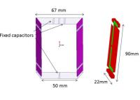
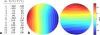
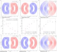
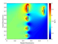
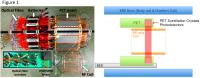
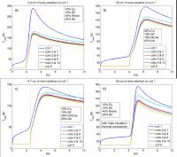
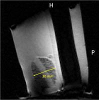
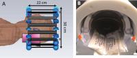
Dipole Array Design Considerations for Head MRI at 10.5T
Jinfeng Tian1, Russell Lagore1, Lance Delabarre1, and J. Thomas Vaughan1
1U. of Minnesota, Minneapolis, MN, United States
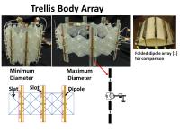
An 8-channel dipole array is a promising structure for human head imaging at 10.5T.  In order to optimize the structure for efficiency and homogeneity over the brain, many variations of the dipole were numerically simulated and compared.  The variations include varying dipole lengths, warping the dipole, adding shielding, adding dielectric padding or dielectric mirrors and including decoupling capacitors.  Compared to a design in use, numerical results predict the RF homogeneity can be greatly improved with a 210 mm dipole array while simultaneously lowering the peak local 1 gram and 10 gram SAR.


3525.   
50 
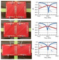
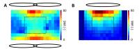
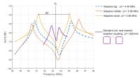
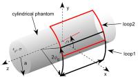
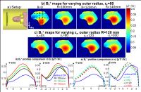
Analytical Modeling of the Coupling within a Human Head Surface Loop Transmit Phased Array at Ultra-High Fields
Nikolai I Avdievich1, Andreas Pfrommer1, Ioannis Giapitzakis1, and Anke Henning1,2
1High-field Magnetic Resonance, Max Planck Institute for Biological Cybernetics, Tübingen, Germany, 2Institute for Biomedical Engineering, UZH and ETH Zurich, Zurich, Switzerland
Decoupling of multi-channel ultra-high field (>7T) transmit and transceiver arrays is a major issue. Analytical modeling of the coupling can facilitate the array optimization. We developed an analytical model describing the impedance matrix for two rectangular loops placed on a cylindrical surface and mimicking the human head array geometry. The developed model was comprehensively validated and allows for the optimization of the geometry and positioning of the loops. The latter enabled simultaneous cancellation of resistive and inductive coupling without additional decoupling circuits. The resulting overlapped array element arrangement improves both transmit and receive performance in comparison to conventional gapped arrays.
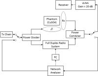


3526.   
51 Optimization of the antenna-subject spacing for transceive surface arrays of dipole antennas at 7T
A.A. Hurshkainen1, I.J. Voogt2, A.A. 
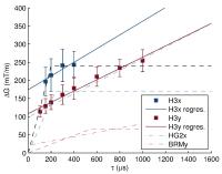
Haghnejad2, D.W. Klomp2, P.R. Luijten2, I.V. Melchakova1, S.B. Glybovski1, 
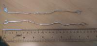
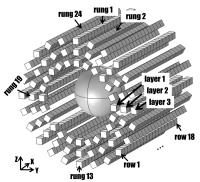
C.A.T. van den Berg2, and A.J.E. Raaijmakers2
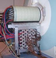
1Department of Nanophotonics and Metamaterials, ITMO University, Saint-Petersburg, Russian Federation, 2Imaging 
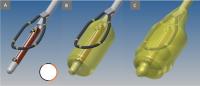
Division, UMC Utrecht, Utrecht, Netherlands
Dipole antennas are being used increasingly for body imaging at 7T. For dipole antennas, SAR levels can be reduced by 
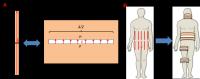
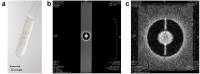
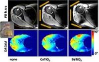
increasing the antenna-subject spacing. However, this will 
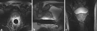
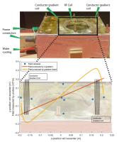
increase inter-element coupling.  In this study we investigate the relationship between antenna-subject spacing, inter-element coupling and maximum local SAR levels for fractionated dipole antennas. We demonstrate that the originally presented antenna-subject spacing (2 cm) can be increased without significant scattering losses. We have realized an 8-element array of fractionated dipole antennas with 4 cm antenna-subject spacing and demonstrate uncompromised imaging performance with 45% lower local SAR levels in comparison to the original design.


3527.   
52 
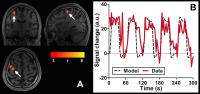
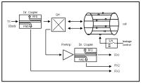
Evaluation of through-time radial GRAPPA for real-time cardiac MR imaging at 7 Tesla
Sascha Brunheim1,2, Sören Johst1, Stefan Maderwald1, Stefan Rietsch1,2, Stephan Orzada1, Marcel Gratz1,2, Juliane Goebel3, Kai Nassenstein3, Nicole Seiberlich4, and Harald H. Quick1,2
1Erwin L. Hahn Institute for Magentic Resonance Imaging, University Duisburg-Essen, Essen, Germany, 2High Field and Hybrid MR Imaging, University Hospital Essen, Essen, Germany, 3Department of Diagnostic and Interventional Radiology and Neuroradiology, University Hospital Essen, Essen, Germany, 4Department of Biomedical Engineering, Case Western Reserve University, Cleveland, OH, United States
Accelerated radial data acquisition of the myocardium in combination with through-time radial GRAPPA offers the opportunity for real-time visualization of cardiac motility without the need for additional ECG or pulse wave synchronization. This is particularly useful in an ultrahigh-field MR environment where conventional gating methods in combination with cardiac dysrhythmia tend to fail. In this work, the performance of through-time radial GRAPPA against an established Cartesian k-space encoding protocol featuring pulse-triggered cine-FLASH has been evaluated and its role as an alternative for real-time cardiac 7-Tesla MR imaging is shown.


3528.   
 
53 Enabling axial diffusion tensor imaging of the human cervical spinal cord at 7T
Aurélien Massire1,2, Pierre Besson1,2, Maxime Guye1,2, Jean-Philippe Ranjeva1,2, and Virginie Callot1,2
1Centre de Résonance Magnétique Biologique et Médicale (CRMBM), UMR 7339, CNRS, Aix-Marseille Université, Marseille, France, 2Centre d'Exploration Métabolique par Résonance Magnétique (CEMEREM), Hôpital de la Timone, Pôle d’imagerie médicale, AP-HM, Marseille, France
MRI at 7T has recently demonstrated its ability to provide high-quality anatomical images of the spinal cord (SC), yet no diffusion tensor imaging (DTI) study was reported so far. Single-shot echo-planar imaging (ss-EPI) is the method of choice for DTI but the sequence is seriously limited by strong susceptibility artifacts. This work demonstrates that a thoughtful implementation of ss-EPI at 7T combined with distortion correction post-processing from two acquisitions with opposed phase-encoding directions can generate high-resolution axial DTI images of the cervical SC with added value compared to lower field standard protocols making SC DTI ready for UHF clinical investigations.


3529.   
54 
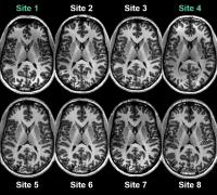
The traveling heads: Qualitative and quantitative evaluation of multicenter brain imaging at 7 Tesla
Maximilian N. Voelker1, Oliver Kraff2, Daniel Brenner3, Astrid Wollrab4, Oliver Weinberger5, Moritz C. Berger6, Simon Robinson7, Wolfgang Bogner7, Christopher Wiggins8, Robert Trampel9, Tony Stöcker3, Thoralf Niendorf5,10, Harald H. Quick2,11, David G. Norris2,12, Mark E. Ladd2,6, and Oliver Speck4,13
1Erwin L. Hahn Institute for Magnetic Resonance Imaging, University Hospital Essen, University of Duisburg-Essen, Essen, Germany, 2Erwin L. Hahn Institute for Magnetic Resonance Imaging, University of Duisburg-Essen, Essen, Germany, 3German Center for Neurodegenerative Diseases (DZNE), Bonn, Germany, 4Otto-von-Guericke-University, Magdeburg, Germany, 5Berlin Ultrahigh Field Facility (B.U.F.F.), Max-Delbrueck-Center for MolecularMedicine, Berlin-Buch, Germany, 6Medical Physics in Radiology, German Cancer Research Center (dkfz), Heidelberg, Germany, 7High Field MR Center, Department of Biomedical Imaging and Image-guided Therapy, Medial University of Vienna, Vienna, Austria, 8ScanNexus, Maastricht, Netherlands, 9Max Planck Institute for Human Cognitive and Brain Sciences, Leipzig, Germany, 10Experimental and Clinical Research Center, a jointcooperation between the Charité Medical Faculty and the Max Delbrück Center for MolecularMedicine, Berlin, Germany,11High Field and Hybrid MR Imaging, University Hospital Essen, University Duisburg-Essen, Essen, Germany, 12Donders Centre for Cognitive Neuroimaging, Nijmegen, Netherlands, 13Leibniz Institute for Neurobiology, Magdeburg, Germany
The “traveling heads” is an experiment started in 2014 to assess the comparability and reproducibility of multicenter human brain imaging at 7T. This is of particular interest as 7T MRI is currently being discussed to become a clinical system in the very near future. The number of installations continues to increase, with currently approximately 60 research sites in operation worldwide. As an advantage, this new technology provides higher SNR, yet the artifact-to-noise ratio is also increased. This can influence the image quality severely and may be different at individual UHF sites, where system hardware differences could diminish reproducibility.


3530.   
55 Very high order B0 Shimming of the human brain at 9.4 T considering Real B0 Shim Fields
Paul Chang1,2, Sahar Nassirpour1,2, and Anke Henning1,3
1Max Planck Institute for Biological Cybernetics, Tuebingen, Germany, 2IMPRS for Cognitive and Systems Neuroscience, Eberhard Karls University of Tuebingen, Tuebingen, Germany, 3Institute for Biomedical Engineering, UZH and ETH Zurich, Zurich, Switzerland
A highly homogeneous B0 field is essential if we are to exploit the advantages of higher field strengths for MR applications. In this work, we model the real field of each shim channel of a 4th order shim system for a 9.4T MR system for in vivo B0 shimming applications. Each shim channel is modelled at a range of frequencies to account for the possibility of amplitude nonlinearities.

By modelling the fields generated by each shim channel, we were able to achieve better shim qualities than if perfect fields were assumed. 


3531.   
56 B1+ homogenization at 7T using an innovative meta-atom
Lisa Leroi1, Alexandre Vignaud1, Pierre Sabouroux2, Elodie Georget1, Benoit Larrat1, Stefan Enoch2, Gérard Tayeb2, Nicolas Bonod2, Alexis Amadon1, Denis Le Bihan1, and Redha Abdeddaďm2
1UNIRS, CEA Saclay - DSV - I2BM - Neurospin - UNIRS, Gif-sur-Yvette, France, 2CNRS, Aix-Marseille Université, Centrale Marseille, Institut Fresnel, UMR 7249, Marseille, France
B1+ heterogeneity at ultra-high field (UHF) can be tackled performing “passive shimming” with High-Dielectric Constant (HDC) pads. Nevertheless, HDC pads have shown structural, manufacturing and composition constraints. Here, we substitute HDC padding with a new meta-atom (MA) structure with a high equivalent dielectric constant, leaving behind the identified limitations. In this work, we compare this MA structure to a classic BaTiO3 pad used in UHF clinical routine. Results demonstrate this solution to strongly impact local B1+ distribution. Implementing multiple MA structures into the coil design might suggest a good potential for brain global B1+ inhomogeneity mitigation.


3532.   
57 
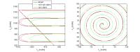
Improving travelling wave efficiency at 7 T using dielectric material placed ”beyond” the region of interest
Rita Schmidt1 and Andrew Webb1
1Radiology, Leiden University Medical Center, Leiden, Netherlands
The concept of traveling-wave MRI has been introduced for ultra-high fields, enabling large field-of-view excitation and physical separation between the antenna and the subject. Several studies have shown that introducing additional materials/structures into the magnet bore or surrounding the subject can improve the efficiency. In this work we explore the use of high permittivity material placed behind the region of interest and show that it can be beneficial for traveling wave efficiency. Separating the region of improved efficiency from that of the dielectric allow positioning of a receive array in the close proximity to the region of interest, physically separate from the dielectric material. 


3533.   
58 
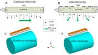
Tuning Microstrip Coil Field Patterns Using Capacitor-Segmented Ground Planes
Xinqiang Yan1,2, John C. Gore1,2,3, and William A. Grissom1,2,3
1Institute of Imaging Science, Vanderbilt University, Nashville, TN, United States, 2Radiology, Vanderbilt University, Nashville, TN, United States, 3Biomedical Engineering, Vanderbilt University, Nashville, TN, United States
 At 7T and higher, the B1 fields of loop and microstrip coils become asymmetric. However, B1 fields of dipole antennas are still symmetric. The different behaviors of dipole and microstrip coils may be explained by the fact that they have similar magnetic-field vectors but different Poynting vectors. We propose to manipulate the Poynting vector and thus the symmetry of the B1 patterns of microstrip coils using capacitor-segmented ground (CSG) planes. This concept has been validated by numerical studies and practical MRI experiments. The CSG method provides additional flexibility for manipulating the shape of the B1 field, which may be advantageous for RF shimming and parallel transmission.


3534.   
59 Control of Excited Modes in Ultra High Magnetic Field MRI with Electrically Hard Surfaces
Patrick Bluem1, Andrew Kiruluta2, Pierre-Francois Van de Moortele3, Gregor Adriany3, and Zoya Popovic1
1Department of Electrical, Computer, and Energy Engineering, University of Colorado at Boulder, Boulder, CO, United States, 2Massachusetts General Hospital, Harvard Medical School, Boston, MA, United States, 3University of Minnesota, Center for Magnetic Resonance Research, Minneapolis, MN, United States
Traditional MRI reactive near-field probe design for B1 field uniformity assumes quasi-static fields. However, for B0>4T, the quasi-static approximation is no longer valid since the wavelength is smaller than the FOV and field wave modes appear, affecting image quality.  This work presents the use of a copper strip waveguide structure combined with a traveling wave excitation at 7T, 10.5T human wide-bore and 16.4T small animal scanners, while observing the effect on a cylindrical distilled water phantom.  A simple flexible copper strip wearable wrap is shown to improve SNR and field distribution in UHF-MRI.


3535.   
60 
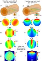
Theoretical and simulation verification of SNR enhancement in traveling wave MRI using free local resonators
Xinqiang Yan1,2 and Xiaoliang Zhang3
1Institute of Imaging Science, Vanderbilt University, Nashville, TN, United States, 2Radiology, Vanderbilt University, Nashville, TN, United States, 3Department of Radiology and Biomedical Imaging, University of California San Francisco, San Francisco, CA, United States
Traveling wave MR is a promising method for large field-of-view imaging at ultrahigh fields. However, a major issue currently faced in traveling wave MR is low transmit efficiency and limited SNR. It was found that the SNR in traveling wave MRI can be significantly improved by using a free local resonator. In this study, we validated this finding in simulation and extended the single loop to a multi-channel array. Based on the simulation results, the SNR on the phantom has a 16-fold gain (56.8 VS 3.6) at near area and 3-fold gain at far area (9.7 VS 3.5) with the help of the free loop. This improvement can be attributed to the secondary magnetic field caused by induced current of the free resonator.


3536.   
61 
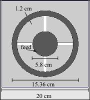
Zeroth-Order Resonator with Stepped Impedance for 7T Magnetic Resonance RF Coil
Vijayaraghavan Panda1,2, Sung-Min Sohn1,2, Thomas J Vaughan1,2, and Anand Gopinath1
1Department of Electrical and Computer Engineering, University of Minnesota, Minneapolis, MN, United States, 2Department of Radiology, Center for Magnetic Resonance Research, Minneapolis, MN, United States
A planar structure based on a metamaterial transmission line Zeroth Order Resonance (ZOR) and Stepped Impedance Structure is designed as a MRI RF coil element. It generates high uniform magnetic field in the 7T Magnetic resonance imaging (MRI) system at 298 MHz, which improves the signal to noise ratio and the quality of the images. The full wave simulation and measurements show comparable results with the microstrip TEM RF coil element [1]. Additionally, it can generate uniform and high H-field intensity for any physical length of the coil as the resonance of the ZORs is independent of its length [2].


3537.   
62 
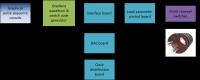
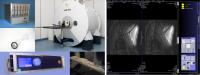
9.4T Animal Scanner for Translation Research with Binary Compatibility to Human Scanner and Clinical UI
Jörg Felder1, Chang-Hoon Choi1, Stefan Schwan1, A. Avdo Celik1, Seong Dae Yun1, Nuno Andre da Silva1, Ana Maria Oros-Peusquens1, and N. Jon Shah1,2
1INM-4, Forschungszentrum Jülich, Jülich, Germany, 22Faculty of Medicine, Department of Neurology, JARA, RWTH Aachen University, Aachen, Germany
In translational research going from animal model to in vivo human it is often desirable to change as few experimental parameters as possible. For this purpose a unique 9.4 T animal scanner has been assembled consisting of a dedicated small bore magnet and being operated with clinical software. Here we demonstrate an initial performance analysis of the system as well as some more advanced image acquisitions.


3538.   
63 
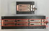
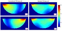
Revised Transmit/Receive Loop Coil for 7T Usage - Permission Withheld
Zhiyong Zhai1 and Michael Morich1
1Philips, Cleveland, OH, United States
At 7T, T/R loop coils have challenges in transmit B1+-field efficiency and receive B1--field sensitivity in a spatial context, due to the increased tissue dielectric/wavelength effect. Here we propose a revised T/R loop coil schema which improves the B1-field non-uniformity at 7T. The proposed modification may make the T/R loop coil construct yet more useful at ultra-high fields.


3539.   
64 
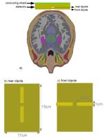
Feasibility of crossed-dipole antenna to excite a circularly-polarized field for human brain imaging at 7T,  A design study
Özlem Ipek1 and Rolf Gruetter2,3,4
1CIBM-AIT, EPFL, Lausanne, Switzerland, 2LIFMET, EPFL, Lausanne, Switzerland, 3Department of Radiology, University of Lausanne, Lausanne, Switzerland, 4Department of Radiology, University of Geneva, Geneva, Switzerland
The aim of this study was to investigate the crossed-dipole antenna by means of electromagnetic simulations and compare it with the surface quadrature head loop and volume head coils in terms of B1+ efficiency for 7T human brain imaging. The crossed-dipole antenna consists of two dipoles placed in a crossed form mounted upon/in an one-side conductor shielded dielectric. This antenna excites the circularly-polarized field and enhances the transmit efficiency in the occipital lobe in a larger FOV compared to the conventional coils. The comparison of the simulated B1+ maps of the coils showed that it is feasible to build it.


3540.   
65 
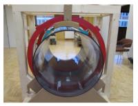
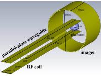
Traveling Wave MRI at 7T Using Dielectric Wave-Guide
Zhiyue J Wang1,2, Alexander Ivanishev1, Keith M Hulsey1, Dah-Jyuu Wang3, and Robert E Lenkinski1
1UT Southwestern Medical Center, Dallas, TX, United States, 2Children's Medical Center Dallas, Dallas, TX, United States, 3Children's Hospital of Philadelphia, Philadelphia, PA, United States
Traveling wave MRI uses a wave-guide for RF transmission.  The metal bore of the scanner magnet serves as a wave-guide and extensions using conductor sheets may be added. Although dielectric materials are frequently introduced into the system, their intended function has been to modify the behavior of the wave-guide. In this work, we show that a dielectric material may be used as a wave-guide by itself, in a fashion similar to optic fibers guiding light transmission. We conducted MRI experiments at 7T using an insulator wave-guide constructed by filling a PVC tube with deionized water.


3541.   
66 Optimizing high permittivity materials for SAR minimization in transmit arrays: influence of the phase distribution of the excitation profile
Gillian G Haemer1,2,3, Manushka V Vaidya1,2,3, Daniel K Sodickson1,2,3, Graham C Wiggins1,2, and Riccardo Lattanzi1,2,3
1The Center for Advanced Imaging Innovation and Research (CAI2R), Department of Radiology, New York University School of Medicine, New York, NY, United States, 2The Bernard and Irene Schwartz Center for Biomedical Imaging, Department of Radiology, New York University School of Medicine, New York, NY, United States, 3The Sackler Institute of Graduate Biomedical Sciences, New York University School of Medicine, New York, NY, United States
Appropriate high-permittivity, low-conductivity materials placed between the RF coil and the sample can provide performance improvement in both transmission and reception. We employed a simulation framework based on dyadic Green’s functions for multi-layered spherical geometries to analyze how HPMs affect the tradeoff between excitation homogeneity and global Specific Absorption Rate (SAR) for RF shimming at 7T using an L-curves analysis. Three target excitation profiles were analyzed, with uniform amplitude and varied phase, to determine the influence that target phase distribution has on the optimal relative permittivity results.


3542.   
67 Evaluation of potential improvements from high permittivity pads for imaging upper extremities at 7 Tesla
Oliver Kraff1, Andrea Lazik-Palm1,2, Wyger M Brink3, Andreas K Bitz4, Mark E Ladd4, and Harald H Quick1,5
1Erwin L. Hahn Institute for MRI, University Duisburg-Essen, Essen, Germany, 2Department of Diagnostic and Interventional Radiology and Neuroradiology, University Duisburg-Essen, University Hospital, Essen, Germany, 3Radiology, Leiden University Medical Center, Leiden, Netherlands, 4Medical Physics in Radiology, German Cancer Research Center (DKFZ), Heidelberg, Germany, 5High Field and Hybrid MR Imaging, University Duisburg-Essen, University Hospital, Essen, Germany
Two sets of dielectric pads with high permittivity (CaTiO3: 110 and BaTiO3: 286) were evaluated for potential improvements in imaging the shoulder joint and upper arm in combination with an 8–channel transmit/receive shoulder coil at 7 Tesla. In vivo images with structural PD TSE and DREAM flip angle maps were obtained and compared to measurements without pads present. Both a fixed RF shim as well as individual RF shimming were applied. For the investigated configurations, no substantial improvements in imaging upper extremities at 7 Tesla were found when applying high permittivity dielectric pads.


3543.   
68 
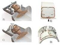
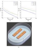
Characterization of a breast gradient insert coil at 7 tesla with field cameras
Tijl van der Velden1, Quincy van Houtum1, Mark W.J.M. Gosselink1, Peter R Luijten1, Vincent O Boer2, and Dennis W.J. Klomp1
1Radiology, UMC Utrecht, Utrecht, Netherlands, 2Danish Research Centre for Magnetic Resonance, Copenhagen University Hospital Hvidovre, Hvidovre, Denmark
In this work a gradient insert coil for breast MRI has been constructed. Its behaviour inside a 7T whole body MR system was characterized using magnetic field cameras. Furthermore, the possibility to correct eddy currents from the gradient insert coil using the built-in gradient set has been investigated.


3544.   
69 Extending the Sensitivity of a Head Coil toward Simultaneous Head and Neck Imaging Using High Permittivity Materials at 7 T
Manushka V. Vaidya1,2,3, Gillian G. Haemer1,2,3, Christopher M. Collins1,2,3, Gang Chen1,2,3, Giuseppe Carluccio1,2, Mary Bruno1,2, Graham C. Wiggins1,2, Daniel K. Sodickson1,2,3, and Riccardo Lattanzi1,2,3
1Center for Advanced Imaging Innovation and Research (CAI2R), Department of Radiology, New York University School of Medicine, New York, NY, United States, 2Bernard and Irene Schwartz Center for Biomedical Imaging, Department of Radiology, New York University School of Medicine, New York, NY, United States, 3Sackler Institute of Graduate Biomedical Sciences, New York University School of Medicine, New York, NY, United States
A standard head-coil may not be sufficient to examine regions inferior to the base of the skull. Previous work demonstrates that the field-of-view of a surface coil can be extended using high permittivity materials (HPM). In this work, we use calcium titanate bags to extend the sensitivity of a commercial head-coil, and demonstrate an increase in the signal-to-noise ratio in the neck muscles, brainstem and superior regions of the spinal cord and cervical vertebrae. Our results indicate that extending the sensitivity of any commercial coil may be possible using appropriately positioned HPMs.   


3545.   
70 7T 8-channel pTx head coil with high B1+ efficiency optimized for MRS
Frank Seifert1, Harald Pfeiffer1, Ralf Mekle1, Patrick Waxmann1, and Bernd Ittermann1
1Physikalisch-Technische Bundesanstalt (PTB), Braunschweig and Berlin, Germany
A 7T 8-channel transmit/receive head volume coil is introduced which is capable to produce transmit fields in the human brain of more than 50 µT necessary for single voxel MRS with acceptable chemical shift artifacts. Key to this good transmit field efficiency was careful design and material selection but also the choice of relatively short coil elements. From the simulation based design process appropriate input power limits were concluded which allow safe operation of the coil in compliance with IEC 60601-2-33.


3546.   
71 On the robustness and reproducibility of spatially selective excitation using parallel transmission at 7T – a multicenter study - Permission Withheld
Maximilian N. Voelker1, Daniel Brenner2, Martina Flöser3, Marcel Gratz4,5, Soeren Johst4, Stephan Orzada4, Tony Stöcker2, Harald H. Quick4,6, Mark E. Ladd3,4, and Oliver Kraff4
1University of Essen, Erwin L. Hahn Institute for Magnetic Resonance Imaging, Essen, Germany, 2German Center for Neurodegenerative Diseases (DZNE), Bonn, Germany, 3Medical Physics in Radiology, German Cancer Research Center (dkfz), Heidelberg, Germany, 4Erwin L. Hahn Institute for Magnetic Resonance Imaging, University of Duisburg-Essen, Essen, Germany, 5High Field and Hybrid MR Imaging, University Hospital Essen, University of Duisburg-Essen, Essen, Germany, 6High Field and Hybrid MR Imaging, University Hospital Essen, University Duisburg-Essen, Essen, Germany
Parallel transmission (pTx) allows the excitation of arbitrarily shaped patterns or reduced field-of-view imaging and is of particular interest in ultra-high field MRI where it is used to diminish artifacts caused by B1 inhomogenities. However, calculation of arbitrarily shaped pulses is not included in standard pTx system procedures, is time consuming, and can only be done with knowledge of additionally acquired transmit B1 fields. To optimize this workflow, it might be advantageous to share pre-calculated pulses between different systems and/or coils. Image patterns were generated and optimized to assess image quality and to evaluate reproducibility and robustness of shared pulses.


3547.   
72 Simulated phase of driving voltage for travelling wave MRI with a parallel-plate waveguide at 7 T - Video Not Available
Fabian Vazquez1, Sergio Solis1, Rodrigo Martin1, and Alfredo O Rodriguez2
1Physics Department, Faculty of Sciences, UNAM, Mexico, DF, Mexico, 2Dep Electrical Engineering, UAM Iztapalapa, Mexico DF, Mexico
Travelling wave magnetic resonance imaging (twMRI) offers to overcome the inhomogeneities due to the standing wave patterns, and the use of coil arrays with multiple coil elements. The excitation of the spins have been commonly done with RF surface coils, dipole and patch antennas, etc. The resonant device should be able to generate an adequate magnetic field to transmit the signal to a distant object using a waveguide. In this paper, we numerically simulated the magnetic field of the principal mode (TM0) as a function of the driving voltage phase.
Exhibition Hall 

16:00 - 17:00

    Computer #

 
3553.   
30 Gradient input optimization based on the measured system frequency response
Signe Johanna Vannesjo1, Christian Vogt1, Lars Kasper1,2, Maximilian Haeberlin1, and Klaas P Pruessmann1
1Institute for Biomedical Engineering, University of Zurich and ETH Zurich, Zurich, Switzerland, 2Translational Neuromodeling Unit, Institute for Biomedical Engineering, University of Zurich and ETH Zurich, Zurich, Switzerland
Accurate gradient time-courses are crucial for MRI, yet actual waveforms generally deviate from ideal. We here propose to treat the task of finding the ideal gradient input for a targeted output gradient waveform as a case-by-case optimization problem, based on an LTI model of the gradient system. The method is aimed to achieve optimal gradient waveform fidelity that a given gradient system is capable of producing, considering both bandwidth and amplitude/slew rate limitations. We perform the optimization for an EPI and a spiral sequence and compare the resulting output to using non-optimized input on measured k-space trajectories.


3548.   
25 A multi-channel gradient driver system for matrix gradient coils
Huijun Yu1, Kelvin Jon Layton1, Sebastian Littin1, Stefan Kroboth1, Feng Jia1, and Maxim Zaitsev1
1Dept. of Radiology, Medical Physics, University Medical Center Freiburg, Freiburg, Germany
The matrix gradient coils are showing its potential in offering high flexibility in generating customized spatial magnetic fields. However, the high demands on the amplifiers might become unacceptable for conventional one-coil one-amplifier drive method. We developed a 12 channel gradient driver system integrated with multi-channel switches for matrix gradient coils to drive a higher number of coil elements. The system was tested successfully with a home-built matrix gradient coil with its channels grouped in 12 clusters.


3563.   
40 Adaptive Integrated Parallel Reception, Excitation, and Shimming (iPRES) with Bipolar Junction Transistors.
Dean Darnell1, Trong-Kha Truong1, and Allen Song1
1Brain Imaging and Analysis Center, Duke University, Durham, NC, United States
An integrated parallel reception, excitation, and shimming coil array with N DC shim loops within each RF coil, termed iPRES(N), improves the shimming of local B0 inhomogeneities relative to the original iPRES design with one loop per coil, but requires N times more power supplies which adds complexity and cost. A new adaptive iPRES design is proposed that uses bipolar junction transistors to generate different DC current paths, and hence different B0 fields for local shimming, within each coil using one power supply per coil, thus maintaining the shimming flexibility of iPRES(N) while reducing the number of power supplies.


3554.   
31 Vibroacoustic Noise Reduction in High Performance Head Gradient Coils Using Ceramic Inserts
Simone Angela Winkler1, Andrew Alejski2, Trevor Wade2, Charles McKenzie2, and Brian K Rutt1
1Dept. of Radiology, Stanford University, Stanford, CA, United States, 2Robarts Research Institute, The University of Western Ontario, London, ON, Canada
We hypothesized that ceramic inserts will reduce sound pressure levels (SPLs) in high performance head gradient coils. We used realistic multi-physics modeling methods (previously validated by experiments) to investigate this hypothesis, and in particular to evaluate vibroacoustic reductions as a function of ceramic insert geometry and frequency of excitation. Averaged over the range 0-3000Hz, we demonstrate a maximum overall SPL reduction of 10.9dB, with a substantially higher reduction in the high frequency regime (2000-3000Hz) of 20.7dB. We show that a uniform 15mm thick cylindrical insert is a practical design that yields the majority of the acoustic reduction benefit.


3557.   
34 Very low field MRI setup for brain imaging
Reina AYDE1 and Claude Fermon1
1SPEC, CEA, CNRS, Université Paris-Saclay, Gif-sur-Yvette, France
Mixed sensors are at present competitive with classical tuned coils for the detection of MRI signals on a very low field range. A very low field head MRI system is developed without pre-polarization technique nor magnetic shielding room. Homogeneity of the system and gradients were characterized. A homogeneity of 115 ppm was achieved in a quasi-open configuration. Linearity of the gradients was verified. The amplitude of each gradient was 100 times lower than at high field but sufficiently high to achieve a resolution of 2 mm x 2 mm x 2 mm.


3564.   
 
41 WITHDRAWN

 
3560.   
37 Evaluation of ZTE localization accuracy for PET/MR hardware attenuation correction
Gaspar Delso1, Mohammad Mehdi Khalighi2, Sabrina Epp3, Felipe de Galiza Barbosa3, Tetsuro Sekine3, Edwin ter Voert3, and Patrick Veit-Haibach3
1GE Healthcare, Zurich, Switzerland, 2GE Healthcare, Stanford, CA, United States, 3University Hospital, Zurich, Switzerland
One challenge of PET/MR imaging is the correction of photon attenuation caused by hardware in the field-of-view.  For the particular case of MR local coil attenuation, a solution based on stored templates is provided by commercially available clinical PET/MR systems. This solution, however, is limited to rigid coils docked in a pre-defined position.  A more general method encompassing other coil types would be desirable to improve the accuracy of PET images. The goal of the present study is to investigate the accuracy of fast ZTE acquisitions for the correction of local coil attenuation.


3550.   
27 Imaging with a high-channel matrix gradient coil
Kelvin Layton1, Huijun Yu1, Stefan Kroboth1, Sebastian Littin1, Feng Jia1, and Maxim Zaitsev1
1Department of Radiology, Medical Physics, University Medical Center Freiburg, Freiburg, Germany
Matrix gradient coils consist of many elements that can be combined to produce a wide variety of magnetic fields for spatial encoding and shimming applications. In this work, first imaging results are presented using a newly constructed 84-channel actively shielded matrix gradient coil. The coil elements are combined in groups and driven by 12 additional gradient power amplifiers to produce approximately linear gradient fields. Images are of comparable quality to the standard clinical system with relatively little calibration of the matrix coil. The integrated system is extremely flexible and enables new research into novel encoding techniques.


3562.   
39 Evaluation of Multileaf Collimator performance in high magnetic fringe fields for MRI-Linac development
Bin Dong1,2, Gary P. Liney1,2,3,4, Kevin K. Zhang1,4, Lois Holloway1,2,3,4,5, Peter E. Metcalfe1,2, and Paul J. Keall1,5
1Ingham Institute for Applied Medical Research, Liverpool, Australia, 2Centre for Medical Radiation Physics (CMRP), University of Wollongong, Wollongong, Australia, 3Department of Medical Physics, Cancer Therapy Centre, Liverpool Hospital, Liverpool, Australia, 4South Western Sydney Clinical School, University of New South Wales, Liverpool, Australia, 5Radiation Physics Laboratory, Sydney Medical School, The University of Sydney, Camperdown, Australia
Hybrid systems combining an MRI with a linear accelerator for image guided radiation treatment are being implemented world-wide. As part of our own development we investigated the effect of increasing magnetic field on the performance of a multi-leaf collimator (MLC) motor and encoder from the treatment head of a linear accelerator. Measurements were made at various locations in the fringe field of a 3T scanner with the MLC positioned to replicate both in-line and perpendicular treatment beam to B0 directions. Results show a static field threshold and speed reduction which is position dependent which can be improved significantly with shielding.


3568.   
45 COSI Magnet: Halbach Magnet and Halbach Gradient Designs for Open Source Low Cost MRI
Lukas Winter1, Antonia Barghoorn1, Peter Blümler2, and Thoralf Niendorf1,3,4
1Berlin Ultrahigh Field Facility (B.U.F.F.), Max Delbrück Center for Molecular Medicine, Berlin, Germany, 2Institute of Physics, University of Mainz, Mainz, Germany, 3Experimental and Clinical Research Center (ECRC), a joint cooperation between the Charité Medical Faculty and the Max Delbrück Center for Molecular Medicine, Berlin, Germany, 4MRI.TOOLS GmbH, Berlin, Germany
Cost effective open source imaging (COSI) is a collaborative initiative currently building an affordable low field open source MR scanner with the technical documentation available at www.opensourceimaging.org. As part of this initiative uniform Halbach magnets have been evaluated in absolute B0, field homogeneity, magnet mass and costs. Halbach quadrupoles are introduced, that produce an adjustable constant gradient field for spatial encoding removing the need for high power gradient amplifiers. Combining these efforts an imaging magnet (B0=0.3T, di=180mm) design is presented that can incorporate a Halbach gradient of 157mT/m. The results are encouraging for low cost low field MR applications.


3555.   
32 Continuous SWIFT on a whole-body 7T system: Initial phantom and in vivo images - Permission Withheld
Florian Maier1, Manuela Rösler1,2, Armin M. Nagel1,3, and Reiner Umathum1
1German Cancer Research Center (DKFZ), Heidelberg, Germany, 2Institute for Biomedical Engineering, University of Zurich and ETH Zurich, Zurich, Switzerland, 3Diagnostic and Interventional Radiology, University Medical Center Ulm, Ulm, Germany
Simultaneous excitation and acquisition is a promising approach to acquire MR images using very low transmission power. This concept is also well-suited for imaging tissues and materials with ultrashort T2* relaxation times since no echo time exists. In this work, a cSWIFT hardware setup, pulse sequence, and reconstruction were designed for and implemented on a whole-body 7T system. Spectroscopy and imaging of phantoms and in vivo imaging of the human forearm were successfully performed. Feasibility of cSWIFT with the implemented setup was demonstrated.


3551.   
28 Analysis and Prediction of Gradient Response Functions under Thermal Load - Permission Withheld
Benjamin E. Dietrich1, Jonas Reber1, David O. Brunner1, Bertram J. Wilm1, and Klaas P. Pruessmann1
1Institute for Biomedical Engineering, University and ETH Zurich, Zurich, Switzerland
Temperature dependent changes of gradient impulse response functions are analyzed by measurements with a recently proposed continuous field camera and the resulting data is used in a locally linear model approach to predict impulse response functions based on temperature data.


3552.   
29 Peripheral Nerve Stimulation Thresholds of a High Performance Insertable Head Gradient Coil - Permission Withheld
Trevor P Wade1,2, Andrew Alejski1, Charles A McKenzie2, and Brian K Rutt3
1Robarts Research Institute, Western University, London, ON, Canada, 2Medical Biophysics, Western University, London, ON, Canada, 3Dept. of Radiology, Stanford University, Stanford, CA, United States

A high performance insertable head gradient was evaluated for peripheral nerve stimulation and found to have PNS thresholds several times higher than body gradients and equal to or exceeding existing head gradients.  The most sensitive axis (Y) had a threshold of ΔGmin = 108 ± 4 mT/m and SRmin = 156 ± 9 T/m/s. This rapidly insertable coil has the hardware capability to reach ?Gmax of 120mT/m single axis and 400mT/m triple axis with presently available clinical gradient amplifiers, meaning extremely high hardware performance but also that PNS thresholds are reached on most axes.


3549.   
26 Variation in strain characteristics for multiscale multi-physics models of a 1.5T conduction cooled MRI system based on a 36 filament MgB2 composite wire
Abdullah Al Amin1, Tanvir Baig2, Robert J. Deissler 2, David Doll3, Michael Tomsic3, Ozan Akkus1, and Michael Martens2
1Department of Mechanical Engineering, Case Western Reserve University, Cleveland, OH, United States, 2Department of Physics, Case Western Reserve University, Cleveland, OH, United States, 3Hyper Tech Research Inc., Columbus, OH, United States
A higher field (>1.5T) MRI magnet system is usually constructed with composite superconducting wire. Usually, modeling of the strain development of this magnet system requires approximation of the material property of composite Magnesium diboride wire using the simple rule of mixture (ROM). The Rule of Mixture (ROM) is a straight forward and simple technique but the underestimation of the strain development may affect the accuracy of the analysis. This study compares the variation in hoop strain development as three different numerical homogenization methods are employed to estimate the material property of the composite wire. 


3558.   
35 Wireless Powering Using MRI Pulse Sequences
Madhav Venkateswaran1, Kevin Johnson2, Daniel van der Weide1, and Sean Fain2
1Electrical Engineering, University of Wisconsin-Madison, Madison, WI, United States, 2Medical Physics, University of Wisconsin-Madison, Madison, WI, United States
We present  models for wirelessly powering implants using the transmit RF excitation for MRI. Detailed models were developed for all hardware components of the transmit chain, including actual MRI sequences. Candidate pulse sequences were compared to predict how wireless powering could operate synchronously with imaging. The methods are useful in designing and optimizing wireless powering inside the MRI scanner.


3559.   
36 ZT-AC:  Zero TE based PET/MR attenuation correction
Florian Wiesinger1, Sandeep Kaushik2, Dattesh Shanbhag2, Venkata Chebrolu2, Vivek Vaidya2, Sangtae Ahn3, Lishui Cheng3, Andrew Leynes4, Jaewon Yang4, Thomas Hope4, and Peder Larson4
1GE Global Research, Munich, Germany, 2GE Global Research, Bangalore, India, 3GE Global Research, Niskayuna, NY, United States, 4University of California San Francisco, San Francisco, CA, United States
We describe a novel PET/MR attenuation correction (AC) method based on zero TE MR imaging that is fast, robust and accounts for bone.  The method, termed ZT-AC, was tested for N=10 PET/MR patients in the head and compared relative to gold-standard CT-AC and atlas-based AC methods.


3561.   
38 Implementation of an 84 Channel Actively Shielded Matrix Gradient Coil
Sebastian Littin1, Feng Jia1, Kelvin J. Layton1, Huijun Yu1, Stefan Kroboth1, and Maxim Zaitsev1
1Radiology, Medical Physics, University Medical Center Freiburg, Freiburg, Germany
In this abstract, we present an 84 channel actively shielded matrix gradient coil. This coil was built and integrated into our 3T MRI scanner. Functionality and characterization measurements such as high voltage tests, eddy current and field map measurements were successfully performed. This system allows for generating spatial encoding fields in a highly flexible fashion, which enables the development of novel imaging techniques.


3556.   
33 Portable Magnet Design Optimization for Brain Imaging without Gradient Coils
Clarissa Zimmerman Cooley1, Melissa Haskell1,2, Jason P Stockmann1, Cristen Lapierre1, Chenoa Schatzki-McClain1, Charlotte Sappo1, Stephen F Cauley1,3, Bastien Guerin1,3, Matthew S Rosen1,3,4, and Lawrence L Wald1,3,5
1A. A. Martinos Center for Biomedical Imaging, Massachusetts General Hospital, Charlestown, MA, United States, 2Biophysics, Harvard University, Cambridge, MA, United States, 3Harvard Medical School, Boston, MA, United States, 4Department of Physics, Harvard University, Cambridge, MA, United States, 5Harvard-MIT Division of Health Sciences Technology, Cambridge, MA, United States
The development of a low-cost portable MRI scanner for brain imaging could address the need for imaging at unconventional sites. We previously presented an appropriate method for 3D imaging in a portable magnet without gradient coils. In-plane image encoding was demonstrated using the natural field variation of a rotating prototype magnet. However, the magnet diameter must be increased for human brain imaging and the built-in encoding field should be tailored to improve image resolution uniformity. We present a method for designing an optimized magnet for this application using the Genetic Algorithm, and evaluate a chosen design via image simulations. 


3565.   
42 Lenz lenses: RF field amplification and increased sensitivity using passive inductive elements
Peter T. While1,2, Nils Spengler3,4, and Jan G. Korvink3
1Department of Radiology and Nuclear Medicine, St. Olav's University Hospital, Trondheim, Norway, 2IMTEK - Laboratory for Simulation, University of Freiburg, Freiburg, Germany, 3Institute of Microstructure Technology, Karlsruhe Institute of Technology, Karlsruhe, Germany, 4IMTEK - Laboratory for Microactuators, University of Freiburg, Freiburg, Germany
Lenz lenses are discrete elements that focus magnetic flux by virtue of their configuration. By the principle of reciprocity, Lenz lenses may be used to increase both B1-field strength and receiver sensitivity in an NMR experiment. We present the theory that describes Lenz lenses and their geometrical optimization, plus some preliminary experimental results that demonstrate a three-fold increase in local SNR.


3566.   
43 A prototype low field dental MRI system
Martyn Paley1, Andreas Hopftgartner2, and Steven Reynolds1
1Immunity, Infection and Cardiovascular Disease, University of Sheffield, Sheffield, United Kingdom, 2University of Wurzburg, Wurzburg, Germany
A prototype dental MRI magnet system has been designed, developed and tested. Initial results show the magnet design agrees well with the predicted field using eletromagnetic modelling.


3567.   
44 Comparison of preemphasis based on a broadband filter vs. a standard exponential model for higher-order dynamic shimming
Signe Johanna Vannesjo1, Yuhang Shi1, Klaas P Pruessmann2, Andrew Dewdney3, Karla L Miller1, and Stuart Clare1
1FMRIB centre, NDCN, University of Oxford, Oxford, United Kingdom, 2Institute for Biomedical Engineering, University of Zurich and ETH Zurich, Zurich, Switzerland, 3Magnetic Resonance, Siemens Healthcare GmbH, Erlangen, Germany
Traditionally, shim preemphasis filters to compensate for eddy current fields have consisted of a sum of exponentially decaying terms. It has recently been proposed to instead implement a non-parametric preemphasis filter based on the shim impulse response function. This can address features that are not captured with the exponential model, such as oscillatory responses. We here compared the two preemphasis approaches for higher-order dynamic shim updating. The non-parametric filter yielded an improved frequency response of the system and about 10 ms faster shim settling, as well as suppression of field oscillations after a shim step.


3569.   
46 Crosstalk between gradient coils
Fangfang Tang1, Fabio Freschi1,2, Maurizio Repetto1,2, Feng Liu1, and Stuart Crozier1
1School of Information Technology and Electrical Engineering, The University of Queensland, Brisbane, Australia, 2Department of Energy, Politecnico di Torino, Torino, Italy
In MRI, gradient coil switching generates eddy currents in surrounding conductors including intra-coil eddy currents in the surrounding gradient coils (crosstalk). In order to investigate the intra-coil eddy currents, a set of gradient coils with differing track widths has been designed for use in a cylindrical MRI system. It was found that the surrounding coils with wide tracks produced significantly larger eddy currents than the cold shield. It is therefore necessary to take into account the crosstalk between coils when evaluating coil performance. We found that the optimal track width to use in gradient coil design is around 14 mm. 


3570.   
47 Theoretical investigation of gradient pulse alterations for acoustic noise reduction in an MRI-LINAC system
Yaohui Wang1, Fangfang Tang1, Yu Li1, Feng Liu1, and Stuart Crozier1
1The University of Queensland, Brisbane, Australia
A theoretical acoustic noise control method was proposed through gradient pulse alterations. This method can remove the resonant-frequency components of the gradient pulse and keep the pulse form. This method is easily implemented without reassembling the MRI system elements. Simulation shows that the noise reduction effect using this method is significant. Imaging quality will be evaluated on future experimental measurements.


3571.   
48 The forgotten planar gradient coil
Hector Sanchez Lopez1
1Department of Electrical Engineering, Universitas Dian Nuswantoro, Semarang, Indonesia
This work present two methods for designing simple and compact planar coils. The length of a constant wire width is minimized using Euclidian and Manhattan distance. Euclidian coils exhibit smooth patterns of slightly lower resistance than that of square-shape coils obtained using Manhattan distance. Manhattan coils show straight conductors which minimizes the force and facilitate the coil manufacturing. The coil sensitivity increases up to 1.8 times when placed in the iron poles but in detriment of the coil slew rate. Compact transverse coils architected using both distance minimization induces a residual eddy current smaller than 0.05% within the DSV.
Exhibition Hall 

17:00 - 18:00

    Computer #

 
3620.   
49 55Mn Fiducial Markers for Automated Coil Localization and Sensitivity Determination for Use With Hyperpolarized 13C MRI
Michael Ohliger1, Cornelius von Morze1, Lucas Carvajal1, Irene Marco-Rius1, Jao Ou1, and Daniel Vigneron1
1Radiology and Biomedical Imaging, University of California San Francisco, San Francisco, CA, United States
RF coil arrays are critically important for hyperpolarized C-13 imaging because of increased sensitivity, coverage, and speed. A major limitation preventing widespread use of coil arrays is the difficulty measuring coil sensitivity profiles due to low C-13 natural abundance. We propose to solve this problem by using fiducial markers filled with 55Mn to determine coil location and then calculate coil sensitivities. We show proof-of-principle using a single RF coil and ethylene glycol phantom. Coil sensitivties derived from fiducial markers and quasitstatic calculations closely match those acquired through experiment.


3621.   
50 “Domino” Feed Board for Receive Coils
Victor Taracila1, Miguel Navarro1, and Fraser Robb1
1GE Healthcare, Aurora, OH, United States
The physical size of the on-coil electronics could be one of the major obstacles in building high density RF coils. First, there is a clear mechanical desire to reduce its size in order to make the coils lighter and thinner; second, bulky electronics placed on small loops can disturb the sensitivity profile; third, for MR-PET applications the RF coils, in addition to the requirements mentioned above, have to be transparent to PET radiation. In order to achieve these critical to quality objectives we propose completely embedding all of the lumped components into a common printed circuit board (PCB).


3622.   
51 Modified Class E Amplifiers Used For Two Channel Digital RF Transmit Array System With Integrated Coil
Redi Poni1,2, Berk Silemek2, Umut Gündogdu2, Taner Demir2, Niyazi Koray Ertan1,2, and Ergin Atalar1,2
1Electrical and Electronics Engineering, Bilkent University, Ankara, Turkey, 2National Magnetic Resoncance Reasearch Center UMRAM, Bilkent University, Ankara, Turkey
In this work we show a two-channel digitally controlled on coil high efficiency modified Class E amplifier for RF excitation easily tunable for 1.5 and 3 T. The amplifiers coupling in dual channel outside the scanner was investigated to show how the operation integrity of nearby amplifiers is affected. MR experiment in a 3 T scanner was conducted in order to test the two-channel system and showed the feasibility of the system for larger number of channels.


3623.   
52 Gradient Amplifier Design with Advanced Power Semiconductor Devices and Simple Topology
Ruxi Wang1, Juan Sabate1, Eladio Delgado1, Xiaohu Liu1, and Fengfeng Tao1
1GE global research center, Niskayuna, NY, United States
Gradient Amplifier is one of the key components in a MRI system to supply the gradient coil with large current (>1000A) and high voltage (>2000V) to achieve strong gradient field and fast slew rate. In this paper, a high efficiency two H-bridge in cascaded gradient driver design with 1700V SiC MOSFET is designed and fabricated. The power module switching frequency is 31.25 kHz and amplifier total output ripple frequency is 125 kHz. The amplifier loss and efficiency (around 99.5%) is verified in experimental results.


3624.   
53 A High Power RF Gated Wireless Power Transfer System
Kelly Byron1, Pascal Stang2, Shreyas Vasanawala3, John Pauly1, and Greig Scott1
1Electrical Engineering, Stanford University, Stanford, CA, United States, 2Procyon Engineering, San Jose, CA, United States, 3Radiology, Stanford University, Stanford, CA, United States
If wireless patient coils could be realized, they would reduce setup time and reduce worry about the liability of the connectors on coils.  Battery powering these coils would limit scan time, so it is desirable to use wireless power transfer (WPT), which uses inductively coupled resonant coils to transmit and receive power at a particular frequency.  With RF gating, imaging while receiving up to 11W is demonstrated and shown to have minimal impact on image quality. 


3625.   
54 gr-MRI: GNU Radio extensions for MRI using software-defined radios
Christopher J Hasselwander1,2 and William A Grissom1,2
1Biomedical Engineering, Vanderbilt University, Nashville, TN, United States, 2Vanderbilt University Institute of Imaging Science, Nashville, TN, United States
Software defined radios have been shown to be a highly configurable, low cost alternative to traditional MRI spectrometers. They are well supported by the open-source GNU Radio software, however there are currently no GNURadio extensions to enable their use for MRI. In this work we describe a library of GNU Radio extensions for MRI, which implements all of the basic capabilities of an MR spectrometer in an SDR. The software will enable users to develop custom, low-cost MR spectrometer systems without requiring custom hardware design. We demonstrate its use via its built-in spin echo, gradient echo, and inversion recovery sequences.


3626.   
55 Magnetic field stability and homogeneity of high-temperature superconducting magnet with REBCO tapes measured using dedicated field camera system
Shin-ichi Urayama1, Taizo Tosaka2, Hiroshi Miyazaki2, Yasumi Ootani2, So Noguchi3, Hiroshi Ueda4, Atsushi Ishiyama5, Shunji Nomura2, Tsutomu Kurusu2, and Hidenao Fukuyama1
1Kyoto University, Kyoto, Japan, 2Toshiba corporation, Tokyo, Japan, 3Hokkaido University, Sapporo, Japan, 4Osaka University, Osaka, Japan, 5Waseda University, Tokyo, Japan
Because of the rapid rise of helium price in recent years, we started a project targeting to produce high-temperature superconducting ultra-high-field MRI systems with REBCO (Rare-earth - Barium - Copper Oxide) tapes and as the first step, we developed a middle-size 1T-REBCO magnet and evaluated its magnetic field stability and homogeneity with dedicated field camera system with 16ch NMR probes. Inhomogeneity was up to 800 ppm and stability with linear trend correction was ±1 ppm, and the causes were investigated. 


3627.   
56 The Study of Quench Propagation for a MgB2 1.5 T MRI Magnet Design at Different Operating Temperatures
Charles R. Poole1, Tanvir Baig1, Robert J. Deissler1, David Doll2, Michael Tomsic2, and Michael A. Martens1
1Department of Physics, Case Western Reserve University, Cleveland, OH, United States, 2Hyper Tech Research Inc., Columbus, OH, United States
To reduce the usage of liquid helium, conduction cooled MRI magnets using high temperature magnesium diboride (MgB2) have been considered. Because the thermal normal zone propagation velocity (NZPV) of MgB2 is much slower compared to NbTi wire, active quench protection is needed. The temperature rise in the magnet design was modeled using Douglas-Gunn method to solve the governing heat equation for operating temperatures ranging from 10 to 18 K. It was shown that the temperature rise is slower for higher operating temperatures, and thus better for quench protection.


3628.   
57 Very-high order shimming in the human spinal cord using a dedicated 24-channel array coil
Ryan Topfer1, Grégoire Germain1, Jason P. Stockmann2, Karl Metzemaekers3, Hoby Hetherington4, Raphaël Paquin5, Piotr Starewicz3, Nikola Stikov1,6, and Julien Cohen-Adad1,7
1NeuroPoly Lab, Institute of Biomedical Engineering, École Polytechnique de Montréal, Montreal, QC, Canada, 2Athinoula A. Martinos Center for Biomedical Imaging, Department of Radiology, Massachusetts General Hospital, Charlestown, MA, United States, 3Resonance Research Inc., Billerica, MA, United States, 4Department of Radiology, University of Pittsburgh, Pittsburgh, PA, United States, 5Siemens Healthcare Ltd., Montreal, QC, Canada, 6Montreal Heart Institute, Université de Montréal, Montreal, QC, Canada, 7Functional Neuroimaging Unit, CRIUGM, Université de Montréal, Montreal, QC, Canada
Pathologies of the spinal cord are a primary cause of functional disability and chronic pain. Although MRI already plays a role in the evaluation of these pathologies, it continues to be hampered by artifacts due to magnetic field inhomogeneity. This study reports the first results applying a specially designed 24-channel shim array to correct magnetic field inhomogeneity in the human spinal cord. Shimming using the custom array improved field homogeneity in the thoracic spinal cord of the two initial subjects by 53.6 % and 31.4 % respectively.


3629.   
58 Dedicated High-Performance, Lightweight, Low-Cryogen Compact 3.0T MRI System for Advanced Brain Imaging - Permission Withheld
Thomas Foo1, Mark Vermilyea1, Minfeng Xu1, Paul Thompson1, Ye Bai1, Gene Conte1, Christopher Van Epps1, James Rochford1, Christopher Immer1, Seung-Kyun Lee1, Ek Tsoon Tan1, Dominic Graziani1, Christopher Hardy1, John Schenck1, Eric Fiveland1, Yunhong Shu2, John Huston III2, Matt Bernstein2, Wolfgang Stautner1, Justin Ricci1, and Evangelos Laskaris1
1GE Global Research, Niskayuna, NY, NY, United States, 2Mayo Clinic, Rochester, MN, United States
A high-performance, lightweight, low-cryogen compact 3.0T MRI system for imaging the brain has been developed. This system has a gradient performance of 80 mT/m and 700 T/m/s, and has a magnet weight of less than 2,000 kg and has a 5 Gauss fringe field area of 24 m2. This novel system has produced images that are equivalent if not better than that encountered in whole-body 3.0T scanners. This was demonstrated in imaging tests in healthy volunteers. Clinical evaluation is scheduled for a patient population.


3630.   
59 A design of a mobile, homogeneous and efficient electromagnet with a large field-of-view for neonatal low field MRI
Steffen Lother1, Steven Schiff2, Thomas Neuberger3, Peter M. Jakob1,4, and Florian Fidler1
1Research Center Magnetic-Resonance-Bavaria (MRB), Wuerzburg, Germany, 2Center of Neural Engineering, Departments of Engineering Science and Mechanics, Neurosurgery, and Physics, Penn State University, University Park, PA, United States, 3High Field MRI Facility, Huck Institutes of the Life Sciences, Penn State University, University Park, PA, United States, 4Department for Experimental Physics 5 (Biophysics), University of Wuerzburg, Wuerzburg, Germany
A mobile and effective electromagnet prototype with a large field-of-view for neonatal magnetic resonance imaging at 23 mT is presented. The efficient implementation succeeded by exploiting the use of steel plates as a housing system. This results in a design that ensures an optimum between large sample volumes, high homogeneity, high B0 field, low power consumption, light weight, without the necessity of a dedicated water cooling system. Simulations and measurements are shown, which illustrate the functionality and quality of this imaging system. There are multiple paths to clinical and medical applications for such low cost devices.


3631.   
60 RF coil configurations for a RF-penetrable PET insert for simultaneous PET/MR
Brian J Lee1,2, Ronald D Watkins2, Alexander M Grant2,3, Chen-Ming Chang2,4, and Craig S Levin2,3,5,6
1Mechanical Engineering, Stanford University, Stanford, CA, United States, 2Radiology, Stanford University, Stanford, CA, United States, 3Bioengineering, Stanford University, Stanford, CA, United States,4Applied Physics, Stanford University, Stanford, CA, United States, 5Physics, Stanford University, Stanford, CA, United States, 6Electrical Engineering, Stanford University, Stanford, CA, United States
Simultaneous acquisition of MRI and PET shows great promise for disease characterization as it enables concurrent collection of complementary molecular and anatomical information. To overcome the limited dissemination of integrated PET/MRI systems due to the high cost, we have developed an RF-penetrable PET insert integrated with custom RF coils that can be inserted into any existing MRI system for simultaneous PET/MRI acquisition. The RF-penetrable PET/RF-receiver insert enables the use of the built-in body coil for more uniform transmit RF field, fast acquisition using parallel imaging, and high SNR MR and PET images.


3632.   
61 A low cost home-built gradient amplifier for matrix gradient coils
Huijun Yu1, Frank Huethe2, Sebastian Littin1, Kelvin J. Layton1, Feng Jia1, Stefan Kroboth1, and Maxim Zaitsev1
1Dept. of Radiology, Medical Physics, University Medical Center Freiburg, Freiburg, Germany, 2Dept. of Clinical Neurology and Neurophysiology, University of Freiburg, Freiburg, Germany
To address the increased cost of high numbers of amplifiers for matrix gradient coils, a low cost home-built 100A/100V gradient amplifier is proposed. The water cooling amplifier consists of several parts: a controller that interfaces to a waveform generator and provides the current control signal; an H-bridge circuit that generates constant current to the load; and a ripple cancellation filter that reduces the inevitable ripple current. The switching frequency and peak ripple current of amplifier is 100 kHz and 300 mA, respectively. The amplifier can be operated with maximum continuous current (100A) for at least 1 min.


3633.   
62 Low-Field Permanent Magnet MR Systems in the Developing World:  A Review of Clinical and Research Applications
Christina Louise Sammet1,2, Godwin Ogbole3, and Steffen Sammet4
1Radiology, Northwestern University, Chicago, IL, United States, 2Medical Imaging, Ann & Robert H. Lurie Children's Hospital of Chicago, Chicago, IL, United States, 3Department of Radiology, University of Ibadan, Ibadan, Nigeria, 4Department of Radiology, University of Chicago, Chicago, IL, United States
Low-field permanent magnet MR systems will be increasingly utilized in resource-limited settings due to their independence from helium and power supply infrastructure. Optimization of available systems for clinical imaging will greatly improve diagnostics and lead to unique opportunities to study health concerns in developing countries. This exhibit summarizes the state-of-the art in permanent magnet MRI to encourage interest in the improvement of existing systems and their utilization for collaborative imaging research in the developing world.


3634.   
63 A simple digital MRI system using a digital oscilloscope
Makoto Tsuda1 and Katsumi Kose1
1University of Tsukuba, Tsukuba, Japan
A digital MRI system was developed using a digital oscilloscope that had a waveform generator function. An RF pulse with 43.85 MHz Larmor frequency was successfully output using this oscilloscope and the NMR signal was captured using the undersampling technique (144 ns dwell time, 6.94 MHz sampling frequency). The acquired a set of 3D MR image using a 1.0 T permanent magnet MRI system demonstrated the validity of our approach.


3635.   
64 An Active Quench Protection System for a 1.5 T MgB2 Conduction-Cooled Magnet
Robert J. Deissler1, Tanvir Baig1, Charles R. Poole1, David Doll2, Michael Tomsic2, and Michael A. Martens1
1Department of Physics, Case Western Reserve University, Cleveland, OH, United States, 2Hyper Tech Research Inc., Columbus, OH, United States
A numerical simulation of the heat equation was performed for a 10 coil persistent mode 1.5 T MgB2 conduction-cooled magnet. A hot spot was initialized by allowing a section of wire to become resistive on one coil.  An active protection system consisting of surface heaters on all the coils was initiated at 100 mV. Using a standard wire, the temperature was found to reach nearly 300 K, which may damage the coil. By increasing the amount of copper, using Glidcop instead of Monel, or increasing the thermal conductivity of the insulation, the temperature is kept within safe limits.


3636.   
65 DETECTION OF MR SIGNAL DURING RF EXCITATION USING FULL DUPLEX RADIO SYSTEM
Maryam Salim1,2, Ali Caglar Ozen3, Michael Bock3, and Ergin Atalar1,2
1Electrical and Electronics Engineering Department, Bilkent University, Ankara, Turkey, 2National Magnetic Resonance Research Center (UMRAM), Bilkent University, Ankara, Turkey, 3Department of Radiology - Medical Physics, University Medical Center Freiburg, Freiburg, Germany
In this work, we demonstrated feasibility of MRI with concurrent excitation and acquisition (CEA) using our decoupling method which is potentially useful for MRI of tissues with ultra-short T2*. We have used the concepts used in the full-duplex radio system to suppress the Tx signal coupled on the Rx coil. We could achieve up to 100 dB decoupling between Tx and Rx coils using the cancellation circuit. 3D pulse radial scan was conducted using CEA. Deconvolved MR signal response from a rubber phantom and image of it in coronal slice orientation is demonstrated as the feasibility of the proposed method.


3637.   
66 Evaluation and Integration of a Dual-Tuned 13C/1H Headcoil into PET/MR Hybrid Imaging
Mark Oehmigen1, Maike E. Lindemann1, Michael Sauer2, Titus Lanz2, and Harald H. Quick1,3
1High Field and Hybrid MR Imaging, University Hospital Essen, Essen, Germany, 2Rapid Biomedical GmbH, Rimpar, Germany, 3Erwin L. Hahn Institute for MR Imaging, University Duisburg-Essen, Essen, Germany
A dual tuned 13C/1H radiofrequency head coil was evaluated towards its potential use in integrated PET/MR hybrid imaging. The amount of PET-signal attenuation due to the head coil was quantified in phantom experiments. A CT-based 3D template µ-map of the hardware components was generated and evaluated for attenuation correction (AC). The head coil homogeneously attenuates the PET signal by 10%. Attenuation correction homogeneously reduced this attenuation bias to below 1%. These results were confirmed in PET/MR measurements of six patients. The RF head coil was successfully integrated into PET/MR hybrid imaging and can now be used for multinucleus hybrid imaging.


3638.   
67 Open Source Imaging Initiative
Lukas Winter1, Haopeng Han1, Antonia Barghoorn2, Werner Hoffmann3, Stefan Hetzer4, Simone Winkler5, Larry Wald6, Andrew Webb7, Peter Blümler8, and Thoralf Niendorf1,9,10
1Berlin Ultrahigh Field Facility (B.U.F.F.), Max Delbrück Center for Molecular Medicine, Berlin, Germany, 2Department of Physics, Technische Universität Berlin, Berlin, Germany, 3Physikalisch Technische Bundesanstalt (PTB), Berlin, Germany, 4Berlin Center for Advanced Neuroimaging, Charité - Universitätsmedizin, Berlin, Germany, 5Department of Radiology, Stanford University, Stanford, CA, United States, 6Athinoula A. Martinos Center for Biomedical Imaging, Harvard Medical School, Massachusetts General Hospital, Charlestown, MA, United States, 7Gorter Center for High Field MRI, Leiden University Medical Center, Leiden, Netherlands, 8Institute of Physics, University of Mainz, Mainz, Germany, 9Experimental and Clinical Research Center (ECRC), a joint cooperation between the Charité Medical Faculty and the Max Delbrück Center for Molecular Medicine, Berlin, Germany, 10MRI.TOOLS GmbH, Berlin, Germany
Regardless public or private healthcare system, MR costs determine healthcare outcomes. There is a high demand for affordable MR technology around the world to improve patient diagnosis and treatment. The aim of the open source imaging initiative (www.opensourceimaging.org) is to collaboratively build an affordable MR scanner and make its technical documentation available according to the standards of open source hardware. Combining innovation and open source allows major reduction of investment and operational costs with the ideal: From the community to the community.


3639.   
68 Decoupling Scheme for a Cryogenic Rx-Only RF Coil for 13C Imaging at 3T
Juan D. Sánchez-Heredia1, Esben Sřvsř Szocska Hansen2,3, Christoffer Laustsen2, Vitaliy Zhurbenko1, and Jan H. Ardenkjćr-Larsen 1
1Department of Electrical Engineering, Technical University of Denmark, Kgs Lyngby, Denmark, 2MR Research Centre, Department of Clinical Medicine, Aarhus University, Aarhus, Denmark, 3Danish Diabetes Academy, Odense, Denmark
In this study we evaluate the different active decoupling schemes that can be used to drive an Rx-only coil, in order to determine the optimal design for 13C MRI at 3T. Three different circuit schemes are studied: two known ones (with regular series and parallel tuning respectively), and a novel one which we found to be optimal for this case. The circuits have been cooled to 77K to reduce coil noise. Preliminary tests with the preamplifier cooled to 77K for reduction of noise figure, are also reported.


3640.   
69 Development of Rotary Magnet MRI for Free Radical Imaging
Ryoma Kobayashi1, Hideo Utsumi1, Atsushi Iikura2, Eiji Sugiyama3, Yuji Ohkubo4, Kazuhiro Ichikawa1, Tatsuya Naganuma5, and Hidenori Kajiwara2
1Innovation Center for Medical Redox Navigation, Kyushu university, Fukuoka, Japan, 2FUJI ELECTRIC CO., LTD., Tokyo, Japan, 3NOEMAX ENGENEERING Co., LTD, Gunma, Japan, 4MEIKO Co., LTD., Yamanashi, Japan, 5Japan Redox Limited, Fukuoka, Japan
poster file


3641.   
70 A hybrid RF shielding for PET inserts using carbon fiber and slotted copper layers - Permission Withheld
Antonio Javier Gonzalez1, Luis Fernando San Sebastian1, Sebastian Stanculovic2, Reineiry Emilio Garcia3, Ralph Wissmann2, Sven Junge2, and Jose Maria Benlloch1
1Institute for Instrumentation in Molecular Imaging, i3M, Valencia, Spain, 2Preclinical Imaging, Bruker BioSpin, Ettlingen, Germany, 3Institute of Design and Manufacture, Valencia, Spain
A novel approach for radio-frequency shielding using hybrid carbon fiber materials and slotted copper layers. A proof of concept has been built and tested in high frecuency MR scanners. The purpose is to use it to shield the MR RF field on PET electronics, allowing simultaneous PET-MR acquisitions.


3642.   
71 Optimization of DWI sequences using a dynamic consumption energy simulation model for large bore Actively Shielded Gradient Coil
Sho Kawajiri1, Naoyuki Furudate1, Yutaka Machii1, Motohiro Miura1, and Masashi Hori1
1MRI development department, Toshiba Medical Systems corp., Otawara, Tochigi, Japan
Estimating the accurate consumption energy of gradient power supply and gradient coil leads to advantage on an optimized power supply for environment and to achieve an economical system. In addition, providing sufficient power supply permits a flexibility in pulse sequences. The gradient power energy consumption depends on the gradient waveform. Because the gradient coil resistance is affected by frequency of gradient waveform, a simulation model is proposed to estimate the dynamic consumption energy with equivalent circuit for the gradient coil and evaluated actual and simulated consumption energy of DWI imaging with various conditions to ensure accuracy of the model.


3643.   
72 Accuracy and long-term stability measurements of a motion estimation sequence for hybrid PET-MR systems
Gaspar Delso1, Mehdi Khalighi2, Dan Rettmann3, Michel Tohme4, and David Goldhaber4
1GE Healthcare, Zurich, Switzerland, 2GE Healthcare, Stanford, CA, United States, 3GE Healthcare, Rochester, MN, United States, 4GE Healthcare, Waukesha, WI, United States
One of the known potential benefits of integrated positron emission and magnetic resonance (PET/MR) scanners is the possibility of using MR data to monitor patient motion.

The present work evaluates the performance of a new pulse sequence designed exclusively for PET/MR head tracking. By assuming that all the required imaging data is provided by the PET subsystem, the MR sequence can be optimized exclusively for the task of providing accurate motion estimates over long periods of time (e.g. 30-60min). Data management, SAR and noise concerns are therefore minimized.


The International Society for Magnetic Resonance in Medicine is accredited by the Accreditation Council for
Continuing Medical Education to provide continuing medical education for physicians.