ISMRM 24th Annual Meeting & Exhibition • 07-13 May 2016 • Singapore

Electronic Poster Session: Interventional

3572 -3595 Interventional 1
3596 -3619 Interventional 2

Exhibition Hall 

17:00 - 18:00

    Computer #

 
3572.   
1 
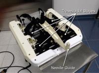
In-Bore MRI-Guided Transperineal Prostate Biopsy using 4-DOF Needle-Guide Manipulator
Junichi Tokuda1, Kemal Tuncali1, Gang 
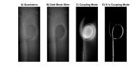
Li2, Nirav Patel2, Tamas Heffter3, Gregory S Fischer2, Iulian I Iordachita4, Everette Clif Burdette 3, Nobuhiko Hata1, and Clare M Tempany1
1Department of Radiology, Brigham and Women's Hospital, Boston, MA, United States, 2Department of Mechanical Engineering, Worcester Polytechnic Institute, Worcester, MA, United States, 3Acoustic MedSystems Inc., Savoy, IL, United States, 4Department Of Mechanical Engineering, Johns Hopkins University, Baltimore, MD, United States
We present the clinical feasibility of our MRI-compatible 4-DOF needle-guide manipulator for in-bore MRI-guided transperineal prostate biopsy. Total 11 men were biopsied in a 3T MRI scanner using this manipulator. All 11 procedures were successfully performed in 102.6±24.5 minutes with targeting errors of 4.9±2.9 mm. The targeting errors were consistent with other clinical studies. Pathology results confirmed prostate cancer with Gleason score ≥ 6 in 5/6 men with previous negative TRUS biopsies, and upgraded 2/5 men on active surveillance to clinically significant cancer with Gleason score 7. In conclusion, In-bore MRI-guided prostate biopsy using the manipulator was feasible.


3573.   
2 
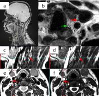
Motion compensated high resolution MR Imaging of Vagus and Recurrent Laryngeal Nerves with Novel Phase-based Navigation Sequences
Ravi Teja Seethamraju1, Jayender Jagadeesan2, Vera Kimbrell2, Aida Faria2, Thomas C Lee2, and Daniel T Ruan3
1MR R&D, Siemens Healthcare, Boston, MA, United States, 2Radiology, Brigham and Women's Hospital, Boston, MA, United States, 3Endocrine Surgery, Brigham and Women's Hospital, Boston, MA, United States
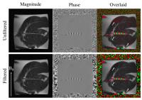
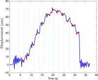
Diagnostic imaging of the recurrent laryngeal (RLN) and vagus nerves (VN) could help in surgical planning and in minimizing the risk of damage to the nerves. However, imaging these nerves is technically challenging due to their size, location, and physiological motions such as breathing and swallowing. The RLN and VN can be visualized on the CISS and T2 TSE, however with a novel phase navigator, the nerves are better delineated on the motion-compensated T2 TSE compared to the CISS which is un-navigated.


3574.   
3 
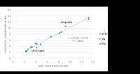
Geometry of Basal Ganglia nuclei in QSM and Histology in Parkinson’s disease brains
Carsten Stueber1,2, Alexey Dimov1, Kofi Deh1, David Pitt2, and Yi Wang1
1Weill Cornell Medical College, New York, NY, United States, 2Yale School of Medicine, Yale University, New Haven, CT, United States
Quantitative susceptibility mapping (QSM) provides a quantitative MRI contrast, which reflects the local iron concentration. Thus, QSM allows to determine the geometries of iron-rich deep grey matter nuclei including substantia nigra (SN) and subthalamic nucleus (STN). These basal nuclei are of particular interest in Parkinson’s disease. However, the measured dimensions need to be validated in histology using post-mortem human brain tissue. In this work, we show the concordance of the geometries measured in QSM and histology using Perls’ iron stain, which opens the door to use QSM as a pre-surgical mapping for deep brain stimulation targeting the STN.


3575.   
4 
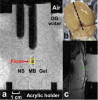
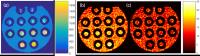
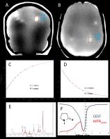
Multi-parametric MRI Characterization of a Polymer Gel Dosimetry Phantom for Non-Invasive 3D Visualization of Radiation Deposition in Gamma Knife Therapy
Ivan E Dimitrov1,2 and Strahinja Stojadinovic3
1Philips Medical Systems, Dallas, TX, United States, 2Advanced Imaging Research Center, UT Southwestern Medical Center, Dallas, TX, United States, 3Radiation Oncology, UT Southwestern Medical Center, Dallas, TX, United States
Radiation therapy aims to maximize dose delivery to tumor areas while minimizing the exposure to healthy tissue. Quality control is required to ensure that the delivered dose closely matches the calculated dose.   We performed a patient-specific quality assurance for cranial radiotherapy using MRI to visualize delivered radiation dose. We utilized an anthropomorphic 3D printed head phantom filled with polymer gel that was scanned before and after exposure to Gamma Knife irradiation. Irradiation changed the polymerization state of the gel and multi-parametric (T1, T2, MR Spectroscopy, CEST) quantitative dose-imaging maps were generated that may lead to optimized patient-specific dose delivery planning.


3576.   
5 TOLD MRI Validation of Reversal of Tumor Hypoxia in Glioblastoma with a Novel Oxygen Therapeutic
Heling Zhou1, David Wilson2, Jason Lickliter3, Jeremy Ruben4, Natarajan Raghunand5, Michael Sellenger6, Ralph P Mason7, and Evan Unger2,8
1UT Southwestern Medical Center, Dallas, TX, United States, 2NuvOx Pharma, Tucson, AZ, United States, 3Nucleus Networks, Melbourne, Australia, 4William Buckland Radiotherapy Centre, Melbourne, Australia, 5Moffitt Cancer Center, Tampa, FL, United States, 6Alfred Hospital, Prahran, Australia, 7Radiology, UT Southwestern Medical Center, Dallas, TX, United States, 8Medical Imaging, The University of Arizona, Tucson, AZ, United States
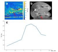
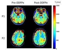
Glioblastoma multiforme (GBM) is known to be a hypoxic tumor and hypoxia adversely affects response to radiation therapy. Dodecafluoropentane emulsion (DDFPe) can improve oxygenation. Tissue oxygen level dependent (TOLD) MRI is an oxygen sensitive imaging technique which is used in this study to assess the improvement of oxygenation after administration of DDFPe. Two different doses were tested and each showed decreased T1 indicating improved oxygenation. 


3577.   
6 
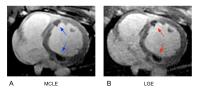
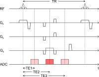
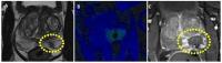
Towards DWI Guidance of Percutaneous Biopsies using Dual Echo Steady State Sequence: Qualitative Assessment in Liver
Elena A Kaye1, Kristin L Granlund2, Stephen B Solomon2, and Majid Maybody2
1Medical Physics, Memorial Sloan Kettering Cancer Center, New York, NY, United States, 2Radiology, Memorial Sloan Kettering Cancer Center, New York, NY, United States
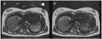
Acquisition of a viable tissue sample is critical to success of a biopsy. DWI could help differentiate between viable and necrotic tissue during the procedure, however, EPI-DWI is not suitable in a percutaneous-biopsy setting due to geometric distortions. DW Dual Echo Steady State (DESS) sequence allows acquisition of 3D undistorted DWI images. This study evaluated the application of DW-DESS during an MR-guided liver biopsy in two patients. Using single breath-hold acquisition, DW-DESS image depicted a liver lesion sharper and less distorted than EPI-DWI. DW-DESS also allowed DWI in the presence of a biopsy needle without distortions of EPI.  


3578.   
7 Scannerless real-time MRI
Frank Preiswerk1, Cheng-Chieh Cheng1, Sanjay S. Yengul1,2, Lawrence P. Panych1, and Bruno Madore1
1Radiology, Brigham and Women's Hospital, Harvard Medical School, Boston, MA, United States, 2Mechanical Engineering, Boston University, Boston, MA, United States
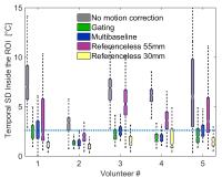
MRI can provide favorable image quality for image-guided interventions, but both magnetic field of the scanner and limited patient access inside the bore impose many limitations on image-guidance endeavors. The aim of this 
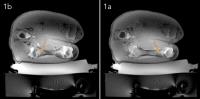
work was to estimate MRI of respiratory organ motion outside the scanner, which allows for a wider range of interventional applications. A single-element ultrasound transducer was used as a surrogate for the MR scanner outside the bore, after the correlation between both signals had been learned in a preceding training phase. Validation of estimated MR images outside the bore was performed using tracked 2D ultrasound.


3579.   
8 
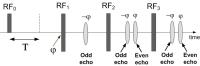
Real-Time Golden Angle Radial iSSFP for Interventional MRI
Samantha Mikaiel1,2, Thomas Boyd Martin1,2, Kyung Sung1,2, and Holden H Wu1,2
1Radiological Sciences, University of California, Los Angeles, Los Angeles, CA, United States, 2Biomedical Physics, University of California, Los Angeles, Los Angeles, CA, United States
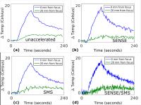
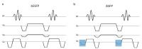
Real-time visualization is crucial to the success of MRI-guided minimally invasive cancer interventions. In this work we combine iSSFP with a golden-angle(GA) ordered radial trajectory and non-Cartesian parallel imaging to create a new real-time MRI sequence with good tissue contrast while suppressing bSSFP banding artifacts. Phantom and volunteer data were acquired and reconstructed using a combined sliding-window SPIRiT algorithm, at different frame rates, showing the capability of the sequence to achieve real-time imaging.  These advantages of GA Radial iSSFP show its potential for improving real-time MRI-guided interventions.


3580.   
9 The Development of Tissue Mimicking Gels
Peter Andrew Hardy1, Christopher J Norsigian2, Walter Witschey3, and Luke H Bradley2
1Radiology, University of Kentucky, Lexington, KY, United States, 2Anatomy & Neurobiology, University of Kentucky, Lexington, KY, United States, 3Smilow Center for Translational Research, University of Pennsylvania, Philadelphia, PA, United States
Developing tissue mimicking materials can be helpful in reducing the cost and duration of experiments which otherwise require animals. We tested a variety of agarose gels of different gel strength as suitable tissue mimicking material for convection enhanced delivery. The results demonstrate a significant difference in infusion volume and we relate that, through MR measurements, to the mechanical stiffness of the gels. 


3581.   
10 
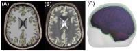
A four-layer boundary element model for MRI-guided transcranial magnetic stimulation
Aapo Nummenmaa1 and Matti Stenroos2
1Athinoula A. Martinos Center for Biomedical Imaging, Department of Radiology, Massachusetts General Hospital, Charlestown, MA, United States, 2Department of Neuroscience and Biomedical Engineering, Aalto University, Espoo, Finland
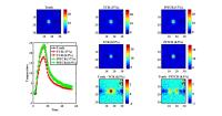
MRI-guided targeting and dosing has the potential increase the consistency and efficacy of transcranial magnetic stimulation (TMS). We propose a boundary element method (BEM) approach for estimating the TMS-induced cortical electric fields (E-fields). The method can be applied based on standard T1/T2-weighted MRI data and can incorporate the cerebrospinal fluid (CSF) as a separate conductivity compartment. Our results show that the CSF layer may increase the estimated E-field amplitudes up to 25%. The effect of the CSF depends on the location and orientation of the TMS coil/target in a rather intricate manner, highlighting the importance of individualized, realistically shaped models.


3582.   
11 
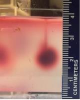
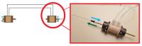
Measurement and Simulation of Susceptibility Artifacts in Variable-TE Radial MRI: Application in an MR-safe Guidewire
Katharina E. Schleicher1, Stefan Kroboth1, Klaus Düring2, Michael Bock1, and Axel Joachim Krafft1,3,4
1Dept. of Radiology - Medical Physics, University Medical Center Freiburg, Freiburg, Germany, 2MaRVis Medical GmbH, Hannover, Germany, 3German Cancer Consortium (DKTK), Heidelberg, Germany,4German Cancer Research Center (DKFZ), Heidelberg, Germany
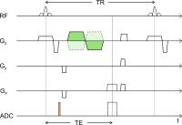
A simulation framework is presented to optimize radial acquisition schemes with variable echo times which are designed to minimize the directional anisotropy of the artifact of an MR-safe guidewire. The simulation results are compared to measurements. We could theoretically and experimentally verify that the homogeneity of the artifact can be improved via the variable-TE method.


3583.   
12 
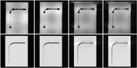
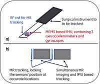
First steps towards concurrent, high rate imaging and MR tracking using an inertial measurement unit (IMU)
Robert Darrow1, Mauricio Castillo-Effen1, Eric Fiveland1, Elizabeth Morris2, and Ileana Hancu1
1GE Global Research Center, Niskayuna, NY, United States, 2Memorial Sloan Kettering Cancer Center, New York City, NY, United States
Tissue motion during MR guided interventional procedures leads to the desire to perform simultaneous high speed tracking of the surgical instrument and imaging. In this work, a novel approach for concurrent tracking and imaging, based on an inertial measurement unit (IMU) and technology from plane/missile tracking, is presented. While using infrequent position updates for the IMU (that could be provided by MR tracking), we have showed fast tracking (166Hz) of the IMU sensor with ~2mm rms error.


3584.   
13 Interventional device visualisation using the coupling mode of a PTx transmit array
Francesco Padormo1, Arian Beqiri1, Joseph V Hajnal1, and Shaihan Malik1
1Division of Imaging Sciences and Biomedical Engineering, King's College London, London, United Kingdom
We propose a novel method to visualise guidewires in interventional MRI procedures using the coupling mode of a PTx array.


3585.   
14 Automatic high temporal and spatial resolution position verification of an HDR brachytherapy source using subpixel localization and SENSE
Ellis Beld 1, Marinus A. Moerland1, Frank Zijlstra2, Jan J.W. Lagendijk1, Max A. Viergever2, and Peter R. Seevinck2
1Department of Radiotherapy, UMC Utrecht, Utrecht, Netherlands, 2Image Sciences Institute, UMC Utrecht, Utrecht, Netherlands
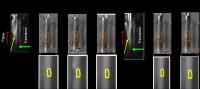
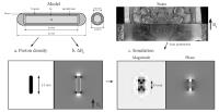
In order to verify the positions of a high-dose-rate (HDR) brachytherapy source during treatment, fast imaging and post-processing are needed. To get high temporal resolutions, the use of lower spatial resolutions in combination with subpixel source localization and the use of parallel imaging were introduced. MR artifacts were simulated and correlated to the experimentally obtained artifacts (by phase-only cross correlation) to determine the position of the HDR source. It was shown that the described method was fast enough for localization of an HDR brachytherapy source in real-time and high accuracy and precision (submillimeter scale) were achieved. 


3586.   
15 Hindered diffusion of Gadolinium-based Contrast Agents in rat brain extracellular micro-environment after ultrasound-induced delivery
Allegra Conti1,2, Rémi Magnin1,3, Matthieu Gerstenmayer1, François Lux4, Olivier Tillement4, Sébastien Mériaux 1, Stefania Della Penna2, Gian Luca Romani2, Erik Dumont3, Denis Le Bihan1, and Benoît Larrat1
1CEA/DSV/I2BM/NeuroSpin, Gif Sur Yvette, France, 2Department of Neuroscience, Imaging and Clinical Sciences, G. D'Annunzio, University of Chieti and Pescara, Chieti, Italy, 3Image Guided Therapy, Pessac, France, 4Université Lyon 1, Lyon, France
We present here a new method to study the diffusion process of Gadolinium-based Contrast Agents within the brain extracellular space after the artificial Blood-Brain Barrier opening induced by ultrasound. Four compounds were tested (MultiHance, Gadovist, Dotarem and AGuIX). By estimating the Free Diffusion Coefficients from in vitro studies, and the Apparent Diffusion Coefficients from in vivo experiments, an evaluation of the tortuosity (λ) in the right striatum of 11 Sprague-Dawley rats has been performed. The values of λ are in agreement with literature and demonstrate that the chosen permeabilization protocol maintains the integrity of brain tissue.


3587.   
16 
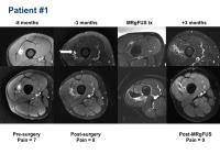
3D Histogram Analysis of Apparent Diffusion Coefficient Maps Predicts Relief of Fibroid Symptoms after MR Imaging–guided High-Intensity Focused Ultrasound Ablation - Permission Withheld
HAO FU1,2, Chenxia Li1, Rong Wang1, Jianxin Guo1, Bilgin Keserci3, and Jian Yang1
1Department of Radiology, the First Affiliated Hospital of Xi'an Jiaotong University, xi'an, China, People's Republic of, 2MR Marketing, Philips Healthcare, xi'an, China, People's Republic of, 3MR Therapy Clinical Science, Philips Healthcare, Seoul, Korea, Republic of
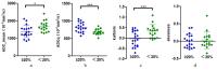
The aim of the study was to investigate the variation among screening fibroids through analysis of ADC histogram, in order to predict fibroids residual NPV proportion (residual NPV%=NPV at 6 months follow up/ NPV immediately after treatment ) and patients Symptom Severity Score (SSS). Thirty five patients who accepted MRgHIFU ablation were divided into group 1 (residual NPV%≥20%) of 19 patients and group 2 (residual NPV%<20%) of 16 patients, respectively. The SSS of patients were obtained at two time-point, screening and 6 months follow up. ADCmean, ADCq, kurtosis and skewness are derived from ADC histogram. The results showed that values of ADCmean, ADCq and kurtosis were significant difference between two groups. The average SSS reduction of group 1 between pre and post treatment was more obvious than that of group 2. Therefore, histogram analysis of ADC maps can provide the quantitative information to predict fibroids ablation outcome and patients symptom relief, which may be indicated as a useful screening tool to guide patients selection for MRgHIFU ablation.


3588.   
17 Real-time MRI-guided interventions using rolling-diaphragm hydrostatic actuators
Samantha Mikaiel1,2, James Simonelli3, David Lu1, Kyung Sung1,2, Tsu-Chin Tsao3, and Holden H Wu1,2
1Radiological Sciences, University of California, Los Angeles, Los Angeles, CA, United States, 2Biomedical Physics, University of California, Los Angeles, Los Angeles, CA, United States, 3Mechanical and Aerospace Engineering, University of California, Los Angeles, Los Angeles, CA, United States
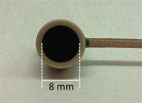
In this work we investigate a new rolling-diaphragm-based hydrostatic actuator design to achieve smooth remote manipulation without fluid leakage for MR-compatible robotic systems. We show that the actuators exhibit negligible impact on MR image fidelity and SNR, the actuator provides a linear displacement response over the fluid lines, and we were able to use the master/slave actuator pair to insert and retract the needle in a phantom with no leakage and no noticeable friction issues. Our new rolling-diaphragm hydrostatic actuators can potentially enable physicians to remotely perform real-time MRI-guided interventions.


3589.   
18 A Simple Scanner Control Technique for Device Localization during MRI-Guided Percutaneous Procedures
Matthew Alexander MacDonald1,2, Adam C. Waspe3,4, Joao Amaral3,4, and Samuel Pichardo1,2
1Electrical Engineering, Lakehead University, Thunder Bay, ON, Canada, 2Thunder Bay Regional Research Institute, Thunder Bay, ON, Canada, 3Medical Imaging, University of Toronto, Toronto, ON, Canada, 4Hospital for Sick Children, Toronto, ON, Canada
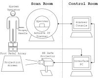
An experiment demonstrates a simple technique for a clinician operator to interactively align scan planes to an interventional device within the scan room during an MRI guided procedure. Input is collected using foot pedal switches to select axial/device intersection points to specify a virtual line of best fit about which auxiliary views are aligned automatically. Volunteer operators position a biopsy needle within an ex vivo porcine specimen while interactively tracking the needle position and measurements are taken to assess the technique's efficiency.


3590.   
19 
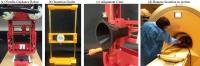
Experimental study of MR Compatible RF Hyperthermia System
Han-Joong Kim1, Jong-Min Kim1, Young-Seung Jo1,2, Suchit Kumar1, Seong-Dae Hong1, Chulhyun Lee2, and Chang-Hyun Lee1
1Electronics and Information Engineering, Korea University, Seoul, Korea, Republic of, 2The MRI Team, Korea Basic Science Institute, Cheongju, Korea, Republic of
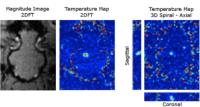
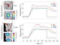
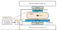
  Many reports suggest that hyperthermia is very effective treatment for tumor therapy. In this work, MR compatible RF hyperthermia system is presented for a 3.0 T MRI. Phantom and animal experiments have been conducted and the results compared with the simulations results for tumor and tissue model. They are in very good coincidence with each other, which confirms the utility and feasibility of the MR compatible RF hyperthermia system with capacitive driving.  


3591.   
20 Tracking of a Robotic Device by Controlling the Visibility of Markers from the Robot Control
Junmo An1, Eftychios G. Christoforou2, Karen Chin3, Jeremy Hinojosa3, Dipan J. Shah3, Andrew G. Webb4, and Nikolaos V. Tsekos1
1University of Houston, Houston, TX, United States, 2University of Cyprus, Nicosia, Cyprus, 3Houston Methodist, Houston, TX, United States, 4Leiden University Medical Center, Leiden, Netherlands
Integrated control system of the manipulator and marker control is important for localization and tracking of multiple optically detunable MR markers on MR-compatible manipulators. Selecting which markers are visible on MR images by the motion of the maneuvering portion of the MR-compatible manipulators allows unambiguous identification of a combination of markers and simplifies both the data acquisition and the post processing. This proposed technique can be employed to track multiple marker positions on interventional devices such as the steerable catheters and the end-effectors of the MR-compatible manipulator.


3592.   
21 Visualization of interventional devices by transient, local magnetic field alterations using bSSFP sequences - Permission Withheld
Frank C Eibofner1, Hansjörg Graf1, and Petros Martirosian1
1University Hospital Tübingen, Tübingen, Germany
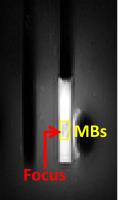
A technique for the visualization of interventional devices by use of transient, local magnetic field alterations and bSSFP sequences is presented. It allows the generation of distinct artifacts with controllable dimension. The instrument is visualized in the phase image obtained in the same scan as the undisturbed anatomical image. Localization is done by subsequent superposition.


3593.   
22 
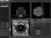
On Demand Reprogramming of MR Sequence Parameters Using MatMRI
Samuel Pichardo1,2, Charles Mougenot3, Steven Engler1,4, Adam C. Waspe5,6, and James Drake5,6
1Thunder Bay Regional Research Institute, Thunder Bay, ON, Canada, 2Electrical Engineering, Lakehead University, Thunder Bay, ON, Canada, 3Philips Healthcare, Toronto, ON, Canada, 4Computer Science, Lakehead University, Thunder Bay, ON, Canada, 5University of Toronto, Toronto, ON, Canada, 6The Hospital For Sick Children, Toronto, ON, Canada
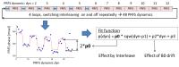
For dynamic studies, it is often desirable to adjust parameters in “real time” depending on decisions made by user-defined algorithms. We performed a modification to the software tool MatMRI to develop a method that allows changing on demand multiple MR sequence parameters. We present results of this new method when applied to the modification of sequence parameters for MR-Acoustic Radiation Force Imaging. Our results demonstrate that it is feasible to reprogram MR sequence parameters dynamically using existing technology for the control of scanners.


3594.   
23 Efficient respiratory navigator-based 4D MRI
Sascha Krueger1 and Tim Nielsen1
1Philips GmbH, Innovative Technologies, Research Laboratories, Hamburg, Germany
4D image data are used in radiation therapy planning to estimate motion of the tumor or regions at risk. Today 4D CT is commonly used for this purpose. Due to better soft tissue contrast and quantitative and functional imaging, a clinical demand for MRI in therapy planning exists. Consequently, there is also a growing interest in 4D MRI techniques. A scan-time-efficient 4D MRI method utilizing the MRI Navigator as motion sensor with high geometric fidelity is proposed.


3595.   
24 MR imaging patterns of Cholangiocarcinoma and post-intervention features: A case-based approach - Video Not Available
Juan C Camacho1, Courtney Moreno1, Peter Harri1, and Pardeep Mittal1
1Department of Radiology and Imaging Sciences, Emory University School of Medicine, Atlanta, GA, United States
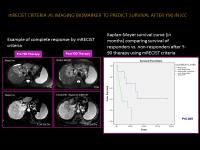
Cholangiocarcinoma may demonstrate typical imaging manifestations and common patterns of organ involvement, guiding diagnosis, and facilitating imaging follow up after therapy. Adequate knowledge of tumoral biology in cholangiocarcinoma and current image-guided therapeutic approaches, along with imaging appearance of cholangiocarcinoma before and after image-guided interventions is crucial for adequate diagnosis and surveillance .MR imaging plays a key role for patient management, assessing therapy response and patient surveillance.
Exhibition Hall 

17:00 - 18:00

    Computer #

 
3596.   
25 Real-Time Monitoring of Focused Ultrasound Induced Inertial Cavitation on Microbubbles by Using Gradient Echo MRI: in Vitro and in Vivo Experiments - Permission Withheld
Chen-Hua Wu1, Shih-Tsung Kang1, Chih-Kuang Yeh1, Wen-Shiang Chen2, and Hsu-Hsia Peng1
1Biomedical Engineering and Environmental Sciences, National Tsing Hua University, Hsinchu City, Taiwan, 2Physical Medicine and Rehabilitation, National Taiwan University Hospital, Taipei, Taiwan
This study aims to real-time monitor inertial cavitation on microbubbles (MBs) while transmitting focused ultrasound (FUS) with various duty cycle and pulse repetitive frequency (PRF) by gradient echo MRI. For in vitro experiments, with increasing duty cycle, more significant signal intensity (SI) changes or prolonged SI drop duration were shown. For in vivo experiment, two SI drop peaks were observed, reflecting the two IC events. In conclusion, FLASH has been proved to be a useful technique for real-time monitoring of IC when transmitting FUS pulses to MBs for in vitro and in vivo experiments.


3597.   
26 Multi-Contrast Late Enhancement Out-Performs Conventional Late Gadolinium Enhancement for Myocardial RFA Lesion Characterization
Philippa Krahn1,2, Haydar Celik3, Venkat Ramanan2, Jennifer Barry2, and Graham A Wright1,2
1Medical Biophysics, University of Toronto, Toronto, ON, Canada, 2Physical Sciences, Sunnybrook Research Institute, Toronto, ON, Canada, 3Sheikh Zayed Institute for Pediatric Surgical Innovation, Washington, DC, United States
In this work we evaluated the quality and accuracy of myocardial RFA lesion visualization using the MCLE sequence. To compare MCLE to the standard LGE, we imaged 29 acute lesions in 11 pigs. Lesion sizes measured from each set of images were highly correlated, yet MCLE consistently achieved higher CNR than LGE. Our relaxometry maps from MCLE suggested a dramatic variation in T1 after contrast injection, which will alter the optimal TI for LGE. Therefore, we demonstrated that MCLE, with the varied TIs in each acquisition, is a more robust sequence for visualizing even the most subtle lesions.


3598.   
27 Improved Thermometry Based on a Fast Spin Echo Sequence
Yuval Zur1
1GE Healthcare, Haifa, Israel
A time shifted Fast Spin Echo (FSE) thermometry sequence which is insensitive to B0 inhomogeneity is used in conjunction with restricted FOV module and parallel imaging to reduce scan time and increase the number of slices. Up to 5 slices are acquired in 3 sec. This sequence works well on a clinical Focused Ultrasound brain system. The temperature signal to noise ratio (TSNR) of this FSE sequence is approximately 2 times higher than the conventional Gradient Echo (GRE) sequence used today. 3 to 5 slices are acquired in 3 sec, rather than 1 slice with GRE.


3599.   
28 Prostate Focal Prostate Laser Ablation under MRI Guidance
Alexander Squires1, Sheng Xu2, Reza Seifabadi2, Yue Chen1, Harsh Agarwal3, Marcelino Bernardo2, Ayele Negussie2, Peter Pinto2, Peter Chokye2, Bradford Wood2, and Zion Tsz Ho Tse1
1College of Engineering, University of Georgia, Athens, GA, United States, 2National Institutes of Health, Bethesda, MD, United States, 3Philips Research North America, Briarcliff Manor, NY, United States
MRI-guided focal laser ablation (FLA) shows promise as an effective treatment for prostate cancer, but presents challenges related to accurate catheter positioning and ablation monitoring. A catheter positioning robot was designed to overcome these problems by positioning the catheter optimally for ablation and monitoring the ablation via MRI thermometry. A remote insertion guide allows the surgeon to perform the procedure without removing the patient from the MRI bore, saving time and increasing accuracy. Treatment planning software iteratively plans optimal catheter positioning. Preliminary tests show the robot can greatly improve efficiency and accuracy without sacrificing image quality.


3600.   
29 PRF Thermometry for Monitoring Small Temperature Changes during Very Long Thermal Therapies: Field Drift Compensation Using FID Navigators
Tetiana Dadakova1, Axel Joachim Krafft1,2,3, Jan Gerrit Korvink4, Stephan Meckel5, and Michael Bock1
1Dept. of Radiology - Medical Physics, University Medical Center Freiburg, Freiburg, Germany, 2German Cancer Consortium (DKTK), Heidelberg, Germany, 3German Cancer Research Center (DKFZ), Heidelberg, Germany, 4Institute of Microstructure Technology, Karlsruhe Institute of Technology, Karlsruhe, Germany, 5Department of Neuroradiology, Neurocenter, University Medical Center Freiburg, Freiburg, Germany
Proton resonance frequency shift thermometry uses phase MR images to calculate temperature change in tissue during thermal therapies. Temporal and spatial changes of local magnetic field influence the phase images and can mimic temperature change. In order to correct for temperature errors due to magnetic field drift with time the FID navigators were used before and after imaging readout. Field-drift related phase slope during each repetition was calculated and used to correct the imaging data. This correction method is especially useful for the long thermometry procedures during which the temperature change is small and high temperature precision is needed.


3601.   
30 MR-guided focal laser ablation as primary treatment for prostate cancer: Preliminary results
Joyce GR Bomers1, Christiaan G Overduin1, Sjoerd FM Jenniskens1, Michiel Sedelaar2, and Jurgen J Futterer1
1Radiology, Radboud University Medical Center, Nijmegen, Netherlands, 2Urology, Radboud University Medical Center, Nijmegen, Netherlands
Focal therapy offers great hopes in terms of cancer control and decreased morbidity (i.e. impotence and incontinence) for patients with localized for low- and intermediate grade prostate cancer (PCa). Transrectal MR-guided focal laser ablation of newly diagnosed PCa was successfully performed in 5 patients. It was technically feasible and safe. PSA level decreased in all patients and follow-up MRI showed no residual or recurrent cancer; indicating local cancer control, without compromising with increased morbidity or a decrease in quality of life. Initial results are promising and more patients have to be included with longer follow-up.


3602.   
31 MR Thermometry-guided Prostate Hyperthermia with Real-time Ultrasound Beamforming and Power Control
Eugene Ozhinsky1, Vasant A. Salgaonkar2, Chris J. Diederich2, and Viola Rieke1
1Radiology and Biomedical Imaging, University of California San Francisco, San Francisco, CA, United States, 2Radiation Oncology, University of California San Francisco, San Francisco, CA, United States
We have developed and evaluated a real-time MR thermometry-guided system for acoustic power output and beam pattern control for prostate hyperthermia therapy. It was designed to be integrated with a commercial focused ultrasound ablation system and supports real-time ultrasound phase pattern switching and MR thermometry for monitoring and automated power adjustment.


3603.   
32 Zero TE based MR thermometry
Silke Lechner-Greite1, Matthew Tarasek2, Desmond Teck Beng Yeo2, and Florian Wiesinger1
1GE Global Research, Munich, Germany, 2GE Global Research, Albany, NY, United States
Monitoring temperature changes in the human skull is important in clinical transcranial MR-guided focused ultrasound, as it can absorb a large amount of energy during sonication and thereby produce heat. We propose to extract relative temperature information from proton density weighted zero TE imaging and additionally calibrate the T1 signal contamination effect.


3604.   
33 Evaluation of common proton resonance frequency shift based MR Thermometry methods in the pancreas
Cyril J Ferrer1, Clemens Bos1, Marijn van Stralen1, Baudoin Denis de Senneville2, Chrit T.W Moonen1, and Lambertus W Bartels1
1University Medical Center Utrecht, Utrecht, Netherlands, 2Mathematical Institute of Bordeaux, Talence, France
MR-guided High Intensity Focused Ultrasound has recently been suggested to provide non-invasive alternative treatment options for pancreatic cancer patients. To successfully apply PRFS MR Thermometry in the pancreas motion correction techniques have to be applied. For other moving organs, several solutions have already been proposed to this end: gating, multibaseline and referenceless. However, due to specific properties of the pancreas, there was still a need to evaluate the performance of these methods in this organ. Our results show that all the evaluated techniques improved thermometry in the pancreas to a level that is sufficient to monitor thermal ablation.


3605.   
34 MR Guided Microwave Thermal Ablation: Guidance and Monitoring for Extending the Physics of Thermal Therapy
Michael Simonson1, Peng Wang2, Christopher Brace1,2,3, and Walter Block1,2,3
1Biomedical Engineering, UW - Madison, Madison, WI, United States, 2Radiology, UW - Madison, Madison, WI, United States, 3Medical Physics, UW - Madison, Madison, WI, United States
Monitoring microwave ablation with thermometry could extend the application of this clinical procedure into territories where greater control is necessary to protect healthy tissue, such as the spine. MRI thermometry could also provide useful insights to generate and validate new thermal models based on the fundamentally different mode of dielectric heating utilized by microwave ablation.  We present here a platform consisting of 1) a high power MR compatible microwave ablation system based on a FDA-approved design, 2) real-time applicator guidance during applicator insertion and 3) high frame rate MR thermometry and tissue monitoring.


3606.   
35 T2-based Temperature Monitoring in Bone Marrow for MR-guided Focused Ultrasound
Eugene Ozhinsky1, Matthew D. Bucknor1, and Viola Rieke1
1Radiology and Biomedical Imaging, University of California San Francisco, San Francisco, CA, United States
Current clinical protocols for bone treatments rely on measurement of the temperature change of adjacent muscle to estimate the temperature of the bone. In this study we have demonstrated for the first time that T2-based thermometry can be used in vivo to measure the heating in the marrow during bone ablation. The ability to monitor the temperature within the bone marrow allowed visualization of the heat penetration into the bone, which is important for local lesion control and treatment of osteoid osteomas.


3607.   
36 Monitoring of Stable Cavitation of Microbubbles by Using MRI - Permission Withheld
Cheng-Tao Ho1, Chen-Hua Wu1, Shih-Tsung Kang1, Chih-Kuang Yeh1, Wen-Shiang Chen2, Hau-Li Liu3, and Hsu-Hsia Peng1
1Biomedical Engineering and Environmental Science, National Tsing Hua University, Hsinchu, Taiwan, 2Physical Medicine and Rehabilitation, National Taiwan University Hospital, Taipei, Taiwan,3Electrical Engineering, Chang-Gung University, Taoyuan, Taiwan
Gas-filled microbubbles (MBs) can be locally cavitated by spatially focused ultrasound (FUS) to trigger stable cavitation (SC) and inertial cavitation (IC) to induce blood–brain barrier (BBB) opening ; therefore, magnetic resonance imaging (MRI) may provide useful imaging information to real-time monitor the process of FUS cavitation on MBs. In this study, a HASTE (Half Fourier Acquisition Single Shot Turbo Spin Echo) sequence was used to observe the signal changes during SC for in vitro experiments. Experiments with different acoustic pressure, pulse repetitive frequency, and duty cycle were performed to clarify the SC effect on MR images.


3608.   
37 The effect of sonication duration on ablation depth during MR-guided focused ultrasound of bone: acute findings with MR in a swine model
Matthew Bucknor1, Rutwik Shah1, Eugene Ozhinsky1, and Viola Rieke1
1Radiology and Biomedical Imaging, University of California, San Francisco, San Francisco, CA, United States
One challenge in magnetic resonance guided focused ultrasound ablation of bone lesions is extending the ablation zone deep to the cortical surface in order to cover the full intramedullary extent of the lesion. Extension of the ablation zone is important for complete treatment of for example, osteoid osteomas, and improved local control of metastatic bone disease. This study used a swine model to compare differences in the depth of the ablation zone between two protocols using short versus longer sonications, respectively, while maintaining the same overall sonication energy (by adjusting acoustic power).


3609.   
38 Assessments of Flow Velocity Changes by Phase-Contrast MRI Near Acoustic Radiation Force Aggregated Bubbles
Zhe-Wei Wu1, Wu Chen-Hua2, Hsu Po-Hong3, Hao-Li Liu3, Yeh Chic-Kuang2, and Peng Hsu-Hsia2
1Biomedical Engineering and Environmental Sciences, Nation Tsing Hua University, Hsinchu, Taiwan, 2Biomedical Engineering and Environmental Sciences, National Tsing Hua University, Hsinchu, Taiwan,3Electrical Engineering, Chang-Gung University, Taoyuan, Taiwan
The aim of this study was to real-time assess flow velocity changes in the neighboring pixels of acoustic radiation force (ARF) aggregated bubbles by using phase-contrast MRI (PC-MRI). The flow velocity increased with the increase of microbubbles (MBs) concentrations, which might be attributed to the increase of aggregated bubbles size, narrowing the chamber diameter and thus presenting higher flow velocity. In conclusion, we verified the feasibility of using PC-MRI to assess flow velocity changes on pixels near ARF aggregated bubbles. In the future, a systematic investigation shall be conducted to comprehend the association between bubble size and the flow velocity.  


3610.   
39 Prior Image based Temporally Constrained Reconstruction for Magnetic Resonance guided HIFU
Jaya Prakash1, Nick Todd2, and Phaneendra K Yalavarthy3
1Institute for Biological and Medical Imaging, Helmholtz Zentrum Munich, Munich, Germany, 2Wellcome Trust Centre for Neuroimaging, UCL Institute of Neurology, University College London, London, United Kingdom, 3Supercomputer Education and Research Centre, Indian Institute of Science, Bangalore, India
Prior image based temporally constrained reconstruction (PITCR) algorithm was found to obtain accurate temperature maps with better volume coverage, and spatial, and temporal resolution than other algorithms using highly undersampled data in magnetic resonance (MR) thermometry. PITCR method is compared with the standard temporally constrained reconstruction (TCR) algorithm using ex-vivo pork muscle sample. It was seen that the PITCR method showed superior performance compared to the TCR approach with highly undersampled datasets. 


3611.   
40 Phase artifact correction for improved ARFI displacement mapping using a short FID navigator
Tetiana Dadakova1, Ali Caglar Özen1, Axel Joachim Krafft1,2,3, Jan Gerrit Korvink4, and Michael Bock1
1Dept. of Radiology - Medical Physics, University Medical Center Freiburg, Freiburg, Germany, 2German Cancer Consortium (DKTK), Heidelberg, Germany, 3German Cancer Research Center (DKFZ), Heidelberg, Germany, 4Institute of Microstructure Technology, Karlsruhe Institute of Technology, Karlsruhe, Germany
MR-guided acoustic radiation force imaging (ARFI) is used for the focal spot localization during high intensity focused ultrasound (HIFU) therapies. The acoustic radiation force related tissue displacement is measured with help of motion encoding gradients (MEG). While MEGs are sensitive enough to detect micrometer-scale displacement, the images acquired using MR-ARFI pulse sequences are often corrupted with artifacts. The method described uses very short FID navigator readout for correction of ARFI images without any manual interaction. ARFI displacement maps corrected by FID navigator have substantially less artifacts, 2.5 times higher SNR, and clearly visible HIFU focal spot.


3612.   
41 TREATMENT OF SMALL VASCULAR MALFORMATIONS USING MR GUIDED HIGH INTENSITY FOCUSED ULTRASOUND: PRELIMINARY RESULTS AFTER FOUR PATIENTS
Pejman Ghanouni1, Sirish Kishore1, Matthew Lungren1, David Mohler2, Raffi Avedian2, and David Hovsepian1
1Radiology, Stanford University, Stanford, CA, United States, 2Orthopaedic Surgery, Stanford University, Stanford, CA, United States
Low flow vascular malformations are typically treated with ultrasound-guided percutaneous sclerotherapy, but not all of these lesions are visible sonographically. MR guided focused ultrasound (MRgFUS) combines the ability of MRI to delineate these tumors with the use of FUS to ablate the tumor non-invasively. We report our technical and clinical results after using MRgFUS to treat four patients with small intramuscular low flow vascular malformations in the lower extremities. 


3613.   
42 First clinical evaluation of real-time cardiac MR thermometry
Valéry Ozenne1, Solenn Toupin1,2, Pierre Bour1, Baudouin Denis de Senneville3, Alexis Vaussy2, Matthieu Matthieu Lepetit-Coiffé2, Pierre Jaïs1,4, Hubert Cochet1,4, and Bruno Quesson1
1Institut Hospitalier Universitaire : LIRYC Institut de Rythmologie et Modélisation Cardiaque, Bordeaux, France, 2Siemens France, Saint Denis, France, 3Mathematical Institute of Bordeaux, Bordeaux, France, 4Centre Hospitalier Universitaire de Bordeaux, Bordeaux, France
Catheter ablation using radiofrequency is commonly used to treat cardiac arrhythmia. However, direct assessment of lesion formation with MRI thermometry during RF delivery may improve safety and efficiency of the therapeutic procedure. Despite recent studies demonstrating the feasibility on volunteers or animal models, there are no reports regarding the possibility of doing cardiac thermometry on patient in the context of arrhythmias. The purpose of the present study was to evaluate the uncertainty of temperature estimate on patients depicting potential arrhythmic episodes, which may alter the precision of the method relying on an ECG-triggered acquisition.


3614.   
43 Using simultaneous multi-slice imaging for PRFS thermometry
Pim Borman1, Clemens Bos2, Sjoerd Crijns1, Bas Raaymakers1, and Chrit Moonen2
1Radiotherapy, UMC Utrecht, Utrecht, Netherlands, 2Imaging Division, UMC Utrecht, Utrecht, Netherlands
PRFS thermometry is important for guidance of thermal therapies. Here we show that the thermometry sequence can be accelerated by the novel parallel imaging technique SMS-CAIPIRINHA and we compare it to an unaccelerated sequence and a SENSE accelerated sequence. Heating was applied by means of HIFU and LITT. A good agreement was seen between temperature curves from SMS-CAIPIRINHA and those from the unaccelerated sequence. Furthermore, the noise level was significantly lower compared to temperature curves from the SENSE accelerated sequence.


3615.   
44 Fully automatic in-vivo localization of LDR brachytherapy seeds for post-implant dosimetry using MR simulations and template matching
Frank Zijlstra1, Job G Bouwman1, Marinus A Moerland2, and Peter R Seevinck1
1Image Sciences Institute, UMC Utrecht, Utrecht, Netherlands, 2Department of Radiotherapy, UMC Utrecht, Utrecht, Netherlands
We present a method which can accurately localize brachytherapy seeds in MRI using fast MR simulations and template matching. In 5 patients we correctly detected and localized 298 out of 307 seeds. Using this method an MRI-only approach to post-implant dosimetry will be possible, which should increase accuracy by not requiring CT to MRI image registration.


3616.   
45 Towards Real-Time In Vivo Volumetric MR Thermometry
Samuel Fielden1, Matthew Geeslin2, Xue Feng1, Wilson Miller2, Kim Butts Pauly3, and Craig Meyer1,2
1Biomedical Engineering, University of Virginia, Charlottesville, VA, United States, 2Radiology & Medical Imaging, University of Virginia, Charlottesville, VA, United States, 3Radiology, Stanford University, Palo Alto, CA, United States
A rapid volumetric MR thermometry sequence, using a stack-of-spirals acquisition, was implemented on a real-time platform in order to support animal model experiments. The performance of the sequence, as measured by the maximum and mean recorded temperature as well as the sequence accuracy, was assessed against a clinically used 2DFT sequence in vivo. The use of efficient spiral trajectories supports rapid generation of volumetric thermometry maps and allows the visualization of the entire insonication as well as correlation with post-ablation imaging of lesion size and location.


3617.   
46 Accuracy of PRFS Temperature Mapping using Fast Interleaved Sequences for MR-HIFU Thermal Therapies
Steffen Weiss1, Jochen Keupp1, and Edwin Heijman2
1Tomographic Imaging, Philips Research, Hamburg, Germany, 2Oncology Solutions, Philips Research, Eindhoven, Netherlands
MR-HIFU therapies require stable PRFS-based temperature monitoring over the treatment duration (>30min). Drift effects may be corrected using independent temperature information e.g. based on T2 or T1 maps and simultaneously obtained using fast interleaved sequences. As interleaves may impact PRFS-accuracy, a dynamic on-off-interleaved sequence pattern is introduced to selectively measure their influence on the PRFS phase. The mechanism of the phase variations is explored using multiple variants of the interleaved sequence. It is shown that eddy currents induced during the interleaved sequences represent the main cause of the phase variation. The spatio-temporal characteristics of the effect are explored as a first step towards compensation.


3618.   
47 Automatic temperature control during MR guided catheter based radiofrequency ablation of the heart
Valery Ozenne1, Thibaud Troadec1, Pierre Bour1, Solenn Toupin1,2, Erik Dumont3, and Bruno Quesson1
1Institut Hospitalier Universitaire : LIRYC Institut de Rythmologie et Modélisation Cardiaque, Bordeaux, France, 2Siemens France, Saint Denis, France, 3Image Guided Therapy, Bordeaux, France
Although catheter-based radiofrequency (RF) ablation is increasingly used to treat cardiac arrhythmia, insufficient temperature increase may lead to incomplete treatment whereas excessive local energy deposition may result in severe adverse effects (e.g. esophageal fistulas or tamponade due to steam pop). Since MR thermometry can monitor in real-time the temperature distribution in the cardiac muscle, automatic regulation of RF energy deposition from temperature images, using well established proportional, integral controller algorithms, may improve safety and efficiency of the therapeutic procedure.


3619.   
48 TREATMENT OF OSTEOID OSTEOMA USING MR GUIDED HIGH INTENSITY FOCUSED ULTRASOUND: PRELIMINARY RESULTS AFTER FOUR PATIENTS
Pejman Ghanouni1, Sirish Kishore1, David Mohler2, Matthew Lungren1, Raffi Avedian2, and Nishita Kothary1
1Radiology, Stanford University, Stanford, CA, United States, 2Orthopaedic Surgery, Stanford University, Stanford, CA, United States
Osteoid osteoma is currently most frequently treated with CT guided radiofrequency ablation (CTgRFA), which is effective, but invasive and exposes the typical young patient population with osteoid osteomas to radiation. Our preliminary results are presented describing MR guided focused ultrasound (MRgFUS) for the treatment of patients with osteoid osteomas. As also reported in two European studies, in our experience, MRgFUS is effective at eliminating pain in these patients. These data form the basis for a proposed clinical trial directly comparing the safety and efficacy of MRgFUS and CTgRFA for treatment of osteoid osteoma.
 

The International Society for Magnetic Resonance in Medicine is accredited by the Accreditation Council for
Continuing Medical Education to provide continuing medical education for physicians.