|
Exhibition Hall 13:30 - 14:30 |
|
|
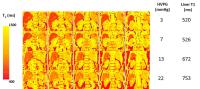
|
Computer # |
|
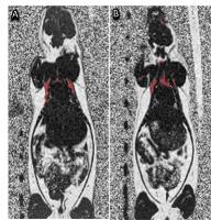
3836.
 |
1 |
Motion insensitive quantification of liver proton density
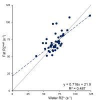
fat-fraction using a single-shot 2D technique 
Jeannine A. Ruby1, Diego Hernando1,
Camilo A. Campo1, Ann Shimakawa2, Karl
K. Vigen1, James H. Holmes3, Kang Wang3,
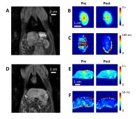
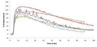
and Scott B. Reeder1,4,5,6,7
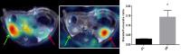
1Radiology, University of Wisconsin-Madison,
Madison, WI, United States, 2Global
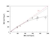
MR Applications and Workflow, GE Healthcare, Menlo Park, CA,
United States, 3Global
MR Applications and Workflow, GE Healthcare, Madison, WI,
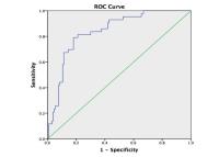
United States, 4Medicine,
University of Wisconsin-Madison, Madison, WI, United States, 5Medical
Physics, University of Wisconsin-Madison, Madison, WI,
United States, 6Biomedical
Engineering, University of Wisconsin-Madison, Madison, WI,
United States, 7Emergency
Medicine, University of Wisconsin-Madison, Madison, WI,
United States
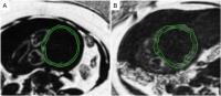
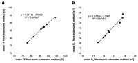
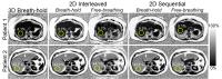
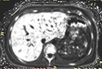
In this study, we developed and validated a “single-shot”
sequential 2D-chemical shift-encoded MRI (CSE-MRI) technique
for motion-insensitive quantification of liver proton
density fat fraction (PDFF). A phantom of 11 vials with
varying PDFF demonstrated equivalent PDFF between 2D- and
3D-CSE-MRI. Fifteen subjects underwent five different CSE-MRI
acquisitions: 3D-single-breathhold (BH), slice-interleaved
2D-single-BH and free-breathing (FB), and sequential
2D-single-BH and FB. PDFF was measured and averaged across
all nine Couinaud liver segments. Good agreement was
observed in PDFF between all 2D-CSE-MRI acquisitions and
3D-CSE-MRI. Qualitative motion artifact evaluation
demonstrated significantly superior scores for
free-breathing “single-shot” sequential 2D-CSE-MRI compared
to free-breathing slice-interleaved 2D-CSE-MRI.
|
 |
3837.
 |
2 |
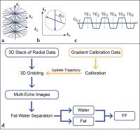
Free-Breathing Liver Fat Quantification Using an Undersampled
Multi-Echo 3D Stack-of-Radial Technique
Tess Armstrong1,2, Isabel Dregely1,3,
Alto Stemmer4, Yutaka Natsuaki5, and
Holden Wu1,2
1Radiological Sciences, University of California,
Los Angeles, Los Angeles, CA, United States, 2Physics
and Biology in Medicine, University of California, Los
Angeles, Los Angeles, CA, United States, 3Biomedical
Engineering, King's College London, London, United Kingdom, 4Siemens
Healthcare, Erlangen, Germany, 5Siemens
Healthcare, Los Angeles, CA, United States
Non-alcoholic fatty liver disease is the leading cause of
chronic liver disease. Multi-echo Cartesian MRI methods can
non-invasively quantify liver fat, but are susceptible to
motion artifacts and limited by breath hold (BH) imaging. We
have developed a new free-breathing (FB) liver fat
quantification method using non-Cartesian 3D stack-of-radial
imaging. To reduce scan time, we undersampled data up to a
factor of R=3. In healthy subjects, mean fat quantification
was statistically equivalent among different R. Initial
comparisons with spectroscopy show good agreement. Our new
technique can potentially achieve accurate whole-liver fat
quantification within a fast 1-minute FB scan.
|
|
3838.
 |
3 |
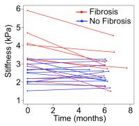
Detection of Reduction in Liver Stiffness as a Result of Weight
Loss Surgery using MR Elastography
Curtis N. Wiens1, Alan B. McMillan1,
Nathan S. Artz1,2, Rashmi Agni3,
Michael Peterson4, Nikolaus Szeverenyi5,
William Haufe5, Catherine Hooker5,
Luke Funk6, Jacob Greenberg6,
Guilherme M. Campos7, Santiago Horgan8,
Garth Jacobsen8, Tanya Wolfson9,
Claude Sirlin5, and Scott B. Reeder1,10,11,12,13
1Radiology, University of Wisconsin, Madison, WI,
United States, 2Diagnostic
Imaging, St. Jude Children's Research Hospital, Memphis, TN,
United States, 3Pathology,
University of Wisconsin, Madison, WI, United States, 4Tacoma
General Pathology, Tacoma, WA, United States, 5Radiology,
University of California, San Diego, CA, United States, 6Surgery,
University of Wisconsin, Madison, WI, United States, 7Virginia
Commonwealth University, Surgery, VA, United States, 8Surgery,
University of California, San Diego, CA, United States, 9Computational
and Applied Statistics Laboratory, University of California,
San Diego, CA, United States, 10Medical
Physics, University of Wisconsin, Madison, WI, United
States, 11Biomedical
Engineering, University of Wisconsin, Madison, WI, United
States, 12Medicine,
University of Wisconsin, Madison, WI, United States, 13Emergency
Medicine, University of Wisconsin, Madison, WI, United
States
This study tracked changes in liver stiffness in morbidly
obese patients undergoing bariatric surgery. 22 patients
undergoing bariatric surgery were recruited for MRI studies
including MR elastography (MRE) at 2 time points: 1-2 days
prior to and 6 months after bariatric surgery. Changes in
liver stiffness as measured by MRE were compared to
intraoperative biopsies which were performed to assess
relevant histological features (steatosis, inflammation and
fibrosis) and their relation to liver stiffness. Follow-up
measurement of liver stiffness 6 months after bariatric
surgery showed statistically significant reductions in liver
stiffness. Patients with biopsy confirmed liver fibrosis,
inflammation and features of NASH exhibited the largest
reductions in liver stiffness.
|
|
3839.
 |
4 |
Is fat a confounding factor for MOLLI T1 measurements in the
liver at 3 Tesla?
Ferenc E Mozes1, Elizabeth M Tunnicliffe1,
Michael Pavlides1,2, and Matthew D Robson1
1RDM Cardiovascular Medicine, University of
Oxford, Oxford, United Kingdom, 2Translational
Gastroenterology Unit, University of Oxford, Oxford, United
Kingdom
The balanced steady-state free precession (bSSFP) sequence
at 3T causes water and fat signals to be out of phase when
TR=2.3 ms. Since the modified Look-Locker inversion recovery
(MOLLI) mapping uses bSSFP readouts, the T1 of voxels that
contain both fat and water is influenced by the choice of
this TR. Simulations, phantom experiments and measurements
collected from patients undergoing bariatric surgery were
used to assess the impact of hepatic lipid content on liver
MOLLI T1 values.
|
|
3840.
 |
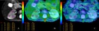
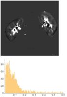
5 |
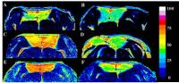
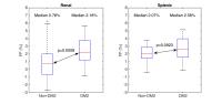
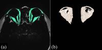
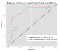
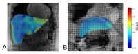
Feasibility of utilizing heterogeneity of hepatic stiffness in
3D MR elastography to improve detection of liver fibrosis in
pediatric patients with nonalcoholic fatty liver disease
Kang Wang1, Paul Manning1, Tanya
Wolfson 2,
Michael S. Middleton1, Jeffrey Schwimmer3,
Kimberley Newton3, Cynthia Behling3,
Janis Durelle3, Melissa Paiz3, Jorge
Angeles3, Meng Yin4, Kevin Glaser4,
Richard Ehman4, and Claude Sirlin1
1Liver Imaging Group, Department of Radiology,
University of California, San Diego, School of Medicine, San
Diego, CA, United States, 2Computational
and Applied Statistics Laboratory, University of California,
San Diego, San Diego, CA, United States,3Department
of Pediatric, University of California, San Diego, San
Diego, CA, United States, 4Departments
of Radiology, Mayo Clinic, Rochester, MN, United States
We evaluated the feasibility of utilizing heterogeneity of
hepatic stiffness in 3D MR elastography to improve detection
of liver fibrosis in a cohort of 70 children with NAFLD.
Children were dichotomized into two classes of fibrosis. We
characterized the heterogeneity of hepatic stiffness by
fitting a bi-Gaussian model to the histogram of hepatic
stiffness. Features from the bi-Gaussian model and the known
class labels were used to develop a support vector machine
(SVM) classification model to predict fibrosis. We
demonstrated that the SVM model has better overall
classification performance than the calculated mean hepatic
stiffness as measured by AUROC.
|
|
3841.
 |
6 |
Proton-density fat-fraction quantification of the liver in the
presence of ferumoxytol at 1.5T and 3T
Camilo A Campo1, Diego Hernando1,
Tilman B Schubert1, Utaroh Motosugi1,2,
Samir D Sharma1, Shane A Wells1, and
Scott B Reeder1,3,4,5,6
1Radiology, University of Wisconsin-Madison,
Madison, WI, United States, 2Radiology,
University of Yamanashi, Yamanashi, Japan, 3Medical
Physics, University of Wisconsin-Madison, Madison, WI,
United States, 4Biomedical
Engineering, University of Wisconsin-Madison, Madison, WI,
United States, 5Medicine,
University of Wisconsin-Madison, Madison, WI, United States, 6Emergency
Medicine, University of Wisconsin-Madison, Madison, WI,
United States
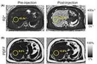
This study evaluated the accuracy of liver proton-density
fat-fraction (PDFF) quantification in the presence of
ferumoxytol. Seven healthy subjects underwent MRI scans
immediately before injection of ferumoxytol and one day
after injection. Our results indicate that
confounder-corrected chemical shift-encoded MRI PDFF
estimates exhibit a small but significant bias in the
presence of ferumoxytol. This bias could be due to
differential shortening in the T2* of water and fat signals.
Therefore, investigators attempting to create human models
of iron and fat within the liver by administering
ferumoxytol to patients with fatty liver should be aware of
this potential source of bias.
|
|
3842.
 |
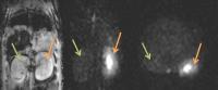
7 |
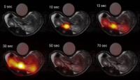
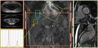
Impaired hepatic arterial buffer response in a rodent model of
chronic liver disease: assessment using caval subtraction
phase-contrast MRI at 9.4T
Manil Chouhan1, Alan Bainbridge2,
Nathan Davies3, Simon Walker-Samuel4,
Shonit Punwani1, Mark Lythgoe4,
Rajeshwar Mookerjee3, and Stuart Taylor1
1UCL Centre for Medical Imaging, University
College London, London, United Kingdom, 2Department
of Medical Physics, University College London Hospitals NHS
Trust, London, United Kingdom, 3UCL
Institute for Liver and Digestive Health, University College
London, London, United Kingdom, 4UCL
Centre for Advanced Biomedical Imaging, University College
London, London, United Kingdom
Total liver blood flow (TLBF) is closely regulated in health
so that reductions in portal venous (PV) flow are buffered
by compensatory rises in hepatic arterial (HA) flow. In
this study we use caval subtraction phase-contrast MRI to
estimate TLBF and HA flow in cirrhotic rats and demonstrate
an impaired HA buffer response after administering
terlipressin, a vasopressin analogue used clinically used to
reduce PV flow in portal hypertension.
|
|
3843.
 |
8 |
Effect of choosing an in-phase vs. a default echo time in 2D MR
elastography to estimate hepatic stiffness
Kang Wang1, William Haufe1, Nikolaus
Szeverenyi1, Alexandra Schlein 1,
Tanya Wolfson 2,
Michael S. Middleton1, Jeffrey Schwimmer3,
Kimberley Newton3, Cynthia Behling3,
Janis Durelle3, Melissa Paiz3, Jorge
Angeles3, Len Lazaro4, Diana De La
Pena4, Carolyn Hernandez4, Rohit
Loomba 4,
Meng Yin5, Kevin Glaser5, Richard
Ehman5, and Claude Sirlin1
1Liver Imaging Group, Department of Radiology,
University of California, San Diego, School of Medicine, San
Diego, CA, United States, 2Computational
and Applied Statistics Laboratory, University of California,
San Diego, San Diego, CA, United States,3Department
of Pediatric, University of California, San Diego, San
Diego, CA, United States, 4NAFLD
Translational Research Unit, Division of Gastroenterology,
University of California, San Diego, San Diego, CA, United
States, 5Departments
of Radiology, Mayo Clinic, Rochester, MN, United States
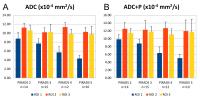
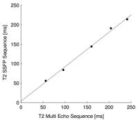
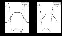
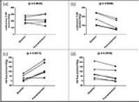
To investigate the effect of different echo times (TE) in 2D
MR elastography (2D MRE) to estimate hepatic stiffness, two
2D MRE scans were acquired in 50 patients using a 3T GE
scanner, one with a default TE value of 20.1 ms
(default-TE), and with a nearest in-phase TE value of 20.6
ms (IP-TE). Wave-image quality of each scan was measured
quantitatively by ROI size. We demonstrated that 2D MRE with
an in-phase TE provides slightly higher wave-image quality
in patients with high PDFF, and potentially may be
advantageous for fibrosis assessment in NAFLD.
|
|
3844.
 |
9 |
Deferiprone has a dose-dependent effect on liver iron
concentration assessed by MRI
Antonella Meloni1, Vincenzo Positano1,
Gianluca Valeri2, Gennaro Restaino3,
Chiara Tudisca4, Paolo Preziosi5,
Elisabetta Chiodi6, Maria Giovanna Neri1,
Stefano Pulini7, Basilia Piraino8,
Petra Keilberg1, and Alessia Pepe1
1Fondazione G. Monasterio CNR-Regione Toscana,
Pisa, Italy, 2Azienda
Ospedaliero-Universitaria Ospedali Riuniti "Umberto
I-Lancisi-Salesi", Ancona, Italy, 3Fondazione
di Ricerca e Cura "Giovanni Paolo II", Campobasso, Italy, 4Policlinico
"Paolo Giaccone", Palermo, Italy, 5Policlinico
“Casilino", Roma, Italy, 6Arcispedale
“S. Anna”, Ferrara, Italy, 7Ospedale
Civile “Spirito Santo”, Pescara, Italy, 8Policlinico
"G. Martino", Messina, Italy
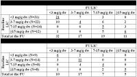
The aim of this multi-centre study was to retrospectively
assess in thalassemia major (TM) if deferiprone (DFP) had a
dose-dependent effect on liver iron concentration (LIC)
assessed by quantitative magnetic resonance imaging (MRI).
We found out that the percentage of patients that worsened
their status was significantly higher in patients with ≤ 75
mg/kg/d than in patients with > 75 mg/kg/d (26.6% vs 7.7%;
P=0.016). So, the worsening in MRI LIC can be prevented by
increasing the dose of deferiprone above the widely used
regimen of 75 mg/kg body weight.
|
|
3845.
 |
10 |
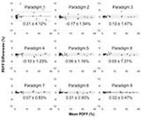
Standardized approach for region-of-interest-based measurements
of proton-density fat-fraction and R2* in the liver
Camilo A Campo1, Diego Hernando1,
Candice Bookwalter1,2, Tilman B Schubert1,
and Scott B Reeder1,3,4,5,6
1Radiology, University of Wisconsin-Madison,
Madison, WI, United States, 2Radiology,
Mayo Clinic, Rochester, MN, United States, 3Medical
Physics, University of Wisconsin-Madison, Madison, WI,
United States, 4Biomedical
Engineering, University of Wisconsin-Madison, Madison, WI,
United States, 5Medicine,
University of Wisconsin-Madison, Madison, WI, United States, 6Emergency
Medicine, University of Wisconsin-Madison, Madison, WI,
United States
This study evaluated the reproducibility of different
region-of-interest (ROI) sampling methods for MRI-based
proton-density fat-fraction (PDFF) and R2* (1/T2*)
measurements in the liver. 53 patient liver MRI datasets
were retrospectively analyzed using ROI sampling methods
that have been previously reported. Patients were not
suspected of having hepatic steatosis or liver iron
overload. Our results demonstrate improved measurement
repeatability when the sampling area of the liver is
increased by using multiple, large ROIs. Therefore,
ROI-based measurements of liver PDFF and R2* should strive
to sample the largest possible area of liver by using ROIs
that are large in size and number.
|
|
3846.
 |
11 |
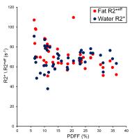
R2* of liver fat and water compared to proton density fat
fraction estimated by 1H MRS
Gavin Hamilton1, Alexandra N Schlein1,
Adrija Mamidipalli1, Michael S Middleton1,
Rohit Loomba2, and Claude B Sirlin1
1Department of Radiology, University of
California, San Diego, San Diego, CA, United States, 2Department
of Medicine, University of California, San Diego, San Diego,
CA, United States
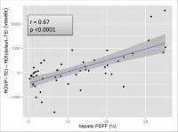
MRI based methods of estimating hepatic proton density fat
fraction (PDFF) measure only one R2* value, as the R2* of
fat and water are assumed to be identical. MRS can estimate
the R2* of both fat and water. Liver MRS spectra were fitted
with constraints derived from those used in MRI, and water
R2* and fat R2*eff (the
effective fat R2* that would be measured by MRI) were
compared to PDFF. We found that water R2* was independent of
PDFF, while fat R2*eff was
weakly and inversely correlated with PDFF.
|
|
3847.
 |
12 |
The Feasibility Study of Liver Cirrhosis Stage Using Quantitative 3D Whole-Liver T1? Mapping at 3.0T
Xin Chen1, Weibo Chen2,3, Guangbin
Wang4, Shanshan Wang4, Tao Gong1,
and Sai Shao1
1Shandong University, Jinan, China, People's
Republic of, 2Shanghai
Key Laboratory of Magnetic Resonance and Department of
Physics, East China Normal University, Shanghai, China,
Shanghai, China, People's Republic of, 3Philips
Healthcare, shanghai, China, Shanghai, China, People's
Republic of, 4Shangdong
Medical Imaging Research Institute, Jinan, China, People's
Republic of
Liver cirrhosis is an abnormal liver condition that the liver would repair it through the deposition of collagen, proteoglycans, and other macromolecules in the extracellular matrix [1]. The risk of liver cancer is greatly increased once cirrhosis develops. A non-invasive method that can objectively and simply assessment and grade the liver fibrosis is clinically required. T1ρ relaxation time has been proven to relevant with the macromolecular composition and proton exchange of tissues[2]. It may play as a non-invasive biomarker to investigate liver fibrosis. The first whole-liver study was carried out on 1.5T MR Scanner [3]. The purpose of our study was to implement the T1ρ method with whole-liver coverage that is breathing-hold free, and to initially apply the method to evaluate severity of whole-liver cirrhosis non-invasively at 3.0T.
|
|
3848.
 |
13 |
Liver parenchymal T1: repeatability and studies in a rodent
model of chronic liver disease at 9.4T
Manil Chouhan1, Rajiv Ramasawmy2,
Adrienne Campbell-Washburn2, Alan Bainbridge3,
Nathan Davies4, Shonit Punwani1,
Rajeshwar Mookerjee4, Simon Walker-Samuel2,
Mark Lythgoe2, and Stuart Taylor1
1UCL Centre for Medical Imaging, University
College London, London, United Kingdom, 2UCL
Centre for Advanced Biomedical Imaging, University College
London, London, United Kingdom, 3Department
of Medical Physics, University College London Hospitals NHS
Trust, London, United Kingdom, 4UCL
Institute for Liver and Digestive Health, University College
London, London, United Kingdom
There is a growing interest in the use of hepatic
parenchymal T1 for the assessment of hepatic fibrosis.
Using Look-Locker T1 measurements at 9.4T in a rat model of
cirrhosis, we demonstrate that these measurements are
repeatable and significantly different in animals with and
without chronic liver disease.
|
|
3849.
 |
14 |
Feasibility of Breath-hold Quantitative Susceptibility Mapping
on Hepatic Iron quantification
Huimin Lin1, Hongjiang Wei2, Chunlei
Liu2, Xu Yan3, Caixia Fu4,
and Fuhua Yan1
1Radiology, Ruijin Hospital, Shanghai Jiao Tong
University School of Medicine, Shanghai, China, People's
Republic of, 2Brain
Imaging and Analysis Center,Duke University, Durham, NC,
United States, 3MR
Collaboration NE Asia, Siemens Healthcare, Shanghai, China,
People's Republic of, 4Siemens
Shenzhen Magnetic Resonance Ltd, Shenzhen, China, People's
Republic of
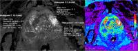
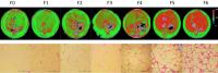
The purpose of this study was to estimate the Quantitative
susceptibility mapping (QSM) in hepatic iron evaluation,
compared with R2 based Liver concentration estimation
(Ferriscan LIC). 7 Patients were scanned on a 1.5 T MR
System using a GRE sequence and a SE sequence, for QSM and
Ferriscan analysis respectively. QSM algorithm provided
susceptibility values estimate. Approximate slices were
selected according to the corresponding cross-section on the
Ferriscan LIC report. Then ROIs were drawn on QSM images
according to LIC maps. Significant positive correlation was
observed between QSM and Ferriscan LIC ( R2 =
0.8).
|
|
3850.
 |
15 |
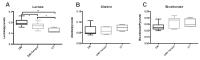
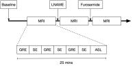
THE EVALUATION OF PORTAL HYPERTENSION USING QUANTITATIVE
MAGNETIC RESONANCE IMAGING (MRI)
Eleanor F Cox1, Naaventhan Palaniyappan2,
Andrew Austin3, Richard O'Neill4, Greg
Ramjas4, Simon Travis4, Hilary White4,
Rajeev Singh3, Peter Thurley3, Indra
Neil Guha2, Guruprasad Padur Aithal2,
and Susan T Francis1
1SPMIC, School of Physics & Astronomy, University
of Nottingham, Nottingham, United Kingdom, 2NIHR
Nottingham Digestive Diseases Biomedical Research Unit,
Nottingham University Hospitals NHS Trust and University of
Nottingham, Nottingham, United Kingdom, 3Royal
Derby Hospital, Derby, United Kingdom, 4Department
of Radiology, Nottingham University Hospitals NHS Trust,
Nottingham, United Kingdom
The hepatic venous pressure gradient (HVPG) is the only
validated measure to assess portal pressure but this is
invasive and not widely available. Here, we use non-invasive
MR parameters as a surrogate for portal pressure.
Longitudinal relaxation time (T1) measures of the liver and
spleen, phase contrast measures of splanchnic and collateral
flow, and ASL measures of perfusion are correlated against
HVPG measures in 30 patients. Liver tissue T1 is shown to be
positively correlated with HVPG (p<0.001). Combining T1
measures with splenic artery velocity using a simple linear
model, it is shown that HVPG can be non-invasively assessed.
|
|
3851.
 |
16 |
MRI Evaluation of Acetaminophen Induced Liver Failure in Mice
using T1 Mapping and Stable Gadoxetate Disodium Administration
Christiane Mallett1, Matthew Latourette1,
Anna Kopec2, James Luyendyk2, and Erik
Shapiro1
1Radiology, Michigan State University, East
Lansing, MI, United States, 2Pathobiology
& Diagnostic Investigation, Michigan State University, East
Lansing, MI, United States
We are developing an MRI method to measure acetaminophen
toxicity in the liver. We obtained T1 maps using the
clinically approved contrast agent gadoxetate disodium
(Eovist). The contrast agent was administered by infusion to
maintain a steady liver concentration throughout the T1
mapping. Mice with acetaminophen toxicity had higher T1 and
heterogeneous gadoxetate disodium uptake compared to healthy
controls. This is a promising method for quantifying drug
induced liver damage in vivo.
|
|
3852.
 |
17 |
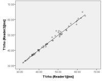
Comparison of R2* of liver water and fat using 1H MRS
Gavin Hamilton1, Alexandra N Schlein1,
Adrija Mamidipalli1, Michael S Middleton1,
Rohit Loomba2, and Claude B Sirlin1
1Department of Radiology, University of
California, San Diego, San Diego, CA, United States, 2Department
of Medicine, University of California, San Diego, San Diego,
CA, United States
To estimate hepatic proton density fat fraction (PDFF), MRI
techniques acquire multi-echo, gradient-echo images,
assuming the R2* of fat and water to be identical. Liver MRS
spectra were fitted with constraints derived from those used
in MRI to examine this assumption. We compared fat R2*eff (the
effective fat R2* that would be measured by MRI) with water
R2* and found that water R2* and fat R2*eff were
correlated. There was no significant difference between
water R2* and fat R2*eff, supporting the
assumption that when measuring PDFF using MRI, fat and water
R2* can be treated as identical.
|
|
3853.
 |
18 |
Iron measurements by quantitative MRI-R2* at 3.0 and 1.5 T
Jin Yamamura1, Sarah Keller1, Regine
Grosse2, Bjoern Schoennagel1, Peter
Nielsen3, Zhiyue Jerry Wang4, Joachim
Graessner5, Hendrick Kooijman6,
Gerhard Adam1, and Roland Fischer3,7
1Diagnostic and Interventional Radiology,
University Medical Center Hamburg-Eppendorf, Hamburg,
Germany, 2Hematology
and Oncology, University Medical Center Hamburg-Eppendorf,
Hamburg, Germany, 3Biochemistry,
University Medical Center Hamburg-Eppendorf, Hamburg,
Germany, 4Radiology,
University of Texas Southwestern Medical Center, Dallas, TX,
United States, 5Siemens
Healthcare AG, Hamburg, Germany, 6Philips
Medical Care, Hamburg, Germany, 7Radiology,
Children’s Hospital & Research Center Oakland, Oakland, CA,
United States
We are investigating the suitability of a 3.0 T imager for
iron measurements over the whole range of possible iron
concentrations in the liver and spleen iron. For liver iron
measurements in severely overloaded patients with LIC > 2400
µg/gliver or > 15 mg/gdry weight, 1.5 Tesla imagers are
better suited than 3.0 Tesla systems.
|
|
3854.
 |
19 |
A prospective study comparing R2* derived Liver iron
concentration(LIC) with noise-corrected post processing of data
against FerriScan reported LIC in patients with liver iron
overload.
Kartik Jhaveri1, Stephan Kannengiesser2,
Nima Sadougi3, Marshall Sussman1,
Hooman Hosseini-Nik3, Leila Zahedi3,
and Richard Ward1
1UHN,University of Toronto, Toronto, ON, Canada, 2Siemens
Germany, Erlangen, Germany, 3UHN,
Toronto, ON, Canada
MRI is currently utilised as a non-invasive method for liver
iron concentration (LIC) estimation and has essentially
replaced liver biopsy. FerriScan derived LIC is considered
the “gold standard” but has associated increased costs and
delay results from required external data transmittal. There
is no universal agreement or standardization of R2* derived
LIC methods. We describe an optimized R2* method with noise
corrected post processing of data for LIC estimation with
simultaneous comparison to FerriScan derived LIC. Our
results show very good correlation between R2* LIC and
FerriScan LIC with potential for substitution of the latter
with our R2* technique.
|
|
3855.
 |
20 |
Agreement between MRE-estimated liver stiffness using 2D GRE and
2D SE-EPI pulse sequences at 3T
Adrija Mamidipalli1, Jonathan Hooker1,
Nikolaus Szevrenyi1, Alexandra Schlein1,
William Hauffe1, Tanya Wolfson2, Gavin
Hamilton1, Michael Middleton1, and
Claude Sirlin1
1Liver Imaging Group, UCSD, San Diego, CA, United
States, 2Computational
and Applied Statistics Laboratory, San Diego Supercomputer,
UCSD, San Diego, CA, United States
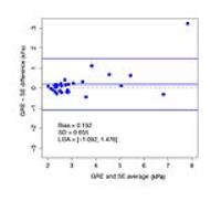
In this study, agreement between MRE-estimated liver
stiffness using 2D GRE and SE-EPI pulse sequences at 3T was
examined in 30 adults with histology-confirmed nonalcoholic
fatty liver disease (NAFLD) enrolled in a research registry.
Results show that liver stiffness values obtained from both
the sequences agree closely across a range of liver
stiffness values for adults with NAFLD, although agreement
tends to diverge at higher stiffness values. Differences at
higher liver stiffness values were not explained by
differences in image wave quality.
|
|
3856.
 |
21 |
Assessment of liver fibrosis by MRI tagging of cardiac-induced
motion: preliminary results
Leonie Petitclerc1,2, Guillaume Gilbert2,3,4,
Claire Wartelle-Bladou5, Giada Sebastiani6,
Bich Nguyen7,8, and An Tang1,2,4
1Centre de recherche du Centre hospitalier de
l'Universite de Montreal, Montreal, QC, Canada, 2Department
of Radiology, Radio-Oncology and Nuclear Medicine,
Universite de Montreal, Montreal, QC, Canada, 3Philips
Healthcare Canada, Montreal, QC, Canada, 4Centre
hospitalier de l'Universite de Montreal, Montreal, QC,
Canada, 5Department
of Gastroenterology and Hepatology, Universite de Montreal,
Montreal, QC, Canada, 6Department
of Medicine, Division of Gastroenterology, McGill University
Health Centre, Montreal, QC, Canada, 7Department
of Pathology, Centre hospitalier de l'Universite de
Montreal, Montreal, QC, Canada, 8Department
of Pathology and Cellular Biology, Universite de Montreal,
Montreal, QC, Canada
Elastography for the staging of liver fibrosis is optimized
for the right liver and requires additional hardware. Using
MRI tagging, the displacement and strain of liver tissue
induced by cardiac motion was quantified with the Harmonic
Phase (HARP) method. Of the four schemes tested for the
extraction of a single measure of strain, one was especially
promising, as it showed high correlation with fibrosis
stages (Spearman’s ρ=-0.913),
as well as a significant p-value
for dichotomized diagnosis of ≥F3 fibrosis (p=0.03).
These preliminary results suggest that strain measurements
could be used as a diagnostic tool for the staging of liver
fibrosis.
|
|
3857.
 |
22 |
Assessment of fibrotic liver using T1rho mapping: a rat model
study
Qihua Yang1, Taihui Yu1, Hui Zhang2,
Hua Guo2, Yingjie Mei3, Ziliang Cheng1,
Jingwen Huang1, and Biling Liang1
1Radiology Dept, Sun Yat-sen Memorial Hospital,
Guangzhou, China, People's Republic of, 2Center
for Biomedical Imaging Research, Department of Biomedical
Engineering, School of Medicine, Tsinghua University,
Beijing, China, People's Republic of, 3Philips
Healthcare, Guangzhou, China, People's Republic of
To find out the relationship among T1rho, liver fibrosis
stages and liver function, MRI including T1rho sequence was
performed in 32 CCl4 induced liver fibrosis SD rat models
and 12 SD rats of control group. Laboratory test related to
liver function were done before execution and liver was
taken out for pathology evaluation. Liver fibrosis was
staged according to staging systems for rodents. Results
showed that T1rho could be used to diagnose early liver
fibrosis (>F2) and correlation was significant between T1rho
values and both liver fibrosis stage and blood serum
parameters.
|
|
3858.
 |
23 |
The effect of long diffusion time on the diffusion measurements
of fibrotic human liver
Hui Zhang1, Pairash Saiviroonporn2, Ed
X Wu3,4, and Hua Guo1
1Center for Biomedical Imaging Research,
Department of Biomedical Engineering, School of Medicine,
Tsinghua University, Beijing, China, People's Republic of, 2Department
of Radiology, Faculty of Medicine Siriraj Hospital, Mahidol
University, Bangkok, Thailand, 3Laboratory
of Biomedical Imaging and Signal Processing, The University
of Hong Kong, Hong Kong SAR, China, People's Republic of, 4Department
of Electrical and Electronic Engineering, The University of
Hong Kong, Hong Kong SAR, China, People's Republic of
To examine whether different diffusion times would yield
different sensitivities in detecting the pathological
alterations in tissue microstructure during liver
fibrogenesis in human livers at 3 T. MRI including
single-shot SE and simulated echo acquisition mode (STEAM)
DWI EPI sequences were performed on 10 healthy volunteers
and 19 liver fibrotic patients. One-way ANOVA with Turkey’s
multiple comparison tests were employed to compare
quantitative measurements between the volunteers and
patients with different diffusion times. Results showed that
diffusion measurements with higher diffusion times will be
more sensitive as a biomarker to detect the pathological
alterations in tissue microstructure in fibrotic patients.
|
|
3859.
 |
24 |
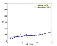
Improving Liver Iron Estimates with a Muscle-based Proton
Density Estimate
Eamon K Doyle, MS1, Andrew Powell, MD2,3,
and John C Wood, MD, PhD4,5
1Biomedical Engineering, University of Southern
California, Sierra Madre, CA, United States, 2Cardiology,
Boston Children's Hospital, Boston, MA, United States, 3Pediatrics,
Harvard School of Medicine, Boston, MA, United States, 4Cardiology,
Children's Hospital of Los Angeles, Los Angeles, CA, United
States, 5Biomedical
Engineering, University of Southern California, Los Angeles,
CA, United States
CPMG-based R2 (1/T2) estimates are traditionally insensitive
to tissue iron load. We show that the application of a
T1-corrected, skeletal muscle-based proton density
constraint increases the sensitivity of R2 for iron
quantitation in phantoms and human subjects. This method
leads to a fundamentally different R2-LIC (liver iron
concentration) calibration curve than has previously been
applied to CPMG fit data.
|
|