|
Exhibition Hall 16:00 - 17:00 |
|
|
|
Computer # |
|
4052.
 |
25 |
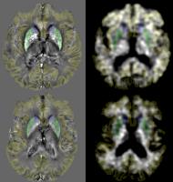
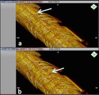
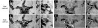
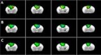
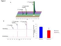
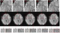
Effect of patient motion on the visibility of small veins in
T2*w imaging: A simulation experiment with implications for the
study of the central-vein-sign (CVS) theory in MS 
Nicola Bertolino1, Michael G Dwyer1,
Paul Polak1, Samuel Daniel Robinson2,
Robert Zivadinov1,3, and Ferdinand Schweser1,3
1Buffalo Neuroimaging Analysis Center, Department
of Neurology,Jacobs School of Medicine and Biomedical
Sciences, The State University of New York at Buffalo,
Buffalo, NY, United States, 2High
Field Magnetic Resonance Centre, Department of Biomedical
Imaging and Image-guided Therapy, Medical University of
Vienna, Vienna, Austria, 3MRI
Molecular and Translational Research Center, Jacobs School
of Medicine and Biomedical Sciences, The State University of
New York at Buffalo, Buffalo, NY, United States
FLAIR* is a fusion of T2-FLAIR and 3D-T2*w images and it is
used to assess the central-vein-sign, a recent promising
research direction in MRI-based study of Multiple Sclerosis.
However in this experiment we show that researchers should
be aware that slight patient movement during acquisition can
produce blurring effect in 3D-T2*w images. This subtle
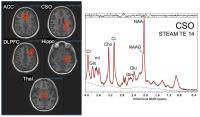
artifact can mask small vessels even in case which the
overall quality of the image is not substantially degraded.
|
|
4053.
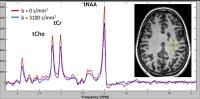
 |
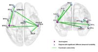
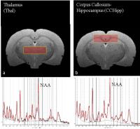
26 |
Combined Anatomic and Functional Connectivity Metric for
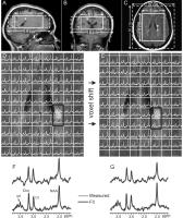
Tracking Disease Progression in MS
Mark J Lowe1, Katherine Koenig1, Erik
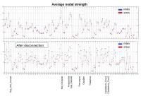
Beall1, Jian Lin1, Ken Sakaie1,
Lael Stone2, and Micheal D. Phillips1
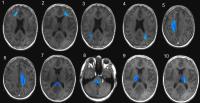
1Imaging Institute, Cleveland Clinic, Cleveland,
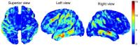
OH, United States, 2Neurologic
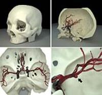
Institute, Cleveland Clinic, Cleveland, OH, United States
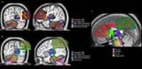
Based on the observation that anatomic and functional
connectivity measures in multiple sclerosis (MS) are
correlated, but not highly correlated, we propose to combine
these metrics into an imaging based measure of disease
progression. We show that this metric is sensitive to
disease progression in a cohort of MS patients over a time
period of one year.
|
|
4054.
 |
27 |
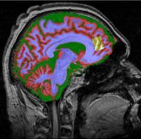
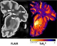
Assessment of ferritin in the multiple sclerosis brain using
temperature induced R2* changes
Christoph Birkl1, Daniele Carassiti2,
Christian Langkammer1, Christian Enzinger1,
Franz Fazekas1, Klaus Schmierer2,3,
and Stefan Ropele1
1Department of Neurology, Medical University of
Graz, Graz, Austria, 2Blizard
Institute (Neuroscience), Queen Mary University of London,
London, United Kingdom, 3Barts
Health NHS Trust, Emergency Care and Acute Medicine Clinical
Academic Group (Neuroscience), London, United Kingdom
Evidence for a possible role of iron in the pathogenesis and
progression of multiple sclerosis (MS) has raised interest
in iron mapping techniques. However, current approaches are
not reliable in white matter because of the diamagnetic
properties of myelin. We recently proposed a new method for
iron mapping which is based on the temperature dependency of
the paramagnetic susceptibility. Here, the temperature
coefficient of R2* (TcR2*) as a
measure of iron content was assessed in three post-mortem MS
brain samples. Validation of TcR2* mapping was
done with immunohistochemistry using cell counting on
ferritin light-chain stains.
|
|
4055.
 |
28 |
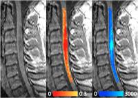
Longitudinal mcDESPOT Shows Contrasting Patterns of Change in
Multiple Sclerosis and Neuromyelitis Optica Cervical Cord
Anna Combes1, Lucy Matthews2, Gareth J
Barker1, Steven CR Williams1, Katrina
McMullen3, Janet Lam4, Anthony
Traboulsee3, David KB Li3,4,
Jacqueline Palace2, and Shannon Kolind3
1Neuroimaging, King's College London, London,
United Kingdom, 2Nuffield
Department of Clinical Neurosciences, University of Oxford,
Oxford, United Kingdom, 3Medicine,
University of British Columbia, Vancouver, BC, Canada, 4Radiology/UBC
MS/MRI Research Group, University of British Columbia,
Vancouver, BC, Canada
Neuromyelitis optica (NMO) severely affects the optic nerves
and spinal cord and shares features with multiple sclerosis
(MS). Ongoing diffuse neurodegeneration, however, is thought
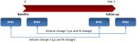
to be absent in NMO between relapses. We collected cervical
cord mcDESPOT at baseline and one-year follow-up in patients
and matched controls. While there were no significant
changes in controls and MS patients, the NMO group showed a
loss of cord volume, decrease in T1 and
increase in myelin water fraction. We hypothesize that
continuing atrophy in lesioned areas reduces the amount of
damaged tissue relative to healthy tissue, and is
responsible for the observed changes.
|
|
4056.
 |
29 |
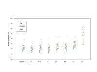
White Matter Water Content in Multiple Sclerosis and
Neuromyelitis Optica
Irene Vavasour1, Sandra Meyers2,
Praveena Manogaran3, Shuhan Xiao3,
Anika Wurl3, Katrina McMullan3, David
Li1, Anthony Traboulsee3, and Shannon
Kolind3
1Radiology, University of British Columbia,
Vancouver, BC, Canada, 2Physics
and Astronomy, University of British Columbia, Vancouver,
BC, Canada, 3Medicine,
University of British Columbia, Vancouver, BC, Canada
Multiple sclerosis (MS) and neuromyelitis optica (NMO) are
both autoimmune diseases of the central nervous system.
Normal appearing white matter is known to be affected by
diffuse tissue damage in MS whereas damage in NMO is thought
to be restricted to acute lesions. Surprisingly, in this
study, water content within whole white matter and white
matter tracts of subjects with neuromyelitis optica (NMO)
was found to be higher than in healthy matched controls and
similar to MS. Both NMO and MS lesions had a higher water
content compared to normal appearing white matter.
|
|
4057.
 |
30 |
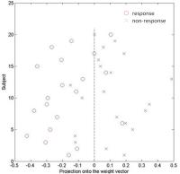
A randomized controlled trial on the efficacy of cognitive
training in MS reveals functional connectivity changes
Ottavia Dipasquale1,2, Jamie Campbell3,
Camila Callegari Piccinin4, Dawn Langdon5,
Waqar Rashid6, and Mara Cercignani3
1IRCCS, Don Gnocchi Foundation, Milan, Italy, 2Department
of Electronics, Information and Bioengineering, Politecnico
di Milano, Milan, Italy, 3Clinical
Imaging Sciences Centre, Brighton and Sussex Medical School,
Brighton, United Kingdom, 4Neuroimaging
Laboratory, University of Campinas, Cidade Universitária,
Campinas, Brazil, 5Psychology
Department, Royal Holloway, University of London, Egham,
United Kingdom, 6Department
of Neurology, Brighton and Sussex University Hospitals NHS
Trust, Brighton, United Kingdom
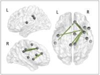
We investigated the resting-state functional connectivity
(FC) changes induced after 6 weeks of computerised,
home-based cognitive rehabilitation in patients with
multiple sclerosis (MS) in a randomized controlled trial.
The intervention and control groups were evaluated at
baseline (T1), after a 6-week intervention period (T2) and a
12-week follow-up period (T3). Out of the 94 regions
investigated, many memory-, attention- and motor-related
areas strengthened their FC at T2 and T3 in the intervention
group. This study supports the hypothesis that this
cognitive rehabilitation is a feasible and effective
approach in patients with MS and confirms that rfMRI is a
useful tool for mapping plastic changes.
|
|
4058.
 |
31 |
Quantitative T2 mapping detects pathology in normal-appearing
brain regions of relapsing-remitting MS patients
Timothy Shepherd1,2, Ivan Kirov1,2,
James S Babb1,2, Mary T Bruno2, Robert
E Charlson3, Jacqueline Smith2, KAI
Tobias Block1,2, Daniel K Sodickson1,2,
and Noam Ben-Eliezer1,2
1Center for Advanced Imaging Innovation and
Research (CAI2R), New York University School of Medicine,
New York, NY, United States, 2The
Bernard and Irene Schwartz Center for Biomedical Imaging,
Department of Radiology, New York University School of
Medicine, New York, NY, United States, 3Department
of Radiology, New York University School of Medicine, New
York, NY, United States
Accurate quantification of T2 values
in vivo is a long-standing challenge hampered by the
inherent inaccuracy associated with rapid multi-SE
sequences. This inaccuracy is, moreover, not constant and
depends on both the pulse sequence scheme and parameter-set
employed, resulting in different vendors or scanners
yielding different results! We used a recently developed
novel T2 mapping
technique, the EMC algorithm,
to quantify T2 changes
in different brain regions of MS patients. Our results
demonstrate that the robustness of the EMC approach allows
the detection of subtle, but statistically significant T2 differences
in normal appearing brain regions for MS patients.
|
|
4059.
 |
32 |
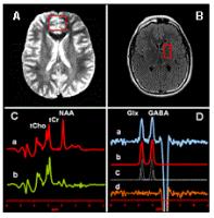
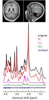
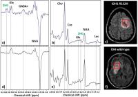
Ultra-high Resolution MRSI of Multiple Sclerosis at 7T
Bernhard Strasser1, Gilbert Hangel1,
Michal Považan1, Stephan Gruber1,
Marek Chmelík1, Assunta Dal-Bianco2,
Fritz Leutmezer2, Siegfried Trattnig1,3,
and Wolfgang Bogner1
1MRCE, Department of Biomedical Imaging and
Image-guided Therapy, Medical University of Vienna, Vienna,
Austria, 2Department
of Neurology, Medical University of Vienna, Vienna, Austria,3Christian
Doppler Laboratory for Clinical Molecular MR Imaging,
Vienna, Austria
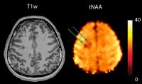
In this study fourteen MS patients were measured with an
FID-based MRSI sequence at 7T with resolutions of 64x64 and
100x100. Metabolic maps of total NAA, total Choline, total
Creatine, and myo-Inositol were compared to FLAIR images.
All patients had lesions with decreased tNAA, and eight had
increased myo-Inositol levels. However, not all lesions
showed decreased tNAA values. Two patients showed decreased
tNAA levels with no visible lesion on the FLAIR image. In
average, a decrease of 26% in tNAA and an increase of 42% in
myo-Inositol were observed in comparison to normal appearing
white matter.
|
|
4060.
 |
33 |
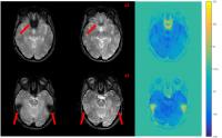
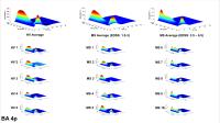
BA 4p BOLD response profile distinguishes low and high MS
morbidity
Adnan A.S. Alahmadi1,2, Matteo Pardini1,3,
Rebecca S. Samson1, Egidio D'Angelo4,5,
Karl J. Friston6, Ahmed T. Toosy1,7,
and Claudia Angela Michela Gandini Wheeler-Kingshott1,5
1NMR Research Unit, Queen Square MS Centre,
Department of Neuroinflammation, UCL Institute of Neurology,
London, United Kingdom, 2Department
of Diagnostic Radiology, Faculty of Applied Medical Science,
KAU, Jeddah, Saudi Arabia, 3Department
of Neurosciences, Ophthalmology and Genetics, University of
Genoa, Genoa, Italy, 4Department
of Brain and Behavioral Sciences, University of Pavia,
Pavia, Italy, 5Brain
Connectivity Center, C.Mondino National Neurological
Institute, Pavia, Italy, Pavia, Italy, 6Wellcome
Centre for Imaging Neuroscience, UCL, Institute of
Neurology, London, United Kingdom, 7NMR
Research Unit, Department of Brain Repair and
Rehabilitation, Queen Square MS Centre, UCL Institute of
Neurology, London, United Kingdom
This study investigates how multiple sclerosis (MS)
selectively affects regional BOLD response to variable grip
forces (GF). It is known that the anterior and posterior BA4
areas are anatomically and functionally distinguishable –
and that in healthy subjects there are linear and non-linear
BOLD response components. After modelling BOLD responses
with a polynomial expansion of the applied GF during task,
we showed that in BA4a MS subjects respond like healthy
subjects. BOLD response in BA4p, instead, was altered in MS,
with those with greatest disability showing the greatest
deviations from the non-linear profile of the healthy
response.
|
|
4061.
 |
34 |
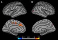
Functional cognitive control load in multiple sclerosis
Paola Valsasina1, Maria Assunta Rocca1,
Laura Vacchi1, Alessandro Meani1,
Mariaemma Rodegher2, Vittorio Martinelli2,
Giancarlo Comi2, Andrea Falini3, and
Massimo Filippi1
1Neuroimaging Research Unit, San Raffaele
Scientific Institute, Vita-Salute San Raffaele University,
Milan, Italy, 2Department
of Neurology, San Raffaele Scientific Institute, Vita-Salute
San Raffaele University, Milan, Italy, 3Department
of Neuroradiology, San Raffaele Scientific Institute,
Vita-Salute San Raffaele University, Milan, Italy
In this study, we investigated behavioral and functional MRI
(fMRI) correlates of a N-back task in 72 patients with
multiple sclerosis (MS). We found a load-dependent
alteration of executive network recruitment, varying
according to the disease phenotype. Increased recruitment of
frontal regions was associated to the early phase of MS.
Conversely, the modulation of regions belonging to the
default mode network was more evident in patients with
long-lasting disease and was related to the global cognitive
profile, suggesting an increased need of cognitive resources
to cope with task-demand.
|
|
4062.
 |
35 |
Formation of transient and persistent multiple sclerosis
lesions: serial follow-up with quantitative MR imaging and
spectroscopy
Ivan Kirov1,2, Shu Liu1,2, Assaf Tal3,
William E. Wu1,2, Matthew S. Davitz1,2,
James S. Babb1,2, Henry Rusinek1,2,
Joseph Herbert4, and Oded Gonen1,2
1Center for Advanced Imaging Innovation and
Research (CAI2R), New York University School of Medicine,
New York, NY, United States, 2Bernard
and Irene Schwartz Center for Biomedical Imaging, New York
University School of Medicine, New York, NY, United States, 3Chemical
Physics, Weizmann Institute of Science, Rehovot, Israel, 4Neurology,
New York University Langone Medical Center, New York, NY,
United States
Using MR imaging and proton spectroscopy, we follow the
evolution of transient and persistent multiple sclerosis
lesions from pre-lesional state to long-term (over 2 years
post-formation) status. The main finding was that the sharp
drop in N-acetyl-aspartate
associated with the formation of an acute lesion was
reversible in resolving, but not in persisting black holes,
substantiating the idea that transient new lesions revert to
pre-lesional axonal state. The additional findings were a
decrease in creatine after the appearance of a persisting
lesion and the lack of metabolic differences between
pre-lesional tissue giving rise to resolving versus
persisting lesions.
|
|
4063.
 |
36 |
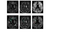
Assessment of grey matter cortical lesions in Multiple Sclerosis
using high resolution ASL at 7T
Richard J Dury1, Molly G Bright1,
Yasser Falah2, Penny A Gowland1, Nikos
Evangelou2, and Susan T Francis1
1Sir Peter Mansfield Imaging Centre, University
of Nottingham, Nottingham, United Kingdom, 2Nottingham
University Hospital, University of Nottingham, Nottingham,
United Kingdom
Grey matter cortical lesions have been associated with
physical disability, cognitive impairment and fatigue in
Multiple Sclerosis. Only one previous study has assessed
cerebral blood flow (CBF) and cerebral blood volume (CBV)
within cortical lesions. Here we use high spatial resolution
7T FAIR TrueFISP ASL to assess the perfusion in grey matter
cortical lesions and compare this to surrounding normal
appearing grey matter. Cortical lesions showed a significant
32% reduction in perfusion signal compared to normal
appearing grey matter. This ASL method can be used to
evaluate longitudinal perfusion changes in new and chronic
cortical lesions.
|
|
4064.
 |
37 |
Energy dysregulation and neuro-axonal dysfunction in multiple
sclerosis measured in-vivo with diffusion-weighted spectroscopy
Benedetta Bodini1, Francesca Branzoli1,2,
Emilie Poirion1, Daniel Garcia-Lorenzo1,2,
Elisabeth Maillart1, Julie Socha1,
Geraldine Bera1, Itamar Ronen3,
Stephane Lehericy1,2, and Bruno Stankoff1
1Brain and Spine Institute, INSERM U1127,
Sorbonne Universités, UPMC, CHU Pitié-Salpêtrière, Paris,
France, 2Brain
and Spine Institute, Center for Neuroimaging Research
(CENIR), CHU Pitié-Salpêtrière, Paris, France, 3C.J.
Gorter Center for High Field MRI, Radiology, Leiden
University Medical Center, Leiden, Netherlands
Diffusion-weighted spectroscopy (DWS), allowing to measure
in-vivo the diffusion properties of endogenous intracellular
metabolites such as total N-acetyl-aspartate (tNAA) and
total creatine (tCr), offers the opportunity to explore the
early phase of neuronal structural damage and energetic
mismatch in multiple sclerosis (MS). We compared the
apparent diffusion coefficient (ADC) of tNAA and tCr in 25
patients with MS and 20 healthy volunteers, and found a
reduced diffusivity of both metabolites in patients, both in
the corona radiate and in the thalami. These results may
reflect an ongoing neuro-axonal damage and a simultaneous
energy dysregulation affecting neurons and/or glial cells in
MS.
|
|
4065.
 |
38 |
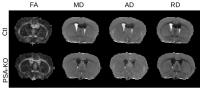
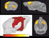
N-acetyl aspartate predicts disease severity in an animal model
of multiple sclerosis (MS)
Amber Michelle Hill1, Mohamed Tachrount2,
David L Thomas3, Kenneth J Smith4,
Xavier Golay5, and Olga Ciccarelli1
1NMR Research Unit, Queen Square MS Centre,
Department of Neuroinflammation, UCL Institute of Neurology,
University College London, London, United Kingdom, 2Department
of Brain Repair and Rehabilitation, UCL Institute of
Neurology, University College London, London, United
Kingdom, 3Leonard
Wolfson Experimental Neurology Centre, UCL Institute of
Neurology, University College London, London, United
Kingdom, 4Department
of Neuroinflammation, Queen Square MS Centre, UCL Institute
of Neurology, University College London, London, United
Kingdom, 5Department
of Brain Repair and Rehabilitation, Queen Square MS Centre,
UCL Institute of Neurology, University College London,
London, United Kingdom
EAE, an animal model of MS, can be investigated with MR to
address the clinical need to understand mechanisms of the MS
disease course. Longitudinal MR studies with EAE are
currently under-explored. This study investigated
longitudinal changes in metabolite concentrations and lesion
development, in relation to neurological deficits in EAE,
using 9.4T MRI and 1H-MRS.
Five time-points of EAE disease progression were assessed.
The results suggest that before visible signs of
neurological deficits, higher [NAA] predicts the severity of
late-stage neurological deficits in EAE. Considering NAA is
predominantly associated with neuronal mitochondria, this
may reflect relevant pathological processes in MS.
|
|
4066.
 |
39 |
Longitudinal follow-up of chronic multiple sclerosis lesions
with quantitative MR imaging and partial volume-corrected proton
MR spectroscopy
Ivan Kirov1,2, Shu Liu1,2, Assaf Tal3,
William E. Wu1,2, Matthew S. Davitz1,2,
James S. Babb1,2, Henry Rusinek1,2,
Joseph Herbert4, and Oded Gonen1,2
1Center for Advanced Imaging Innovation and
Research (CAI2R), New York University School of Medicine,
New York, NY, United States, 2Bernard
and Irene Schwartz Center for Biomedical Imaging, New York
University School of Medicine, New York, NY, United States, 3Chemical
Physics, Weizmann Institute of Science, Rehovot, Israel, 4Neurology,
New York University Langone Medical Center, New York, NY,
United States
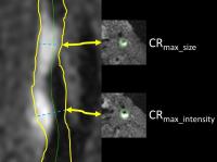
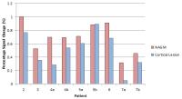
We describe the evolution of chronic multiple sclerosis
lesions from a quantitative MR imaging and spectroscopy
perspective. Metabolite concentrations were obtained along
with measures of lesion T1-hypointensity and size.
Moderately hypointense lesions were more metabolically
active than severely hypointense lesions, driving an
increase in the glial marker myo-inositol. Correlational
analyses revealed that lesion size is a better predictor of
axonal health than T1-hypointensity, with lesions larger
than 1.5 cm3 exhibiting
terminal axonal injury. A positive correlation between
changes in choline and in lesion size in moderately
hypointense lesions implied that changes in lesion size are
mediated by chronic inflammation.
|
|
4067.
|
40 |
2D Localised Correlated Spectroscopy (L-COSY): A potential tool
for identifying biochemical changes in Multiple Sclerosis
Jameen ARM1, Scott Quadrelli2, Karen
Ribbons3, Jeanette Lechner-Scott3, and
Saadallah Ramadan4
1Imaging, HMRI, Newcastle, Australia, 2Imaging,
TRI Brisbane, Brisbane, Australia, 3Neurology,
John Hunter Hospital, Newcastle, Australia, 4Faculty
of Health and Medicine, University of Newcastle, Newcastle,
Australia
1H Magnetic Resonance Spectroscopic techniques
(1H-MRS) have been utilised to assess inflammatory nature of
both acute and chronic lesions as well as normal appearing
brain tissue to understand the neuro degenerative
irreversible component of multiple sclerosis (MS) from early
stages1-3. However, due to high spectral overlap
in one-dimensional (1D) 1H-MRS,
it has been challenging to establish a standard specific
spectral pattern in plaques or normal appearing brain
tissues. L-COSY might provide the needed spectral dispersion
|
|
4068.
 |
41 |
Multiple Sclerosis: Assessment of normal-appearing white matter
hypoperfusion with DCE MRI
Michael Ingrisch1, Steven Sourbron2,
Moritz Schneider1, Sina Herberich3,
Tania Kümpfel4, Reinhard Hohlfeld4,
Maximilian Reiser3, and Birgit Ertl-Wagner3
1Josef-Lissner-Laboratory for Biomedical Imaging,
Institute for Clinical Radiology,
Ludwig-Maximilians-University Munich, Munich, Germany, 2Leeds
Institute of Cardiovascular and Metabolic Medicine,
University of Leeds, Leeds, United Kingdom, 3Institute
for Clinical Radiology, Ludwig-Maximilians-University
Munich, Munich, Germany, 4Institute
for Clinical Neuroimmunology, Ludwig-Maximilians-University
Munich, Munich, Germany
Several studies have reported diffuse hypoperfusion in
normal-appearing white matter(NAWM) in patients with
relapsing-remitting multiple sclerosis(RR-MS). Here, we
investigate this issue using dynamic contrast-enhanced
(DCE)MRI. The statistical power of a DCE-MRI acquisition to
reveal hypoperfusion was estimated for n=16 patients at 96%
using a Monte-Carlo simulation. 24 patients with RR-MS and
16 healthy controls underwent a DCE-MRI examination and
cerebral blood flow (CBF), cerebral blood volume (CBV) and
permeability-surface area product (PS) were quantified in
NAWM, revealing no significant differences between groups.
This indicates that, in our patient cohort, NAWM
hypoperfusion is much less pronounced than in previous DSC
studies.
|
|
4069.
 |
42 |
Structural connectivity in multiple sclerosis and simulation of
disconnection
Elisabetta Pagani1, Maria Assunta Rocca1,2,
Ermelinda De Meo1, Bruno Colombo2,
Mariaemma Rodegher2, Giancarlo Comi2,
Andrea Falini3, and Massimo Filippi1,2
1Neuroimaging Research Unit, San Raffaele
Scientific Institute, Vita-Salute San Raffaele University,
Milan, Italy, 2Department
of Neurology, San Raffaele Scientific Institute, Vita-Salute
San Raffaele University, Milan, Italy, 3Department
of Neuroradiology, San Raffaele Scientific Institute,
Vita-Salute San Raffaele University, Milan, Italy
Aim of the study was to quantify structural connectivity
integrity in multiple sclerosis (MS) patients with different
clinical phenotypes, to simulate a disconnection due to T2
visible lesions and to test its effect on network based
measures. Diffusion tensor MRI was obtained from 239 MS
patients and 131 healthy controls; connectivity matrices
were produced and then artificially disconnected based on T2
visible lesion distribution. Global and nodal network
metrics were calculated for both cases. Crucial nodes of the
network were found to be different in strength between MS
phenotypes. Disconnection simulation highlighted the role of
T2 lesions in determining structural connectivity
abnormalities.
|
|
4070.
 |
43 |
Preliminary Experience Using Magnetic Resonance Fingerprinting
in Multiple Sclerosis
Anagha Deshmane1, Kunio Nakamura2,
Deepti K Guruprakash2, Yun Jiang 1,
Dan Ma3, Jar-Chi Lee 4,
Elizabeth Fisher 5,
Richard A. Rudick 5,
Jeffrey A. Cohen6, Mark J. Lowe6,
Daniel Ontaneda6, Mark A. Griswold1,3,
and Vikas Gulani1,7
1Biomedical Engineering, Case Western Reserve
University, Cleveland, OH, United States, 2Biomedical
Engineering, Cleveland Clinic, Cleveland, OH, United States, 3Radiology,
Case Western Reserve University, Cleveland, OH, United
States, 4Quantitative
Health Sciences, Cleveland Clinic, Cleveland, OH, United
States, 5Biogen,
Boston, MA, United States, 6Mellen
Center for Multiple Sclerosis Research and Treatment,
Cleveland Clinic, Cleveland, OH, United States, 7Radiology,
University Hospitals, Cleveland, OH, United States
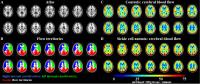
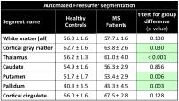
Magnetic Resonance Fingerprinting (MRF) is used to
simultaneously map T1, T2, and spin
density in the normal appearing white matter and normal
appearing grey matter of multiple sclerosis patients and
healthy controls. Relaxation parameters measured by MRF are
found to be significantly different between MS subjects and
healthy controls, to distinguish between relapsing remitting
MS and secondary progressive MS in certain brain structures,
and to correlate with clinical measures of function and
disability.
|
|
4071.
 |
44 |
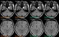
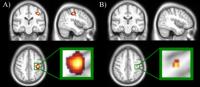
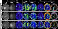
QSM is sensitive to myelin changes just beyond the boundaries of
conventional T2 lesion detection
Sneha Pandya1, Yan Zhang1, Thanh
Nguyen1, Yi Wang1, Susan A Gauthier2,
and Sneha Pandya1
1Department of Radiology, Weill Cornell Medicine,
New York, NY, United States, 2Department
of Neurology, Weill Cornell Medicine, New York, NY, United
States
MRI-derived measures of lesion accrual and tissue loss have
acquired a central role in the understanding of MS disease
evolution, pathogenesis of symptoms, and prediction of
clinical outcome. Conventional MRI imaging is highly
sensitive for detection of MS lesions, which are
characteristically hyperintense on a T2 weighted images,
however this technique lacks pathological specificity. QSM
can help identify myelin and iron content changes during an
MS lesion’s lifetime.
|
|
4072.
 |
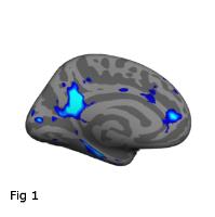
45 |
Influence of cognitive impairment and depression on cortical
thinning in patients with multiple sclerosis
Paola Valsasina1, Maria Assunta Rocca1,
Emanuele Pravatà1,2, Gianna Riccitelli1,
Giancarlo Comi3, Andrea Falini4, and
Massimo Filippi1
1Neuroimaging Research Unit, San Raffaele
Scientific Institute, Vita-Salute San Raffaele University,
Milan, Italy, 2Department
of Neuroradiology, Neurocenter of Southern Switzerland,
Lugano, Switzerland, 3Department
of Neurology, San Raffaele Scientific Institute, Vita-Salute
San Raffaele University, Milan, Italy, 4Department
of Neuroradiology, San Raffaele Scientific Institute,
Vita-Salute San Raffaele University, Milan, Italy
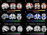
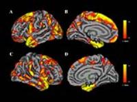
In this study, we investigated cortical thickness
abnormalities associated with cognitive impairment and
depression in 126 patients with multiple sclerosis (MS).
Compared with controls, MS patients exhibited a widespread
bilateral cortical thinning involving all brain lobes. While
cognitive impairment was associated with atrophy of regions
located in the fronto-parietal lobes (including the middle
and superior frontal gyrus, the inferior parietal lobule and
the precuneus), depression was linked to atrophy of the
orbitofrontal cortex. This study shows that cortical
thickness analysis was able to detect specific effects of
clinical symptoms on cortical atrophy in MS.
|
|
4073.
 |
46 |
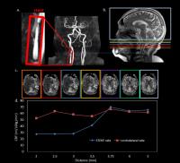
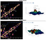
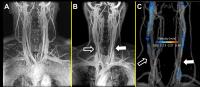
CE-MRV with concordant 4D flow MRI and ultrasound reveals no
internal jugular venous outflow obstruction in multiple
sclerosis
Eric Schrauben1, Sarah Kohn2, Samuel
Frost2, Oliver Wieben2,3, and Aaron
Field2
1Centre for Advanced MRI, University of Auckland,
Auckland, New Zealand, 2Radiology,
University of Wisconsin - Madison, Madison, WI, United
States, 3Medical
Physics, University of Wisconsin - Madison, Madison, WI,
United States
Contrast-enhanced MR venography scoring in internal jugular
veins is performed and compared with 4D flow MRI and
ultrasound assessment in patients with multiple sclerosis,
patients with other neurological disorders and healthy
controls. Narrowing assessment is shown to be more variable
in flow MRI and ultrasound.
|
|
4074.
 |
47 |
Multimodal Characterization of Grey Matter Alterations in
Neuromyelitis Optica - Video Not
Available
Yaou Liu1, Yunyun Duan2, Huiqing Dong2,
Tianyi Qian2, Frederik Barkhof3,
Jinhui Wang4, and Kuncheng Li2
1Xuanwu Hospital,Capital Medical University,
Beijing, China, People's Republic of, 2Beijing,
China, People's Republic of, 3Amsterdam,
Netherlands, 4Hanzhou,
China, People's Republic of
Combining
double-inversion-recovery (DIR), high-resolution structural
MRI, diffusion tensor imaging (DTI) and resting-state
functional MRI (rs-fMRI), this study systematically
investigated structural and functional alterations in
grey-matter (GM) structures in thirty-five neuromyelitis
optica (NMO) patients compared with healthy controls. We
demonstrated that NMO exhibits both structural and
functional alterations of GM in the cerebrum and cerebellum.
Multimodal MRI techniques complementary worked to capture
NMO-related GM abnormalities. GM alterations, especially
diffusion abnormalities, correlated with cognitive
impairment in NMO. These findings have important
implications for understanding the roles of GM damage and
also for highlighting multimodal MRI techniques as objective
biomarkers in NMO.
|
|
4075.
 |
48 |
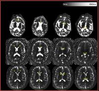
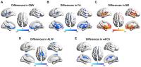
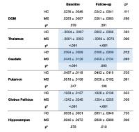
A longitudinal assessment of brain iron using quantitative
susceptibility mapping (QSM) in multiple sclerosis (MS) over 2
years
Ferdinand Schweser1,2, Nicola Bertolino1,
Michael G Dwyer1, Jesper Hagemeier1,
Paul Polak1, Niels P Bergsland1,3,
Andreas Deistung4, Bianca Weinstock-Guttman5,
Jürgen R Reichenbach4,6, and Robert Zivadinov1,2
1Buffalo Neuroimaging Analysis Center, Department
of Neurology, Jacobs School of Medicine and Biomedical
Sciences, The State University of New York at Buffalo,
Buffalo, NY, United States, 2MRI
Molecular and Translational Research Center, Jacobs School
of Medicine and Biomedical Sciences, The State University of
New York at Buffalo, Buffalo, NY, United States, 3MR
Research Laboratory, IRCCS Don Gnocchi Foundation ONLUS,
Milan, Italy, 4Medical
Physics Group, Department of Diagnostic and Interventional
Radiology, Jena University Hospital - Friedrich Schiller
University Jena, Jena, Germany,5Department of
Neurology, Jacobs School of Medicine and Biomedical
Sciences, The State University of New York at Buffalo,
Buffalo, NY, United States, 6Michael
Stifel Center for Data-driven and Simulation Science Jena,
Friedrich Schiller University Jena, Jena, Germany
Quantitative susceptibility mapping (QSM) is the most
sensitive technique currently available to study brain iron in
vivo. The
technique opens the door to a longitudinal
assessment of brain iron, bearing the potential to
understand and disentangle factors resulting in the large
scatter of reported iron concentrations in later decades of
life. In the present work, we investigated longitudinal
changes of brain magnetic susceptibility in a cohort of 40
healthy controls (HCs) and 160 multiple sclerosis (MS)
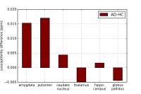
patients over a period of 2 years. |
|