|
Exhibition Hall 11:30 - 12:30 |
|
|
|
Computer # |
|
4316.
 |
49 |
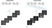
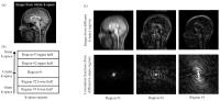
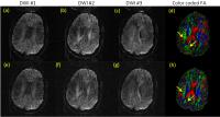
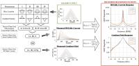
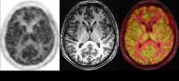
A combinatorial model approach for feature selection from
multimodal MRI data 
Xiaowei Zhuang1, Virendra Mishra1,
Karthik Sreenivasan1, Charles Bernick1,
Sarah Banks1, and Dietmar Cordes1,2
1Cleveland Clinic Lou Ruvo Center for Brain
Health, Las Vegas, NV, United States, 2Department
of Psychology and Neuroscience, University of Colorado
Boulder, Boulder, CO, United States
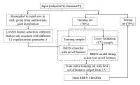
Clinical applications of brain abnormality detection with
supervised machine learning techniques are limited due to
less and unbalanced sample sizes as compared to rich feature
sets in patient population. We proposed a new combinatorial
model approach, fs-RBFN, involving sampling from
multivariate joint distribution, LASSO feature selection,
RBFN cross validation, and inverse probability weighting to
solve this problem. The proposed approach was validated
against a ground truth phantom and further tested on a
multimodal MRI dataset for cognitively impaired and
non-impaired professional fighters. Our results suggest
superior performance of this technique over several other
out-of-the-bag feature selection algorithms.
|
|
4317.
 |
50 |
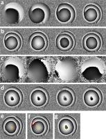
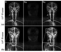
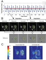
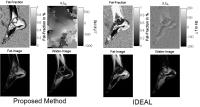
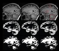
Three-dimensional lung tumour motion tracking using an advanced
template matching technique: Texture Reformatted Angle
Correlation (TRAC)
Kevin K. Zhang1,2, Shivani Kumar2,3,
Robba Rai3, Armia George3, Bin Dong1,4,
and Gary P. Liney1,2,3,4
1Ingham Institute for Applied Medical Research,
Sydney, Australia, 2South
Western Sydney Clinical School, University of New South
Wales, Sydney, Australia, 3Department
of Medical Physics, Cancer Therapy Centre, Liverpool
Hospital, Sydney, Australia, 4Centre
for Medical Radiation Physics (CMPR), University of
Wollongong, Sydney, Australia
Real-time lung tumour tracking and motion analysis is
important in MRI-based radiotherapy planning to inform
treatment margins and to permit accurate delivery for
developing MR-Linac technology. This work describes a
template matching approach to provide 3D motion assessment
of lung tumours from real-time 2D images. Compared to
previous work the TRAC technique utilises a multi-angled
correlation analysis of the target region to correctly
identify the tumour position. Results in both a moving
phantom and in lung cancer patients show that the technique
is feasible, accurate and can be easily adopted in widely
used single plane cine imaging.
|
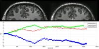
|
4318.
 |
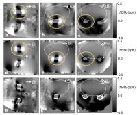
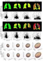
51 |
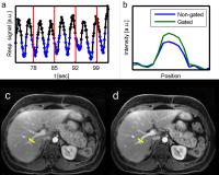
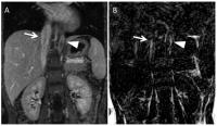
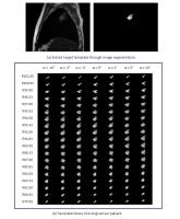
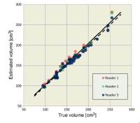
Renal segmentation from non-contrast T1-weighted MR images
Nicole Wake1, Jeremy C Lim2, Artem
Mikheev1, Jas-mine Seah3, Elissa
Botterill2, Shawna Farquharson4, Henry
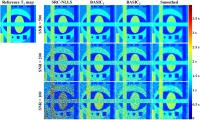
Rusinek1, and Ruth P Lim2,5
1Bernard and Irene Schwartz Center for Biomedical
Imaging, Center for Advanced Imaging Innovation and
Research, Department of Radiology, New York University
School of Medicine, New York, NY, United States, 2Department
of Radiology, Austin Health, Melbourne, Australia, 3Department
of Endocrinology, Austin Health, Melbourne, Australia, 4Florey
Neuroscience Institute, Melbourne, Australia,5The
University of Melbourne, Melbourne, Australia
A semi-automatic renal segmentation technique for
non-contrast T1-weighted MR images was developed. Renal
segmentation and volumetric analysis was tested in ten
healthy volunteers and ten Type I diabetic patients. We
found that this segmentation tool is fast, reliable, and
requires minimal user interaction. Upon further validation,
this method has clinical potential for monitoring renal
status in appropriate patient populations.
|
|
4319.
 |
52 |
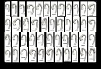
Exploring abnormal arch shape patterns using CMR-based
hierarchical 3D shape clustering: Application to a generic
imaging population of repaired coarctation of the aorta
Jan L Bruse1, Abbas Khushnood1,
Tain-Yen Hsia1, Andrew M Taylor1,
Vivek Muthurangu1, and Silvia Schievano1
1Centre for Cardiovascular Imaging, UCL Institute
of Cardiovascular Science & Great Ormond Street Hospital for
Children, London, United Kingdom
We present a novel method for hierarchical 3D shape
clustering of aortic arch shape models segmented and
reconstructed from CMR imaging data. We apply the method to
a cohort of 45 patients post aortic coarctation repair in
order to explore previously unknown arch shape patterns that
may relate to clinical outcome. Exploring a pathologic shape
population using data mining and statistical shape modeling
techniques can provide novel insight for improved diagnosis
and treatment strategies and can thereby assisst in clinical
decision making when analysing complex cases.
|
|
4320.
 |
53 |
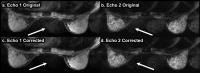
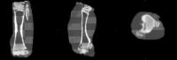
Direct CT conversion from a single ultra-short echo sequence
Soumya Ghose1, Jason Dowling1, Robba
Rai2, Benjamin Schmitt3, and Gary
Liney2,4,5,6
1eHealth, CSIRO, Brisbane, Australia, 2Liverpool
Cancer Therapy Centre, Liverpool, Australia, 3Siemens
Healthcare Pty Ltd, Macquarie Park, Australia, 4Medical
Physics, Ingham Institute, Liverpool, Australia, 5UNSW
Australia, Liverpool, Australia, 6University
of Wollongong, Wollongong, Australia
Accurate conversion of MRI into attenuation correction maps
is of current interest in PET-MR and MR-only radiotherapy
planning in particular, where electron density calculation
is particularly demanding and usually derived from CT. MRI
methods to date have usually involved building a patient
atlas and/or use of multiple imaging sequences and are time
intensive. We propose a new single sequence approach based
on ultra-short echo time to identify tissue classes of air,
bone and soft-tissue in combination with a dynamic
clustering regression based model that provides a direct CT
conversion which is both efficient and accurate.
|
|
4321.
 |
54 |
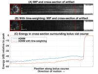
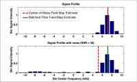
Non-invasive estimation of arterial input function for imaging
of cerebral blood flow on a PET/MR scanner
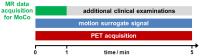
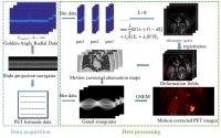
Mohammad Mehdi Khalighi1, Audrey Peiwen Fan2,
Gaspar Delso3, Praveen K. Gulaka2, Bin
Shen4, Aileen Hoehne4, Prachi Singh2,
Jun-Hyung Park4, Dawn Holley2,
Frederick T. Chin2,4, and Greg Zaharchuk2,4
1Applied Science Lab, GE Healthcare, Menlo Park,
CA, United States, 2Radiology
Department, Stanford University, Stanford, CA, United
States, 3Applied
Science Lab, GE Healthcare, Zurich, Switzerland,4Molecular
Imaging Program, Stanford University, Stanford, CA, United
States
Accurate measurement of Arterial Input Function (AIF) is
essential in quantitative analysis of cerebral blood flow
(CBF) using 15O-H2O
PET imaging. The time-of-flight enabled Signa PET/MR scanner
(GE Healthcare, Waukesha, WI, USA) provides quality PET
images during the arrival of 15O-H2O
tracer to the brain arteries, which can be used for carotid
artery segmentation. The optimal time frame to segment these
brain arteries for image-based AIF, is found by binning the
PET list file every second and plotting the total number of
true and scatter coincident events over time.
|
|
4322.
 |
55 |
Partial volume correction of quantitative susceptibility maps
for oxygen extraction fraction measurements.
Phillip G. D. Ward1,2, Audrey P. Fan3,
Parnesh Raniga1, David G. Barnes2,4,
David L. Dowe2, and Gary F. Egan1,5
1Monash Biomedical Imaging, Monash University,
Clayton, Australia, 2Faculty
of Information Technology, Monash University, Clayton,
Australia, 3Lucas
Center for Imaging, Department of Radiology, Stanford
University, Stanford, CA, United States, 4Monash
eResearch Centre, Monash University, Clayton, Australia, 5ARC
Centre of Excellence for Integrative Brain Function,
Melbourne, Australia
Partial volume effects impede the use of quantitative
susceptibility maps for assessing small veins. Oxygen
extraction fraction measures are particularly sensitive to
these effects. We propose a geometric technique for
calculating partial volume from binary venograms. The
technique is able to calculate accurate partial volume maps,
and vessel geometry, on simulated veins of sub-voxel radius.
These partial volume maps are used to adjust for partial
volume effects in estimating venous magnetic susceptibility.
|
|
4323.
 |
56 |
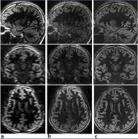
Improving Tissue Segmentation of Brain MRI through
Sparsity-guided Super-resolution Imaging
Jean-Christophe Brisset1, Louise E Pape1,
Ricardo Otazo1, and Yulin Ge1
1Radiology, New York University School of
Medicine, New York, NY, United States
Since human gray matter cortex is a relatively thin
structure and has a complex folding pattern blended with
white matter and cerebrospinal fluid (CSF), partial volume
effect is always considered a challenging issue for precise
tissue segmentation. Super-resolution (SR) is a common
method that is often used in the picture world to recover a
high-resolution image from low-resolution images. This study
was performed to test whether a newly developed
sparsity-guided SR algorithm can be adapted on standard
clinical MRI images to improve brain tissue segmentation by
decreasing partial volume effect.
|
|
4324.
 |
57 |
Data and cluster-extent based thresholding to analyze
statistical parametric maps in the study of knee articular
cartilage biochemical composition.
Allison B Randolph V1, Valentina Pedoia1,
Lorenzo Nardo1, and Sharmila Majumdar1
1Radiology & Biomedical Imaging, UCSF, San
Francisco, CA, United States
Voxel-based relaxometry (VBR) allows for MR relaxtion time
analysis without the sometimes deletorious assumtions of
traditional ROIs. However, VBR introduces potentially new
analysis issues, such as noise and map heterogeneity. In
this study we propose to use VBR significance thresholding
in conjunction with cluster-extent based thresholding to
define data-driven regions of interest (ROIs) that include
the most critical information in Statistical Parametric Maps
(SPM), controlling the aforesaid issues. The results
suggests that the data driven voxel cluster ROIs and
predefined traditional ROIs have unique, separate anatomical
locations, and that the data-driven clusters perform better
when correlated to osteoarthritis (OA) disease markers.
|
|
4325.
 |
58 |
Automatic organ-specific localization and quantification of fat
in abdominal chemical shift encoding-based water-fat MRI:
application to weight-loss in obesity
Jun Shen1, Thomas Baum2, Christian
Cordes2, Beate Ott3, Claudia Eichhorn3,
Thomas Skurk3,4, Hendrik Kooijman5,
Ernst J Rummeny2, Hans Hauner3,4,
Bjoern H Menze1, and Dimitrios C Karampinos2
1Department of Computer Science, TU Munich,
Munich, Germany, 2Department
of Radiology, TU Munich, Munich, Germany, 3Else
Kröner Fresenius Center for Nutritional Medicine, TU Munich,
Munich, Germany, 4ZIEL
Research Center for Nutrition and Food Sciences, TU Munich,
Munich, Germany, 5Philips
Healthcare, Hamburg, Germany
The accumulation and regional distribution of abdominal
adipose tissue and organ fat plays an important role in
several diseases including obesity, metabolic syndrome and
diabetes. The present work proposes a fully automatic method
for abdominal organ segmentation and adipose tissue
classification and measurement based on chemical shift
encoding-based water-fat MR images. The results from the
automatic method showed very good agreement with the
manually created references. The developed automatic
algorithm allowed the detection of regional differences in
changes of adipose tissue depots in a study of 20 obese
women undergoing a calorie restriction intervention.
|
|
4326.
 |
59 |
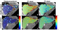
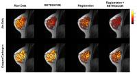
Semi-Automatic Comparison of Myocardial Tissue Injury using a
Non-Rigid Registration Method in patients with non-ischemic
disease
Leili Riazy1, Simone Fritzschi2,3,
Arthur Stötzner2,3, Fabian Mühlberg2,3,
Luisa Schmacht2,3, Matthias Dieringer1,2,4,
Florian von Knobelsdorff-Brenkenhoff2,3, Thoralf
Niendorf1,2, and Jeanette Schulz-Menger2,3
1Berlin Ultrahigh Field Facility (B.U.F.F.),
Max-Delbrueck Center for Molecular Medicine, Berlin,
Germany, 2Working
Group on Cardiovascular Magnetic Resonance, Experimental and
Clinical Research Center (ECRC), Berlin, Germany, 3Department
of Cardiology and Nephrology, HELIOS Klinikum Berlin Buch,
Berlin, Germany, 4Siemens
Healthcare GmbH, Erlangen, Germany
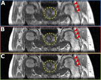
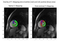
Late Gadolinium Enhancement (LGE) is the noninvasive gold
standard for focal fibrosis, parametric mapping with and
without contrast-media enable detection of diffuse fibrosis.
We developed a non-rigid registration method to superimpose
LGE images and T1-Maps allowing for pixel-wise comparison of
LGE extent and abnormal T1 times. We observed significantly
larger regions of ECV, T1 native and post-contrast
abnormalities than LGE positive areas. However, LGE was not
always completely covered by abnormalities of any of the
mentioned parameters.
|
|
4327.
 |
60 |
MRI-guided PET image denoising using a non-local means filter
Marie Anne Richard1, Réjean Lebel1,
Jérémie P. Fouquet1, and Martin Lepage1
1Centre d'imagerie moléculaire de Sherbrooke,
Université de Shebrooke, Sherbrooke, QC, Canada
Positron emission tomography (PET) images suffer from
statistical noise, especially in short time frames. For
applications requiring high temporal resolution, such as
dynamic studies, efficient edge-preserving denoising
algorithms such as the non-local means filter (NLMF) are
needed. Because this filter relies on structural data,
coregistered MR images were used to guide the NLMF. This
novel method was compared to conventional PET-guided NLMF
and proved superior in terms of increased
contrast-to-background ratio and signal-to-noise ratio in a
phantom model. It also increased small structure resolution
in a rat model.
|
|
4328.
 |
61 |
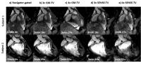
THOMAS: Thalamus Optimized Multi-Atlas Segmentation at 3T
Jason Su1, Thomas Tourdias2,
Manojkumar Saranathan3, Pejman Ghanouni4,
and Brian Rutt4
1Electrical Engineering, Stanford University,
Stanford, CA, United States, 2Neuroradiology,
Bordeaux University Hospital, Bordeaux, France, 3Radiology,
University of Arizona, Tucson, AZ, United States,4Radiology,
Stanford University, Stanford, CA, United States
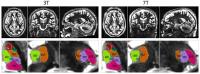
The efficacy of the Thalamus Optimized Multi-Atlas
Segmentation (THOMAS) algorithm for segmentation of thalamic
nuclei with white-matter-nulled MP-RAGE images is studied in
3T and 7T variants of the image contrast. 5 subjects are
evaluated at both field strengths and ground truth manual
delineations of nuclei are performed on the 7T images. We
demonstrate that the algorithm performs as well on 3T images
as on 7T within a dice coefficient of ±0.1 as evaluated
against the ground truth. This indicates that THOMAS can now
reach a much wider audience of interested groups.
|
|
4329.
 |
62 |
Evaluation of feature-driven clustering of dynamic contrast
enhanced and oxygen enhanced MRI data to assess tumour
microenvironment heterogeneity
Adam K Featherstone1,2, James P B O'Connor2,3,
Ross A Little1, Yvonne Watson1, Sue
Cheung1, Kaye J Williams2,4, Julian C
Matthews1,2, and Geoff J M Parker1,2,5
1Centre for Imaging Sciences, The University of
Manchester, Manchester, United Kingdom, 2CRUK
& EPSRC Cancer Imaging Centre in Cambridge and Manchester,
Cambridge and Manchester, United Kingdom, 3Institute
of Cancer Sciences, The University of Manchester,
Manchester, United Kingdom, 4School
of Pharmacy, The University of Manchester, Manchester,
United Kingdom, 5Bioxydyn
Ltd., Manchester, United Kingdom
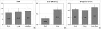
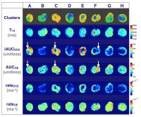
DCE-MRI and OE-MRI scans were performed on 8 preclinical U87
tumour xenografts. Heuristic features (area-under-curve and
rate-of-enhancement) were calculated from tumour voxel
enhancement curves for each imaging modality. Clustering
algorithms (k-means clustering and Gaussian mixture
modelling) were applied to these features and native tissue
T 1 to
investigate their utility in characterising physiological
heterogeneity in tumours. Efficacy in identifying large
regions where there is agreement between features is shown.
Further optimisation is needed to optimise the approach to
characterise smaller, and potentially important, regions
where there is a lack of concordance between features.
|
|
4330.
 |
63 |
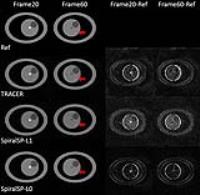
Automatic sodium maps reconstruction using PatchMatch algorithm
for phantom detection
Ferran Prados1,2, Bhavana S Solanky2,
Patricia Alves Da Mota2, Manuel Jorge Cardoso1,
Wallace J Brownlee2, Niamh Cawley2,
David H Miller2, Xavier Golay3,
Sebastien Ourselin1, and Claudia Angela Michela
Gandini Wheeler-Kingshott2,4
1Translational Imaging Group, Medical Physics and
Biomedical Engineering, University College London, London,
United Kingdom, 2NMR
Research Unit, Queen Square MS Centre, Department of
Neuroinflammation, UCL Institute of Neurology, University
College London, London, United Kingdom, 3Brain
Repair & Rehabilitation, UCL Institute of Neurology,
University College London, London, United Kingdom, 4Brain
Connectivity Center, C. Mondino National Neurological
Institute, Pavia, Italy
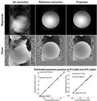
Quantitative sodium magnetic resonance imaging (23Na-MRI)
enables the non-invasive measurement of in vivo total 23Na
concentration (TSC) in the human brain. This involves a
complex process of reconstructing datasets acquired to
calculate a TSC map. Quantitative TSC map calibration relies
on external reference phantoms with known concentration for
linear calibration. This commonly involves manually
segmenting the phantoms by trained raters, hindering
automatic image analysis, and presenting a bottleneck in the
TSC computation. We propose to substitute the manual
segmentation by OPAL, a novel, fast, robust and reliable
technique for segmenting sodium phantoms that allows
fully-automatic reconstruction of TSC maps.
|
|
4331.
 |
64 |
Dynamic Whole-Brain Connectivity underlying Abnormal Brain
States in Late-onset Depression
Mingze Xu1,2, Shiyang Chen2, Bing Ji2,3,
Jiuquan Zhang4, Huaiqiu Zhu1, Yi Zhang5,
Yonggui Yuan6, Jiahong Gao1, Yijun Liu1,
and Xiaoping Hu2
1Biomedical Engineering, Peking University,
Beijing, China, People's Republic of, 2Biomedical
Engineering, Emory University & Georgia Institute of
Technology, Atlanta, GA, United States, 3University
of Shanghai for Science & Technology, Shanghai, China,
People's Republic of, 4Department
of Radiology, Southwest Hospital, Third Military Medical
University, Chongqing, China, People's Republic of,5School
of Life Science and Technology, Xidian University, Shaanxi,
China, People's Republic of, 6Department
of Psychosomatics and Psychiatry, ZhongDa Hospital, School
of Medicine, Southeast University, Nanjing, China, People's
Republic of
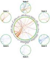
We conducted dynamic whole-brain connectivity analysis in
Late-onset depression (LOD) to investigate alterations in
brain networks. All subjects’ ROI-to-ROI dynamic FC were
explored using a data-driven method to obtain the most
explanatory states. Each state indicate a particular
ROI-to-ROI FC pattern. The property of each state were
determined based on its scores across time. Besides
decreased FC in normal state, we found LOD patients switch
between brain states more frequently and tend to enter
LOD-risk states, due to and its high states variance and
dominating increased FC in LOD-risk states. These results
suggest neural mechanisms of disorder from dynamic
perspective.
|
|
4332.
 |
65 |
Application of Partial Least Squares regression for Fast and
Robust Dictionary Matching for Magnetic Resonance Fingerprinting
Shivaprasad Ashok Chikop1, Vimal Chandran2,
Imam Shaik1, Rashmi Rao1, Mauricio
Antonio Reyes Aguirre2, and Sairam Geethanath1
1Medical Imaging Research Center, Dayananda Sagar
Institutions, Bangalore, India, 2Institute
of Surgical Technology and Biomechanics, University of Bern,
Bern, Switzerland
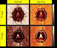
The step size of the parameters used for simulation of
dictionary determines the parameters being determined.
Partial Least squares (PLS) can be used as a general frame
work for fast and robust dictionary matching. Regression
co-efficient matrix obtained from PLS can be used for
localizing the different brain tissue types thus avoiding
iterative searching. The increase in contrast between the
grey matter and white matter can be attributed to the
intermediate values generated by PLS based matching. PLS
matches comparatively better at low SNR images compared to
the straight forward dot product method.
|
|
4333.
 |
66 |
Cluster Analysis of Dynamic Contrast-Enhanced MRI
Pharmacokinetic Parameters for Prostate Cancer Risk
Stratification: a Step towards Practical Translation
Saba N Elias1, Guang Jia2, Firas G
Petros3, Huyen Nguyen1, Debra L Zynger4,
Zarine K Shah5, Ronney Abaza6, and
Michael V Knopp1
1Radiology/Wright Center of Innovation, The Ohio
State University, Columbus, OH, United States, 2Department
of Physics & Astronomy, Louisiana State University, Baton
Rouge, LA, United States,3Urology, The Ohio State
University, Columbus, OH, United States, 4Pathology,
The Ohio State University, Columbus, OH, United States, 5Radiology,
The Ohio State University, Columbus, OH, United States, 6Robotic
Urologic Surgery, OhioHealth Dublin Methodist Hospital,
Dublin, OH, United States
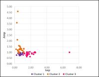
Feasibility of classifying PCa into clusters based on
microcirculatory features has the potential to predict
outcome and assist in
therapeutic treatment of PCa.
|
|
4334.
 |
67 |
Pulmonary Imaging Biomarkers of COPD for Personalized Treatment
and Better Outcomes
Dante PI Capaldi1, Anthony Lausch2,
Khadija Sheikh1, Fumin Guo1, David G
McCormack3, and Grace Parraga1
1Robarts Research Institute, The University of
Western Ontario, London, ON, Canada, 2Credit
Valley Hospital, Mississauga, ON, Canada, 3Department
of Medicine, The University of Western Ontario, London, ON,
Canada
In this proof-of-concept demonstration, we developed and
generated multimodal-parametric-response-mapping (mPRM) from
CT and MRI pulmonary measurements to phenotype chronic
obstructive pulmonary disease (COPD). We performed
principal component analysis of the voxel distribution
generated from co-registered inspiration or expiratory CT
with 3He MRI SV cluster maps and 3He
MRI ADC maps for ex-smokers with and without COPD. Further
work is necessary to determine the appropriate combination
of imaging biomarkers generated from MRI and CT to provide
useful information in deeply phenotyping COPD.
|
|
4335.
 |
68 |
Brain Connectivity Analysis of Parkinson's Disease and “Scans
Without Evidence for Dopaminergic Deficit" Patients
Tiago Constantino1,2,3, André Santos Ribeiro4,
Ricardo Maximiano3, John Mcgonigle4,
David Nutt4, and Hugo Alexandre Ferreira3
1Lisbon School of Health Technology-ESTeSL,
Lisbon, Portugal, 2Spitalzentrum
Biel, Biel, Switzerland, 3Institute
of Biophysics and Biomedical Engineering, Faculty of
Sciences of the University of Lisbon, Lisbon, Portugal, 4Centre
for Neuropsychopharmacology, Imperial College London,
London, United Kingdom
In this work we propose a comparison study between “Scans
Without Evidence for Dopaminergic Deficit" (SWEDD) and
Parkinson’s Disease (PD) patients against healthy subjects
using the MIBCA toolbox. Here, we studied the difference in
imaging and connectivity metrics obtained from anatomical
(T1-weighted) and structural (Diffusion Tensor Imaging) data
between the three groups. Results showed increased mean
diffusivity in the frontal pole, rostral middle frontal
gyrus and superior frontal gyrus between SWEDD and PD
patients, which can be related with the dopaminergic
mesocortical pathway degeneration in PD. These preliminary
results help clarify the differences between SWEDD and PD
patients.
|
|
4336.
 |
69 |
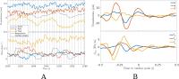
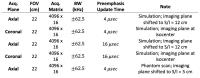
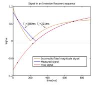
Resolving ambiguity in T1 mapping using complex MRI data
Kees M. van Hespen1, Dirk H.J. Poot1,2,
Harm A. Nieuwstadt1, and Stefan Klein1
1Departments of Medical Informatics and
Radiology, Erasmus MC, Rotterdam, Netherlands, 2Imaging
Science and Technology, Delft University of Technology,
Delft, Netherlands
We have recently developed an optimized T1 mapping
protocol for carotid atherosclerotic plaque imaging, using a
combination of inversion and recovery prepared acquisitions.
This protocol requires less images to be taken (and thus
shorter acquisition time) for precise T1 estimation
than conventional inversion-prepared or saturation-prepared
acquisition schemes. However, estimating T1 from
magnitude data, acquired with the optimized settings, causes
bimodality of T1 estimates,
due to the ambiguity in sign of the inversion prepared
magnitude images. Simulations and experiments on a hardware
phantom and a volunteer show that the ambiguity resolves
when we fit a complex-valued model to the complex data.
|
|
4337.
 |
70 |
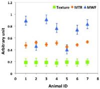
Comparing MRI texture heterogeneity with MTR and myelin water
fraction as measures of myelin integrity
Tim Luo1, Shrushrita Sharma2, Mark
Polivchuk3, Peng Zhai4, and Yunyan
Zhang4
1Bachelor of Health Sciences, University of
Calgary, Calgary, AB, Canada, 2Biomedical
Engineering Program, University of Calgary, Calgary, AB,
Canada, 3Computer
Science, University of Calgary, Calgary, AB, Canada, 4Radiology
and Clinical Neurosciences, University of Calgary, Calgary,
AB, Canada
Changes in myelin integrity are associated with many
neurological diseases. We acquired 9.4T MRI from healthy
mouse brain to evaluate the utility of texture heterogeneity
in T2-weighted MRI for assessing myelin integrity, in
comparing with proposed measures including magnetic transfer
ratio and myelin water fraction. Measurements were focused
on the corpus callosum, with both anatomical (genu, body,
splenium) and hemispheric (left, center, right) locations
evaluated. All 3 methods showed the uniformity of myelin in
corpus callosum between hemispheres, and no significant
differences between anatomical locations were detected.
Texture heterogeneity showed the best consistency between
animals and deserves further verification.
|
|
4338.
 |
71 |
Color mapping in medical imaging - you're (probably) doing it
wrong
Jan-Gerd Tenberge1
1University of Münster, Münster, Germany
Some imaging software packages do not accurately display
datasets due to difficulties in color mapping. We show some
of the shortcomings an three of the most widely used tools
(FSL, SPM, FreeSurfer) and provide an easy fix that can be
applied to correct the images output by these tools.
|
|
4339.
 |
72 |
T1 Mapping through Bayesian Analysis with Spatial Information
Collaboration (BASIC) using Steady-State-Based Imaging Data -
Permission Withheld
Mustapha Bouhrara1 and
Richard G. Spencer1
1NIA, NIH, Baltimore, MD, United States
We introduce two Bayesian-based analyses that use spatial
information as a prior to improve the quality of
voxel-by-voxel T1-mapping from spoiled
gradient recalled echo (SPGR) imaging data. These
approaches, called BASIC, combine voxel-by-voxel fitting
with region-of-interest (ROI) parameter estimation. ROI
parameters act as a constraint, while voxel fitting
mitigates blurring and detail loss. The results were
compared with those derived using a conventional nonlinear
least-squares-based algorithm. Estimation of T1 from
SPGR imaging data was markedly improved through use of the
BASIC methods.
|
|